The Blog

Building a Future Free of Age-Related Disease

Cancerous Tumor

The Fibroblasts That Protect Ovarian Cancer

In Aging, researchers have described a subpopulation of fibroblasts that nurture ovarian cancer tumors and shield them from harm.

Cancer’s natural defenders

Ovarian cancer is particularly dangerous, both in death rates and in recurrence [1]. Normally, chemicals that contain platinum are effective, but this sort of cancer develops resistance to such treatments; after that, prognoses are usually very poor, even with modern drugs [2].

This cancer’s tumors, however, are not just made up of cancer cells: cancer-associated fibroblasts (CAFs) support its progression [3] and that of other cancers. These cells are in constant intercellular communication with the cancer, sending a steady stream of signals back and forth [4] and remodeling the extracellular matrix (ECM) around the tumor, which makes drug delivery uniquely difficult [5]. Even though the tumor microenvironment is highly inflammatory, which can be used to trigger immunotherapies [6], one particuar compound in these tumors, biglycan, impedes the effectiveness of such therapies [4].

CAFs are heterogenous, coming from multiple sources. Most of them began as resting fibroblasts, but others started out as muscle cells or structural cells of the blood vessels or other organs [7]. These cells do not even share the same differentiation, as some of them have stem cell origins [8].

Prior work has made efforts to describe these subpopulations of cells in order to more precisely target them [9]. Such categorizations include their origins, their development, their inflammatory status and antigens, and how they can be targeted in therapies.

dCAF is not good for you

After finding appropriate biomarkers for identification, this study divided fibroblasts into three of these categories: myofibroblastic (myCAFs), inflammatory (iCAFs), and desmoplastic (dCAFs), the last of which has been associated with poor prognoses. These three different types have very distinct clusters, both in gene expression and in physical distribution.

As expected, myCAFs express genes related to muscle function and iCAFs express inflammatory factors, while dCAFs express collagen-forming genes. While the iCAFs, as expected, communicated quite a bit, predominantly through their inflammatory factors, it was the dCAFs that communicated the most, using the ECM to do so.

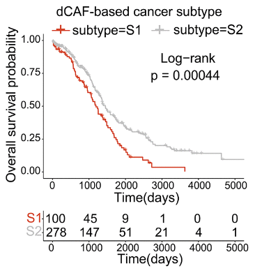
Low-dCAF cancers were found to be far less dangerous than high-dCAF cancers. Genes associated with platinum resistance were much more upregulated in the high-dCAF group, and they showed signs of being resistant to multiple other drugs. As dCAF is correlated with collagen production, it is no surprise that the high-dCAF group’s tumors were richer in collagen, and therefore stiffer and tougher, than the low-dCAF group’s.

The researchers were able to use gene expression analysis to construct a prognostic test through a machine learning algorithm. Patients who had more dCAF genes expressed were, indeed, more likely to die of cancer under modern treatment conditions.

dCAF Survival

While this paper’s authors did not get into how dCAFs could be specifically targeted and eliminated, they hold that this is a promising therapeutic strategy. If these very particular cells are found to be vulnerable to a novel therapy, the prognosis of people with ovarian cancer or many other cancers may significantly improve.

We would like to ask you a small favor. We are a non-profit foundation, and unlike some other organizations, we have no shareholders and no products to sell you. All our news and educational content is free for everyone to read, but it does mean that we rely on the help of people like you. Every contribution, no matter if it’s big or small, supports independent journalism and sustains our future.

Literature

[1] Siegel, R. L., Miller, K. D., Wagle, N. S., & Jemal, A. (2023). Cancer statistics, 2023. CA: a cancer journal for clinicians, 73(1), 17-48.

[2] Gaillard, S., Oaknin, A., Ray-Coquard, I., Vergote, I., Scambia, G., Colombo, N., … & Lorusso, D. (2021). Lurbinectedin versus pegylated liposomal doxorubicin or topotecan in patients with platinum-resistant ovarian cancer: a multicenter, randomized, controlled, open-label phase 3 study (CORAIL). Gynecologic oncology, 163(2), 237-245.

[3] Rimal, R., Desai, P., Daware, R., Hosseinnejad, A., Prakash, J., Lammers, T., & Singh, S. (2022). Cancer-associated fibroblasts: Origin, function, imaging, and therapeutic targeting. Advanced drug delivery reviews, 189, 114504.

[4] Zheng, S., Liang, J. Y., Tang, Y., Xie, J., Zou, Y., Yang, A., … & Lin, Y. (2023). Dissecting the role of cancer‐associated fibroblast‐derived biglycan as a potential therapeutic target in immunotherapy resistance: A tumor bulk and single‐cell transcriptomic study. Clinical and Translational Medicine, 13(2), e1189.

[5] Guo, J., Zeng, H., & Chen, Y. (2020). Emerging nano drug delivery systems targeting cancer-associated fibroblasts for improved antitumor effect and tumor drug penetration. Molecular pharmaceutics, 17(4), 1028-1048.

[6] Feig, C., Jones, J. O., Kraman, M., Wells, R. J., Deonarine, A., Chan, D. S., … & Fearon, D. T. (2013). Targeting CXCL12 from FAP-expressing carcinoma-associated fibroblasts synergizes with anti–PD-L1 immunotherapy in pancreatic cancer. Proceedings of the National Academy of Sciences, 110(50), 20212-20217.

[7] Huang, J., Tsang, W. Y., Li, Z. H., & Guan, X. Y. (2023). The origin, differentiation, and functions of cancer-associated fibroblasts in gastrointestinal cancer. Cellular and Molecular Gastroenterology and Hepatology.

[8] Kanzaki, R., & Pietras, K. (2020). Heterogeneity of cancer‐associated fibroblasts: opportunities for precision medicine. Cancer science, 111(8), 2708-2717.

[9] Öhlund, D., Handly-Santana, A., Biffi, G., Elyada, E., Almeida, A. S., Ponz-Sarvise, M., … & Tuveson, D. A. (2017). Distinct populations of inflammatory fibroblasts and myofibroblasts in pancreatic cancer. Journal of Experimental Medicine, 214(3), 579-596.

Neuronal connections

Neuronal Reprogramming Alleviates Alzheimer’s in Mice

Scientists have shown that long-term intermittent reprogramming limited to hippocampal neurons increases their fitness and improves cognitive function in a mouse model of Alzheimer’s disease [1].

Targeted in time and space

Partial cellular reprogramming is one of the hottest directions in longevity research for a reason: it allows to rejuvenate cells without driving them all the way to pluripotency, where they lose their identity. One of the ways that partial reprogramming can be achieved is through intermittent administration (“pulsing”) of reprogramming factors. This approach has produced increased lifespan and healthspan in various animal models [2].

Modern technologies now allow reprogramming to be limited not only by time but also by specific organs and cell types. For example, David Sinclair’s group demonstrated that partial reprogramming of retinal ganglion cells could restore vision after optic nerve injury [3]. However, the nervous system remains underexplored in this context. A new study by scientists at the University of Barcelona, published in Cell, aims to close this gap.

Reprogramming in utero

The researchers began by investigating the effects of partial cellular reprogramming on brain development by administering OSKM to pregnant mice and limiting the expression to the nervous system.

The offspring of treated mice developed significantly larger brains, up to double the normal weight. To refine their approach, the researchers adjusted the protocol by using a lower dose of the inducing compound (doxycycline), which allowed them to preserve brain morphology and survival, even though the mice’s brains were still larger than those of the control group.

Professor del Toro, a leading author on the study, explains that “when Yamanaka’s factors are introduced during the developmental phase, more neurons are generated, and the brain is more voluminous. This translates into better motor and social activity in the adult stages.”

“These results,” he adds, “are explained by the fact that we made it possible for all brain cells to express these factors, including stem cells. It was very surprising to discover that, if we control the expression of these factors very precisely, we can also control the process of cell proliferation and obtain brains with a larger cerebral cortex without losing the correct structure and functions.”

Fitter neurons, better cognition

However, the main goal was to assess whether partial reprogramming in neurons could alleviate neurodegeneration. The researchers created a mouse model of Alzheimer’s disease with the ability to conditionally express Yamanaka factors in hippocampal neurons.

From 12 to 35 weeks of age, these mice followed an intermittent reprogramming protocol, with factor expression activated for three days each week. At eight months, a stage when this strain of mice typically displays severe Alzheimer’s-like symptoms, the researchers conducted behavioral, histological, and molecular tests.

They found that hippocampal neurons in the treated mice were healthier, with better dendritic spines and synapses compared to controls. Remarkably, the size and number of amyloid beta plaques, a key hallmark of Alzheimer’s, were greatly reduced. However, other Alzheimer’s-related markers, such as neuroinflammation and stress responses, remained unchanged.

The epigenetic age of neurons was substantially lower in the treated mice compared to controls. Most importantly, the treatment improved some cognitive functions, including cognitive flexibility and spatial memory.

Making smarter babies?

Professor Albert Giralt, another leading author, explains: “In this case, we induced the expression of Yamanaka factors only in mature neurons. As these cells do not divide, their number does not increase, but we identified many markers that indicate a process of neuronal rejuvenation.”

He adds: “In these rejuvenated neurons, we detected that the number of synaptic connections increases, the altered metabolism is stabilized, and the epigenetic profile of the cell is also normalized. All these changes have a very positive effect on their functionality as neurons.”

This study, which includes renowned geroscientist Manuel Serrano among its authors, suggests that partial reprogramming could offer a viable preventative strategy against Alzheimer’s disease. The findings related to reprogramming during brain development are equally intriguing, hinting at the possibility of enhancing the cognitive abilities of offspring in utero.

In this study, we demonstrate that transient reprogramming with YFs not only safely increases neural proliferation during mouse brain development but also prevents the development of AD-related features in adulthood. The increased proliferation leads to more neurons and glial cells, expanding the cortex and improving behavioral performance. At adult stages, we found that principal neurons in the hippocampus tolerate transient YF expression for several months. Instead, the expression of YFs prevented the development of several AD-related hallmarks and ameliorated some of the cognitive deficits in the 5xFAD mouse model. These findings enhance our understanding of YFs as a tool to modulate neural proliferation and highlight their potential use in brain disorders.

We would like to ask you a small favor. We are a non-profit foundation, and unlike some other organizations, we have no shareholders and no products to sell you. All our news and educational content is free for everyone to read, but it does mean that we rely on the help of people like you. Every contribution, no matter if it’s big or small, supports independent journalism and sustains our future.

Literature

[1] Shen, Y. R., Zaballa, S., Bech, X., Sancho-Balsells, A., Rodríguez-Navarro, I., Cifuentes-Díaz, C., … & Del Toro, D. (2024). Expansion of the neocortex and protection from neurodegeneration by in vivo transient reprogramming. Cell Stem Cell.

[2] Macip, C. C., Hasan, R., Hoznek, V., Kim, J., Lu, Y. R., Metzger IV, L. E., … & Davidsohn, N. (2024). Gene therapy-mediated partial reprogramming extends lifespan and reverses age-related changes in aged mice. Cellular Reprogramming, 26(1), 24-32.

[3] Lu, Y., Brommer, B., Tian, X., Krishnan, A., Meer, M., Wang, C., … & Sinclair, D. A. (2020). Reprogramming to recover youthful epigenetic information and restore vision. Nature, 588(7836), 124-129.

Liver sepsis

A Senolytic Treatment for Liver Sepsis

A paper published in Aging Cell offers evidence for the idea that senolytics might be a treatment for acute liver sepsis.

A deadly condition linked to a hallmark of aging

Sepsis, an inflammatory condition most commonly known as blood poisoning, is the most common cause of mortality in intensive care units. The World Health Organization has made dealing with sepsis a priority [1]. Previous work has found that senescence is closely related to the suppression of the immune system [2], and immune dysregulation is the key feature of sepsis [3]. The liver is one of the first organs to fail during sepsis, and senescence has also been directly linked to liver diseases, including cirrhosis [4].

While previous research had directly linked liver senescence to sepsis [5], there was no treatment component involved. Here, the researchers decided to see if anything could be done about acute sepsis using senolytics, drugs that are normally only considered for long-term conditions such as age-related diseases.

Senolytics are effective in this model

A day after sepsis was induced in mice, the researchers noted an immediate increase in inflammatory factors associated with the SASP, including inflammatory cytokines and TNF-α, along with the key senescence marker SA-β-gal. There was also an increase in the senescence markers p53 and p21 but not p16. Many of these results were confirmed at both the protein and RNA levels. Overall, the researchers concluded that the phenotype of liver tissues quickly became one of senescence.

At the single-cell level, p21 was mostly expressed in the basic functional cells of the liver (hepatocytes), cells that line blood vessels (endothelial cells), and immune cells. While only 3% of the total cells were found to express p21 during sepsis, a full three-fifths of macrophages in the liver expressed it. While SASP markers were upregulated with RNA, a great many other genes were downregulated.

Administering the senolytic combination of dasatinib and quercetin before sepsis was induced had an immense effect on survival in these mice: within four days, all 20 of the mice that had induced sepsis were dead, while 6 of the 20 mice given the senolytic combination were still alive at day 10. The severity of sepsis in the treatment group was, as expected, dramatically decreased a day after it had been induced.

Senolytic sepsis survival and severity

SASP biomarkers, including cytokines, were also significantly reduced by the senolytic combination, as were many other biomarkers of senescence. ALT, the most well-known marker of liver damage, was also reduced by the treatment, as were markers of oxidative stress.

Senolytic sepsis biomarker effects

A question of treatment

Acute sepsis is a life-threatening condition for which it is difficult to generate a clinical trial, and it must be noted both that senolytics are often more effective on mice than people and that the senolytics in this case were administered before the sepsis was induced. However, these results are very strong, in both biomarker analysis and survival rates. If senescence occurs in the same way in people during sepsis as it does in mice, senolytics or other drugs that affect senescent cells may become a staple of the ICU.

We would like to ask you a small favor. We are a non-profit foundation, and unlike some other organizations, we have no shareholders and no products to sell you. All our news and educational content is free for everyone to read, but it does mean that we rely on the help of people like you. Every contribution, no matter if it’s big or small, supports independent journalism and sustains our future.

Literature

[1] Reinhart, K., Daniels, R., Kissoon, N., Machado, F. R., Schachter, R. D., & Finfer, S. (2017). Recognizing sepsis as a global health priority—a WHO resolution. New England Journal of Medicine, 377(5), 414-417.

[2] Monneret, G., Gossez, M., & Venet, F. (2021). Sepsis and immunosenescence: closely associated in a vicious circle. Aging Clinical and Experimental Research, 33, 729-732.

[3] Singer, M., Deutschman, C. S., Seymour, C. W., Shankar-Hari, M., Annane, D., Bauer, M., … & Angus, D. C. (2016). The third international consensus definitions for sepsis and septic shock (Sepsis-3). Jama, 315(8), 801-810.

[4] Sanfeliu-Redondo, D., Gibert-Ramos, A., & Gracia-Sancho, J. (2024). Cell senescence in liver diseases: pathological mechanism and theranostic opportunity. Nature Reviews Gastroenterology & Hepatology, 1-16.

[5] He, K., Zhou, D., Pu, Z., Chen, S., Shen, Y., Zhao, S., … & Xu, X. Cellular Senescence in Acute Liver Injury: What Happens to the Young Liver?. Aging and disease.

Skin collagen

HAPLN1 in Blood Found to Rejuvenate Skin

Investigating the factors involved in skin rejuvenation processes, researchers have identified the role of hyaluronan and proteoglycan link protein 1 (HAPLN1) in restoring collagen and hyaluronic acid in aged skin [1].

The most noticeable sign of aging

Skin aging, one of the most visible forms of aging, is influenced by both intrinsic and extrinsic factors. The extrinsic ones, such as sunlight exposure, air pollution, and cigarette smoke, are well known. However, the intrinsic ones, such as age-induced hormonal changes, are less well-researched and understood.

On a molecular level, some of the most prominent changes are to the skin’s extracellular matrix (ECM). The ECM is a structure built from such components as collagen, hyaluronic acid (HA), and proteoglycans. During aging, collagen and hyaluronic acid levels decrease. This decrease impacts the integrity of the ECM.

The claim of slowing down skin aging by promoting collagen synthesis is a common feature of cosmetics advertising. However, so far, “no effective agents for preventing or restoring aged skin have been identified.”

While reversing the signs of skin aging has aesthetic value, this study’s authors argue that their research on this topic can be a stepping stone to a better understanding of whole-body aging and therapies to address it.

Sharing circulation

To investigate skin aging, the researchers used a parabiosis experiment in which two animals are connected following surgery to share the bloodstream. In this experiment, the researchers connected the circulation of young and old animals, along with control groups of connected young animals and connected old animals.

The researchers examined the animals’ hyaluronic acid and collagen levels after three weeks after surgery. They observed that the levels of hyaluronic acid of old animals connected to young ones “were almost completely restored.” This treatment also increased newly synthesized collagen and the gene activity of some of the procollagen types in the deep skin (dermis) of old mice connected to the bloodstream of the young mice were also increased compared to the old mice.

They also tested the thickness of the dermis 10 weeks after surgery, observing improvement in dermal thickness. The dermis thickness of the old animals that shared the circulation system with the young ones was similar to that of the young control animals.

The authors suggested that blood-borne factors contribute to the restoration of the dermis in old animals. Therefore, they tested the differences in the composition of proteins in these animals’ plasma. After identifying several differences, they focused on one protein: HAPLN1.

HAPLN1 plasma and skin levels were decreased in aged animals, but parabiosis led to its increase. HAPLN1 plays a structural role in ECM architecture, as it connects two proteins, aggrecan and hyaluronic acid. It also plays a signaling role.

The authors note that this protein was not previously linked to aging. However, their investigation suggests a rejuvenating effect of HALPN1 and a possible role in depositing collagen and hyaluronic acid in aged skin.

Multiple molecular processes involved

As their next step, the researchers specifically tested HALPN1’s role in skin rejuvenation. Injection of recombinant HAPLN1 significantly increased newly synthesized collagen and total hyaluronic acid levels in skin tissues. They also observed an increase in versican, another molecule important in ECM remodeling. These results suggest that HAPLN1 has a positive role in reversing aging-induced changes, although the authors suggest future testing using different doses.

The authors also aimed to understand the molecular mechanisms behind these changes, so they treated human dermal fibroblasts with recombinant HAPLN1. The results of this experiment suggest the involvement of TGF-β in HAPLN1-induced collagen and hyaluronic acid restoration. The results also suggest the involvement of HAPLN1 in protecting “against oxidative stress-induced degradation of TGF-β R2.”

This participation in antioxidant pathways also prompted an investigation into intertwined pathways related to inflammation and senescence, and the results here suggest that HAPLN1 has possible effects against both. The authors summarize that HAPLN1 “could negatively regulate cellular senescence, suggesting its contribution to reversing skin aging that occurs during oxidative stress and the inflammatory process.”

The authors stress that this study is the first to discuss the role of HAPLN1 in skin rejuvenation processes. They also suggest that their findings can help improve anti-aging skin treatments. However, since this research has been done on mice and cell cultures, further testing on humans is still required.

We would like to ask you a small favor. We are a non-profit foundation, and unlike some other organizations, we have no shareholders and no products to sell you. All our news and educational content is free for everyone to read, but it does mean that we rely on the help of people like you. Every contribution, no matter if it’s big or small, supports independent journalism and sustains our future.

Literature

[1] Fu, Z., Yang, G., Yun, S. Y., Jang, J. M., Ha, H. C., Shin, I. C., Back, M. J., Piao, Y., & Kim, D. K. (2024). Hyaluronan and proteoglycan link protein 1 – a novel signaling molecule for rejuvenating aged skin. Matrix biology : journal of the International Society for Matrix Biology, S0945-053X(24)00111-2. Advance online publication.

Oxford Grant Award

Grant Awarded for Heart Aging at the University of Oxford

The Longevity Science Foundation (LSF), a nonprofit organization dedicated to funding research aimed at extending the healthy human lifespan, is proud to announce a grant award to researchers at the University of Oxford’s Department of Physiology, Anatomy, and Genetics, within the Medical Sciences Division, for the study “Metabolic Mechanisms of Ageing in the Heart – Novel Dietary Interventions for Enhancing Healthspan.” The research is led by Pawel Swietach, Professor of Physiology, and co-supervised by Richard Siow, Visiting Professor in the same Department at Oxford and Director of Ageing Research at King’s College London (ARK). The Foundation’s grant, which forms a critical part of the study’s overall budget, is being provided over a three-year period, beginning in 2024.

The research undertaken by Prof. Swietach’s team focuses on improving cardiac function to extend the human healthspan by studying the chemical modifications to molecules that regulate the heart. Since the heart’s ability to regenerate is limited, damage accumulates over time: sadly, about one in three deaths are related to heart disease. This study explores how metabolism, particularly changes in methylation related to diet, influences heart aging. By using genetically-altered experimental models, the team investigates how elevated methionine accelerates age-related heart dysfunction. They are testing whether diet changes can reverse these effects and studying its impact on cardiac function, metabolism, and methylation patterns to identify potential therapies. The LSF is funding critical aspects of the study which will advance the understanding of heart aging. The funding also provides training opportunities for students and fellows, fostering the next generation of leaders in aging and longevity research. As the LSF plays a crucial role in the success of this research project, we look forward to ongoing collaboration in developing a center of excellence in longevity research at the University of Oxford, as well as fostering additional partnerships at this global leading institution.

“We eagerly anticipate the outcomes of the research Pawel Swietach’s team will make to the scientific field,” said Joshua C. Herring, the President & CEO of LSF. “We are proud to have the Department of Physiology, Anatomy and Genetics at the University of Oxford as partners, and we look forward to the mutual impact we will create. As the Foundation seeks and sources substantial additional capital through donations, we plan to continue reverberating through the community via our granting to the best research teams available”

Pawel Swietach, Professor of Physiology and principal investigator says: “We are excited that we can implement an element of longevity research into our portfolio, thanks to support from the LSF. The research we plan is ambitious, innovative and important, but expensive to run. Every contribution to this effort makes a massive difference to us, and we hope it brings tangible benefits to the wider community.”

“This groundbreaking research will contribute to a better understanding of metabolic changes in the heart during aging and the mechanisms by which dietary interventions can prevent heart disease. We are grateful to the Longevity Science Foundation for enabling this groundbreaking research at the University of Oxford and look forward to contributing to the development of novel interventions to enhance healthy longevity,” said Dr. Richard Siow.

​​Press Contact

Lev Dvornik

press@longevity.foundation

About the Longevity Science Foundation

The Longevity Science Foundation (LSF) is a nonprofit organization advancing human longevity by funding research and development of medical technologies to extend the healthy human lifespan. The long-term mission of the Longevity Science Foundation is to help make longevity-focused care accessible to everyone, no matter their background, by bringing cutting-edge science on aging out of the laboratory and into the mainstream. To learn more, visit www.longevity.foundation.

About the Department of Physiology, Anatomy and Genetics, University of Oxford

The Department is home to internationally-renowned teams of scientists addressing major questions in biomedicine, the answers to which will have a profound effect on modern biology, working to provide outstanding opportunities for graduate students and postdoctoral workers to gain a research training of the very highest quality, which will enable them to become the leaders of tomorrow and to communicate and employ their research in the wider world. The University of Oxford has been ranked number one in the world by the Times Higher Education (THE) World University Rankings for nine consecutive years, from 2017 to 2025, and the Anatomy and Physiology at Oxford has been ranked #1 in the QS World University Rankings by subject from 2020 to 2024. To learn more, visit https://www.dpag.ox.ac.uk/

We would like to ask you a small favor. We are a non-profit foundation, and unlike some other organizations, we have no shareholders and no products to sell you. All our news and educational content is free for everyone to read, but it does mean that we rely on the help of people like you. Every contribution, no matter if it’s big or small, supports independent journalism and sustains our future.
Pancreas structure

Autodigestion From Gut Enzymes May Drive Aging

A new study links damage caused by digestive enzymes escaping from the gut to several hallmarks of aging. The researchers dubbed this effect “autodigestion.” [1]

Don’t eat yourself up!

To live, we must eat, which involves breaking down compounds that we ourselves consist of, such as fat and proteins. Evolution has developed ingenious ways to prevent our bodies from consuming themselves. Digestive enzymes are produced by the pancreas and are only activated in the gut. However, once activated, they hardly differentiate between ingested food and the body’s own biomolecules, and they happily chomp on both.

What prevents them from harming us is the protective mucosal barrier in the gut. Unfortunately, like any bodily system, this one gets dysregulated with age [2]. In a new study, scientists from the University of California San Diego investigated this process in depth and ended up proposing what they call “a previously unexplored mechanism of aging due to autodigestion.”

The rogue enzymes

In young rats, the researchers found low levels of the protein-breaking enzyme trypsin, although the tiny folds (villi) of the small intestine and the lungs contained more of this molecule. However, trypsin levels were much higher in old animals.

“High densities are on sections of the intestine, liver, and lung, organs that are in the pathway of digestive enzymes leaking from the small intestine,” the paper says. Elevated levels of other digestive enzymes (elastase, lipase, and amylase) were also detected throughout the body.

These findings suggest that pancreatic enzymes find their way out of the intestine and into vital organs. In further experiments, the researchers focused on trypsin. They report that a two-week treatment with an oral trypsin inhibitor resulted in a significant reduction in trypsin accumulation in internal organs and the skin.

Moreover, the treatment alleviated some of the damage ostensibly caused by trypsin. This includes collagen degradation and fragmentation, which is a significant aspect of aging. The researchers call the extensive cleavage of collagen in organs “a key finding” of their study.

Digestive proteases are also suspected of cleaving various receptors on cellular membranes, causing further damage. This includes insulin receptors, which participate in insulin signaling and must work properly to prevent metabolic problems.

The researchers found that the density of the receptors’ extracellular domains was decreased in aged mice, which coincided with an increase in plasma glucose levels. Trypsin inhibition partially rescued receptor density and lowered blood glucose. The researchers hypothesize that cleavage of insulin receptors by rogue trypsin might be linked to increased diabetes prevalence in old age.

Inhibiting trypsin partially restored the integrity of the mucosal barrier, which is under constant attack by digestive enzymes, even in young organisms. The treatment attenuated the accumulation of not just trypsin but also another digestive enzyme, amylase, in the intestinal wall.

Relevant to aging

“This research brings to light that whereas life is only possible with digestion (of the food we eat), there is a price to pay in the form of autodigestion (of one’s own tissue) due to pancreatic digestive enzymes,” said Geert Schmid-Schönbein of the University of California San Diego, the corresponding author on the study, to lifespan.io. “Autodigestion is consistent with end-of-life multi-organ failure.”

The study has numerous potential implications. For instance, the non-specificity of trypsin can partially explain the heterogeneity of the loss of function in various cells (i.e., trypsin cleaves various receptors, causing cells to stop working properly).

The researchers also suggest that this enzyme leakage might be a cause of the pervasive age-related inflammation known as inflammaging. Inflammation is a sign of tissue repair, which might be a reaction to tissue damage caused by digestive enzymes.

Thus, digestive enzymes’ leakage might directly impact at least two hallmarks of aging: inflammation and extracellular matrix degradation. While no inflammation markers were investigated in this study, others have found that enteral blockades of digestive proteases attenuate even severe acute inflammation [3].

We are what, when, and how much we eat

The levels of digestive enzymes in the gut can be affected by the amount and type of food as well as by meal frequency. This might be particularly relevant for older people whose mucosal barriers are already thinned out.

The researchers hypothesize that this effect might explain some of the benefits of caloric restriction and time-restricted eating. “Prolonging the periods between meals may enhance the reconstitution of the microvilli and the epithelial/mucin barrier and thereby minimize autodigestion,” the paper says.

“We only have general ideas at the moment, no specific data,” said Schmid-Schönbein, “but the amount of food we eat is likely to influence the damage to the intestine: exacerbate it with more food consumption versus alleviate it with less food consumption.”

With regard to food types, Schmid-Schönbein mentioned a high-fat diet: “It leads to excessive generation of free fatty acids by pancreatic lipase. Free fatty acids can damage the intestinal epithelium and break the mucosal barrier.”

Alcohol consumption is probably not a good idea either. “Ethyl alcohol,” Schmid-Schönbein said, “can dissolve lipid membranes and thus also damage the mucosal barrier.”

The researchers admit that inhibiting digestive enzymes is a balancing act that “needs to be nuanced to block autodigestion but not digestion.” Asked whether long-term trypsin inhibition or other similar treatments are plausible in humans, Schmid-Schönbein said, “Yes, to some degree, and we are working on that.”

We would like to ask you a small favor. We are a non-profit foundation, and unlike some other organizations, we have no shareholders and no products to sell you. All our news and educational content is free for everyone to read, but it does mean that we rely on the help of people like you. Every contribution, no matter if it’s big or small, supports independent journalism and sustains our future.

Literature

[1] DeLano, F. A., & Schmid-Schönbein, G. W. (2024). Aging by autodigestion. PloS one, 19(10), e0312149.

[2] Sovran, B., Hugenholtz, F., Elderman, M., Van Beek, A. A., Graversen, K., Huijskes, M., … & Wells, J. M. (2019). Age-associated impairment of the mucus barrier function is associated with profound changes in microbiota and immunity. Scientific reports, 9(1), 1437.

[3] DeLano, F. A., Hoyt, D. B., & Schmid-Schönbein, G. W. (2013). Pancreatic digestive enzyme blockade in the intestine increases survival after experimental shock. Science translational medicine, 5(169), 169ra11-169ra11.

LCC Hackathon

Solve Aging and Enhance Brains at MIT This Week!

It might take a second renaissance bringing polymaths across science to solve aging and enhance human mental capacity to groundbreaking heights. This week’s Longevity, Computing, and Cognition Research Hackathon at MIT tackles this initiative. From 10/25 to 10/27, Ekkolápto, Augmentation Lab, and Meditation Artifacts will unite interdisciplinary minds to explore how emerging computational paradigms can address the age-old inscrutability of aging and consciousness.

Building on MIT’s reputation as the global epicenter of innovation, this hackathon features special guest speakers:

Details:

  • Win Prizes: Winners can walk away with free Apple Watches, AirPods, a Meta Quest VR Headset, a ticket to the 2024 Biomarkers of Aging Conference, and more!
  • Networking: Participants and spectators will connect with leading researchers, industry professionals, and peers passionate about advancing longevity, cognitive augmentation, and computing.

Key Themes:

  • Could the physics and computational properties of aging—such as information loss—inspire new ways of enhancing the human brain and building AI? Participants will delve into cutting-edge fields such as reservoir computing, optical computing, and biocomputing to develop solutions that could revolutionize our understanding of aging, consciousness, and the future of human-machine interfaces.
  • Longevity Breakthroughs: solve aging through new biological paradigms (e.g., calorie restriction, senolytics, fasting, exercise, Free Radicals, How Life Works, Antagonistic Pleiotropy, Information Theory of Aging, etc.)
  • Unconventional Computing: build innovations in computing methods (e.g., quantum computing, biological computing, chemputation, neuromorphic computing, planetary or mortal computation, etc.)
  • Cognitive Breakthroughs: advance our understanding of cognition (e.g., Levin’s TAME, Friston’s FEP, The Book of Minds, memory sports, plant cognition, lucid dreaming, meditation, cessation, psychedelics, QRI’s Neural Annealing, Patterns in Nature, etc.).

Registration and Additional Information

Registration for the Longevity, Computing, and Cognitive Research Hackathon is now open. For more up-to-date details on registration, schedule, and speakers, please visit the official event website. If interested in speaking in-person or remotely, or in sponsoring this event, please contact us here!

About Ekkolápto: A research community of polymaths rigorously trying to understand and improve reality, building new foundations in cognition and aging through our polyMath conferences and cognitiveHackathons. Follow Ekkolápto on Instagram and X (Twitter).

About Augmentation Lab: A hacker collective dedicated to developing practices, systems, and technologies that augment our minds and bodies. We work trans disciplines and deeply integrate ethics into human engineering, building systems that encourage flourishing and protect people from malicious behavioral hijacking. Follow Augmentation Lab on Instagram and X (Twitter).

About Meditation Artifacts: Through his research, founder Luca Del Deo focuses on the cognitive and phenomenological science of meditation in American populations to innovate multicultural services for meditation learning. Find Luca on LinkedIn.

Special Thanks to James Hale from lifespan.io and our friends Dr. Will Hahn, Misha Klopukh, and Michael Ostroff at the innovative Rubin Gruber Sandbox in Florida Atlantic University.

Skin cancer

The Reason Why the p16 Senescence Pathway Exists

Researchers publishing in Aging Cell have investigated the biology of skin cells taken from people who don’t produce the senescence-related compound p16.

A necessary evil

The aging community has published a lot of papers on p16INK4a (p16) and its SASP-associated dangers, as excessive SASP production is a key driver of aging. However, p16 is a protein that must be coded for by the genome, so what happens if a mutation deprives that gene (CDKN2A) of function? There is already a name for this condition: familial melanoma syndrome (FMS) [1], and it leads to both melanoma and pancreatic cancer [2].

The relationship beween senescence and cancer is well-known. While excessive amounts of senescence can drive cancer, it serves a protective function under normal circumstances [3]. As cellular senescence is highly heterogenous, the removal of one of its compounds affects some cells more than others. As expected, correlating with its associated cancers, CDKN2A mutations primarily affect the skin.

While previous work has been done to investigate what such mutations do, that research is somewhat dated [4]. These researchers decided to take a modern look at the problem, investigating skin cells taken from people with CDKN2A mutations and seeing how they behave.

The benefits of less p16 don’t outweigh the risks

This study used cells derived from 16 patients aged 18 to 68 recruited in London. The majority of these patients had missense mutations that reduced the binding ability of the created proteins. These cells did not produce no p16 at all; rather, compared to healthy controls, the number of p16-expressing cells was the same, but the amount of this protein in each cell was significantly reduced. While there were only three male patients, there were no sex differences discovered. Unlike the control group, older people with these FMS mutations did not have more p16 than similar younger people did.

Pigment-producing melanocytes were found to be more prone to DNA damage than skin fibroblasts in both the control and FMS groups. Interestingly, the FMS group had slightly less senescence-related DNA damage than the control group, as measured by the co-location of the damage marker H2AX with telomeres. This was particularly noticeable in the melanocytes. Also interestingly, cellular proliferation, as measured by the marker ki67, was not different in skin cells between the groups.

Dermal fibroblasts could be driven senescent by chemicals or radiation. Under both methods, this senescence occurred through the p21 pathway in the FMS group, while the control group used p16. However, melanocytes were not able to produce as much p21; they are more reliant on the p16 pathway, which corroborates previous research [5].

When left to replicate themselves into senescence, fibroblasts derived from FMS patients did go senescent over time, but they divided more and for longer, even when the cells were derived from older people. Replicative senescence occurred in all of the normal cells in less than a year; all of the FMS cells took longer than a year to become senescent. The cells still accumulated some DNA damage as measured by H2AX; it simply did not stop them from proliferating as quickly.

This was accompanied by less immune activity. The number of active immune cells, including lymphocytes and T cells, was significantly less in the skin of FMS patients than in the control group. These immune cells, isolated and examined, were found to behave similarly; it was the lack of SASP expression that discouraged them from appearing in the skin at normal levels in the FMS group.

In total, while a lack of p16 appears to have significant benefits, none of them outweigh the increased risk of cancer. Therefore, anyone developing a therapy that involves affecting the SASP, or anyone seeking a drug that reduces the SASP, should keep research such as this in mind.

We would like to ask you a small favor. We are a non-profit foundation, and unlike some other organizations, we have no shareholders and no products to sell you. All our news and educational content is free for everyone to read, but it does mean that we rely on the help of people like you. Every contribution, no matter if it’s big or small, supports independent journalism and sustains our future.

Literature

[1] Potrony, M., Badenas, C., Aguilera, P., Puig-Butille, J. A., Carrera, C., Malvehy, J., & Puig, S. (2015). Update in genetic susceptibility in melanoma. Annals of translational medicine, 3(15).

[2] Mize, D. E., Bishop, M., Resse, E., & Sluzevich, J. (2009). Cancer Syndromes.

[3] Campisi, J. (2013). Aging, cellular senescence, and cancer. Annual review of physiology, 75(1), 685-705.

[4] Sviderskaya, E. V., Gray-Schopfer, V. C., Hill, S. P., Smit, N. P., Evans-Whipp, T. J., Bond, J., … & Bennett, D. C. (2003). p16/cyclin-dependent kinase inhibitor 2A deficiency in human melanocyte senescence, apoptosis, and immortalization: possible implications for melanoma progression. Journal of the National Cancer Institute, 95(10), 723-732.

[5] Sviderskaya, E. V., Hill, S. P., Evans-Whipp, T. J., Chin, L., Orlow, S. J., Easty, D. J., … & Bennett, D. C. (2002). p16Ink4a in melanocyte senescence and differentiation. Journal of the National Cancer Institute, 94(6), 446-454.

Bacteria and cancer

Scientists Create a Potent Bacterial Anti-Cancer Vaccine

A new study describes a novel anti-cancer vaccine based on antigen-producing bacteria that can tackle solid and metastatic cancers [1].

Invading an invader

Years ago, scientists discovered that bacteria can colonize tumors [2]. Some bacteria are drawn to the tumor microenvironment due to factors such as necrotic tissue, hypoxia, and nutrient availability. For example, Clostridium species prefer anaerobic conditions and have been explored in tumor-targeting therapies. Salmonella and E. coli strains have also shown an affinity for tumors [3].

This led to the idea of a microbial anti-cancer vaccine: using tumor-targeting bacteria to improve an organism’s immune response to cancer. However, this task has proven to be difficult. In a new study published in Nature, researchers from Columbia University report creating a sophisticated bacterial vector that is effective against solid tumors, which are considered particularly tough targets.

Takes out cancer, including metastatic

It takes several mutations to turn a healthy cell into a cancerous one. The products of those mutations, which might include full-length mutated proteins and truncated protein chains, can elicit an immune response (become antigens).

The researchers identified several such “neoantigens” in a type of colorectal carcinoma and genetically engineered E. coli to produce them in large quantities. Mice were then inoculated with cancer cells. After tumors developed, the bacterial vaccine was injected directly into the tumor microenvironment.

The bacteria readily colonized the tumors but not healthy tissues. As expected, tumor antigens produced by the bacteria elicited a strong, multi-faceted immune response. A single injection effectively prevented tumor growth, with three out of seven mice exhibiting a complete response (full tumor eradication).

The researchers then complicated the task: the mice were inoculated with cancer cells on both sides of the body, leading to the appearance of two tumors. Only one tumor was treated with the bacterial vaccine to see if this could produce a systemic response.

As the researchers hoped, the treatment produced a sustained systemic immune response. The bacteria were only found in the treated tumors, but the untreated tumors also came under attack by the immune system.

Injecting a drug directly into the tumor can be complicated, so the researchers tried intravenous delivery. They found that the bacteria were quickly cleared away from healthy tissues but successfully colonized the tumor and produced results comparable to intratumoral administration.

Another tough test was metastases. Metastatic (stage 4) cancers are virtually incurable. In this study, lung metastases were created by injecting carcinoma cells into the bloodstream. After the engraftment, the bacterial vaccine was also injected intravenously. Amazingly, all the treated animals survived past day 50 of the experiment, while all the animals in the other groups succumbed to cancer much earlier.

Bacterial Tumor Mouse Survival

The researchers then tackled a more aggressive tumor cell type (B16F10 melanoma). Of course, they had to engineer a new strain of bacteria carrying melanoma-specific antigens. The treatment produced strong results with localized tumors, blocking their growth almost completely. With metastatic melanoma, the survival rate was 60% in the treatment group versus zero in the control group.

A bespoke treatment

According to the authors, their modified bacteria “recruit and activate dendritic cells, stimulate both neoantigen-specific and broad adaptive immunity, and reduce immunosuppression within the tumor microenvironment.” The researchers also predict that their system might produce a synergistic effect when combined with other treatments.

The new therapy would have to be tailored not only to a specific type of cancer but to each patient. “The time to treatment will first depend on how long it takes to sequence the tumor,” said Tal Danino, associate professor of biomedical engineering at Columbia’s School of Engineering and a leading author on the study. “Then we just need to make the bacterial strains, which can be quite fast. Bacteria can be simpler to manufacture than some other vaccine platforms.”

However, there’s an upside to that level of personalization. Since the vaccine is based on several tumor-specific antigens, the cancer will hardly be able to evade it by rapidly mutating. “Because our platform allows us to deliver so many different neoantigens, it theoretically becomes difficult for tumor cells to lose all those targets at once and avoid the immune response,” said another leading author, Nicholas Arpaia, associate professor of microbiology and immunology at Columbia University’s Vagelos College of Physicians and Surgeons.

We would like to ask you a small favor. We are a non-profit foundation, and unlike some other organizations, we have no shareholders and no products to sell you. All our news and educational content is free for everyone to read, but it does mean that we rely on the help of people like you. Every contribution, no matter if it’s big or small, supports independent journalism and sustains our future.

Literature

[1] Redenti, A., Im, J., Redenti, B., Li, F., Rouanne, M., Sheng, Z., Sun, W., Gurbatri, C. R., Huang, S., Komaranchath, M., Jang, Y., Hahn, J., Ballister, E. R., Vincent, R. L., Vardoshivilli, A., Danino, T., & Arpaia, N. (2024). Probiotic neoantigen delivery vectors for precision cancer immunotherapy. Nature, 1-9.

[2] Yu, Y. A., Zhang, Q., & Szalay, A. A. (2008). Establishment and characterization of conditions required for tumor colonization by intravenously delivered bacteria. Biotechnology and bioengineering, 100(3), 567-578.

[3] Pawelek, J. M., Low, K. B., & Bermudes, D. (1997). Tumor-targeted Salmonella as a novel anticancer vector. Cancer research, 57(20), 4537-4544.

Hippocampus

A Potential Target for Post-Surgery Cognitive Impairment

In Aging Cell, researchers have identified a receptor in the brain that appears to be responsible for cognitive problems after surgery, particularly in older people.

Surgery can cause cognitive problems

Neurological symptoms such as postoperative cognitive dysfunction [1] and postoperative delirium [2] are common after surgery, particularly when the surgery is intensive or the patient is older. These symptoms, known collectively as postoperative neurocognitive disorders (pNCDs), have been associated with inflammation but are largely poorly understood.

This research is focused on Nogo-66 receptor 1 (NgR1), a receptor that naturally restricts neuroplasticity [3], the ability of neurons to change shape and form new memories. Previous research has implied that it is a core reason why childhood trauma is difficult to forget for adults [4]. Other work has found that this protein instigates the spread of the disease in Alzheimer’s model mice [5] and that it interacts with amyloid beta [6].

The brains of healthy organisms can learn when and how to become more plastic [7]. This metaplasticity is regulated in part by receptors known as AMPARs, which are composed of four separate subunits [8]. Actin, specifically the ratio of F-actin to G-actin, affects how neurons grow and develop, regulates the function of AMPARs, and is key to neuroplasticity [9]. These researchers, therefore, hypothesized that NgR1 has effects in this area.

An increase in anxiety

In the first experiment, aged mice (20-22 months) were anaesthetized and had their abdominal regions opened (laparotomy). Compared to the control group, these mice had consistently greater levels of NgR1 in the hippocampus for a week, but this did not hold true for other brain regions. Its co-receptors, which are necessary for its function, were also similarly upregulated.

This was accompanied by several behavioral changes. Mice that had been subjected to surgery had increases in marble burying and grooming behaviors, and when given a choice between closed and open areas, spend less time in open areas than mice not subjected to surgery. This represents increased anxiety and fear memory. However, when a peptide known to be antagonistic to NgR1 (NEP1-40) was also administered to block its function, these behavioral changes were diminished to be indistinguishable from a control group given no surgery at all.

Potential protection on multiple levels

This behavioral protection was accompanied by protection for the synapses as well. In mice given surgery, PSD95, a marker of synaptic activity, was significantly reduced. However, mice given NEP1-40 with the surgery did not have this marker reduced. As expected, NEP1-40 reduced both NgR1 and the related compound NogoA.

The researchers found that this chemical protection has physical effects. Many aspects of CA1 pyramidal neurons in the hippocampus, including total length, intersections, and branching, were affected neither by surgery nor by NEP1-40. However, these neurons’ dendrites were significantly affected; the number of thin and stubby dendritic spines was unchanged, but the surgical group had fewer mature and healthy spines than the control group. NEP1-40 also reversed this change.

This was found to be directly related to changes to the F-actin/G-actin ratio, a benefit that was recapitulated in a cellular experiment. Multiple enzymes and proteins related to actin, both downstream and upstream, were affected by surgery and restored by NEP1-40. Likewise, AMPAR activity, specifically in the expressions of Glu1 and Glu2, was reduced by surgery and restored by NEP1-40. Calcium responses were found to be similarly affected.

Neuroplasticity is a long-known problem in the world of aging, not just in the context of surgery and trauma but in the learning ability of older people more generally. If NgR1 can be affected by interventions, it may be possible to restore some degree of learning and memory retention to older people, particularly those recovering from serious injuries. Much more work will need to be done, including drug discovery, to determine if this is the case.

We would like to ask you a small favor. We are a non-profit foundation, and unlike some other organizations, we have no shareholders and no products to sell you. All our news and educational content is free for everyone to read, but it does mean that we rely on the help of people like you. Every contribution, no matter if it’s big or small, supports independent journalism and sustains our future.

Literature

[1] Alam, A., Hana, Z., Jin, Z., Suen, K. C., & Ma, D. (2018). Surgery, neuroinflammation and cognitive impairment. EBioMedicine, 37, 547-556.

[2] Jin, Z., Hu, J., & Ma, D. (2020). Postoperative delirium: perioperative assessment, risk reduction, and management. British journal of anaesthesia, 125(4), 492-504.

[3] Akbik, F. V., Bhagat, S. M., Patel, P. R., Cafferty, W. B., & Strittmatter, S. M. (2013). Anatomical plasticity of adult brain is titrated by Nogo Receptor 1. Neuron, 77(5), 859-866.

[4] Bhagat, S. M., Butler, S. S., Taylor, J. R., McEwen, B. S., & Strittmatter, S. M. (2016). Erasure of fear memories is prevented by Nogo Receptor 1 in adulthood. Molecular psychiatry, 21(9), 1281-1289.

[5] Wang, J., Qin, X., Sun, H., He, M., Lv, Q., Gao, C., … & Liao, H. (2021). Nogo receptor impairs the clearance of fibril amyloid‐β by microglia and accelerates Alzheimer’s‐like disease progression. Aging Cell, 20(12), e13515.

[6] Zhao, Y., Sivaji, S., Chiang, M. C., Ali, H., Zukowski, M., Ali, S., … & Wills, Z. P. (2017). Amyloid beta peptides block new synapse assembly by nogo receptor-mediated inhibition of T-type calcium channels. Neuron, 96(2), 355-372.

[7] Toyoizumi, T., Kaneko, M., Stryker, M. P., & Miller, K. D. (2014). Modeling the dynamic interaction of Hebbian and homeostatic plasticity. Neuron, 84(2), 497-510.

[8] Diering, G. H., & Huganir, R. L. (2018). The AMPA receptor code of synaptic plasticity. Neuron, 100(2), 314-329.

[9] Gu, J., Lee, C. W., Fan, Y., Komlos, D., Tang, X., Sun, C., … & Zheng, J. Q. (2010). ADF/cofilin-mediated actin dynamics regulate AMPA receptor trafficking during synaptic plasticity. Nature neuroscience, 13(10), 1208-1215.

An image of a blueberry.

Blueberry Extract Aids Cognition in People With Inflammation

A reanalysis of data from a previous study identified a difference between people with low and high levels of inflammatory biomarkers. The cognitive performance of people with high levels of inflammation improved after they consumed anthocyanin supplements [1].

Inflammation and dementia

Dementia is a complex disorder that involves multiple biological processes, including neuroinflammation. Previous research has found that in some susceptible individuals, neurodegeneration and cognitive decline might be accelerated by exposure to persistent inflammation [2]. The researchers believe that understanding this interaction might offer some therapeutic options.

Anthocyanins are flavonoids present in many fruits and vegetables, such as red and purple berries, grapes, apples, plums, and cabbage [3], and are known for having antioxidant and anti-inflammatory properties [4]. The authors of this paper believe that these anthocyanins are a possible intervention to reduce inflammation, thus slowing down the progression of dementia.

Taking another look at its effects

The authors of the paper recently conducted a Phase 2 24-week placebo-controlled randomized study addressing “the impact of anthocyanins on cognitive function in older individuals at increased risk of dementia.” Their original study did not find statistically significant effects. However, they made some observations that could indicate benefits in subgroups of participants [5].

The study enrolled 100 men and 101 women aged 60 to 80 years, and 98.5% of participants completed the study. 65 participants had mild cognitive impairment (MCI), and 136 had cardiometabolic disorders (CMDs). None of the participants was diagnosed with dementia during the enrollment. 103 of the participants received the treatment, and 98 received placebo. The participants had three follow-ups at 4, 12, and 24 weeks.

The participants in the intervention group took capsules “containing 80 mg of naturally purified anthocyanins from blueberry (Vaccinium myrtillus) and black currant (Ribes nigrum).” Participants took four capsules daily, divided into two doses (320 mg of anthocyanins per day). Identical capsules were given to the placebo group but didn’t have active ingredients. Dosing was established based on a previous study [6].

The participants’ cognitive abilities were assessed monthly using ten tests, including cognitive domains such as attention, memory, and cognitive speed.

Two clusters with different effects

The authors analyzed the original study’s data, focusing on participants’ inflammatory profiles and finding that they could be grouped into two distinct clusters. In Cluster 1, participants had higher inflammatory biomarkers and “higher body mass index, the prevalence of diabetes, medication usage, and leukocyte counts.” In Cluster 2, inflammatory biomarkers were lower and HDL (“good”) cholesterol levels were higher.

When those clusters were analyzed separately, the researchers noted significant improvement in the cognitive performance of Cluster 1 participants compared to placebo. Cluster 2 didn’t show significant improvements.

The authors suggest that the anthocyanins’ anti-inflammatory properties helped improve cognition, but this is just speculation as they did not investigate the molecular mechanisms behind this observation.

Personalized approaches

Previous studies addressed different supplements as possible treatments for dementia prevention. Those studies looked at omega-3 supplements, Mediterranean diets, vitamin E, and anthocyanins [7, 8], but most did not see positive effects. The authors suggest that a personalized approach that considers inflammation alongside genetic, environmental, and lifestyle differences may be more effective. They believe that “anthocyanins can be a promising intervention for individuals at risk of dementia and with elevated inflammation.”

People with higher levels of inflammation demonstrated significantly better cognitive function after anthocyanin treatment compared to placebo at 24 weeks. Additionally, these individuals had higher BMI, a greater prevalence of diabetes, increased medication usage, and lower HDL cholesterol levels compared to those with lower inflammatory profiles. The anti-inflammatory and antioxidant properties of anthocyanins suggest they could be a promising intervention, warranting future prospective trials in individuals with elevated inflammation levels.

Strengths and limitations

This analysis was not planned in the original research. Thus, the small cohort size is not powered enough to calculate effect size and sub-group analysis. The researchers also do not have the data regarding participants’ dietary changes during the study that could impact anthocyanin absorption.

The cohort included a population from Norway with homogeneous habits and similar diets. This makes the study participants similar but limits the generalizability of results to different populations. The cohort also varied because it included people with MCI and CMD, which might make it more difficult to detect the impact of the treatment on one group, but it can also be viewed as a strength since it can broaden clinical applicability to different populations.

The authors believe that future studies should focus on populations with higher levels of inflammation and employ larger sample sizes.

We would like to ask you a small favor. We are a non-profit foundation, and unlike some other organizations, we have no shareholders and no products to sell you. All our news and educational content is free for everyone to read, but it does mean that we rely on the help of people like you. Every contribution, no matter if it’s big or small, supports independent journalism and sustains our future.

Literature

[1] Borda, M. G., Barreto, G. E., Baldera, J. P., de Lucia, C., Khalifa, K., Bergland, A. K., Pola, I., Botero-Rodríguez, F., Siow, R. C., Kivipelto, M., Zetterberg, H., Ashton, N. J., Ballard, C., Aarsland, D., & NJ FINGER (2024). A randomized, placebo-controlled trial of purified anthocyanins on cognitive function in individuals at elevated risk for dementia: Analysis of inflammatory biomarkers toward personalized interventions. Experimental gerontology, 196, 112569.

[2] Langworth-Green, C., Patel, S., Jaunmuktane, Z., Jabbari, E., Morris, H., Thom, M., Lees, A., Hardy, J., Zandi, M., & Duff, K. (2023). Chronic effects of inflammation on tauopathies. The Lancet. Neurology, 22(5), 430–442.

[3] Mattioli, R., Francioso, A., Mosca, L., & Silva, P. (2020). Anthocyanins: A Comprehensive Review of Their Chemical Properties and Health Effects on Cardiovascular and Neurodegenerative Diseases. Molecules (Basel, Switzerland), 25(17), 3809.

[4] Reis, J. F., Monteiro, V. V., de Souza Gomes, R., do Carmo, M. M., da Costa, G. V., Ribera, P. C., & Monteiro, M. C. (2016). Action mechanism and cardiovascular effect of anthocyanins: a systematic review of animal and human studies. Journal of translational medicine, 14(1), 315.

[5] Aarsland, D., Khalifa, K., Bergland, A. K., Soennesyn, H., Oppedal, K., Holteng, L. B. A., Oesterhus, R., Nakling, A., Jarholm, J. A., de Lucia, C., Fladby, T., Brooker, H., Dalen, I., & Ballard, C. (2023). A Randomised Placebo-Controlled Study of Purified Anthocyanins on Cognition in Individuals at Increased Risk for Dementia. The American journal of geriatric psychiatry : official journal of the American Association for Geriatric Psychiatry, 31(2), 141–151.

[6] Zhang, H., Xu, Z., Zhao, H., Wang, X., Pang, J., Li, Q., Yang, Y., & Ling, W. (2020). Anthocyanin supplementation improves anti-oxidative and anti-inflammatory capacity in a dose-response manner in subjects with dyslipidemia. Redox biology, 32, 101474.

[7] Castellanos-Perilla, N., Borda, M. G., Aarsland, D., & Barreto, G. E. (2024). An analysis of omega-3 clinical trials and a call for personalized supplementation for dementia prevention. Expert review of neurotherapeutics, 24(3), 313–324.

[8] Guerchet, M., Prince, M., Prina, M., 2014. Nutrition and dementia: a review of available research. Published online February 11. https://www.alzint.org/resource/nutrition-and-dementia/.

Astronaut

Heart Organoids Flown to Space Show Signs of Dysfunction

Scientists have found that human heart tissue is harmed by even a short stint in orbit. This might have implications for future space travel [1].

Fly me to the Moon

Space exploration is cool, but the human body was obviously not made for it. All its systems have evolved to function under normal gravity and the protection of Earth’s atmosphere against cosmic radiation. Since the early days of space flight, scientists have known that it alters human biology in troubling ways. Now, thanks to novel technologies, we are closer to understanding how and why.

Cardiac function is known to be altered by microgravity. In a twin study, an astronaut who spent almost a year in space exhibited changes associated with deconditioning, probably because the heart muscle doesn’t have to work as hard when there’s no gravity [2].

Another study documented cardiac arrhythmias during long space flights in otherwise notoriously healthy astronauts [3]. Yet another found decreased cardiac output and cardiac muscle atrophy [4]. However, since not many humans go to space, the sample sizes are small, so scientists have to look for other research modalities.

Adding a dimension

Heart muscle cells (cardiomyocytes) have already traveled to space, but a two-dimensional cell culture is not the best model of heart function. In this new study, published in Proceedings of the National Academy of Sciences, scientists used more sophisticated 3D organoids that are better at imitating a real heart.

These tiny patches of cardiac muscle were built in an intricate process, using scaffolds made of decellularized myocardial extracellular matrix and an electroconductive synthetic material to better recreate the contractile function. Each organoid was placed between two posts to allow it to contract freely, and tiny magnets were used to measure contractions.

Less twitchy, less healthy

After twelve days of space flight on board the International Space Station (ISS), the organoids began showing a decline in cardiomyocyte twitch forces, which produce contraction, compared to both baseline values and a control group of organoids that remained on Earth. The twitch force values continued to decline for the whole month of the flight and remained low until the end of the nine-day follow-up period.

Sarcomeres are the smallest functional contractile units inside muscle cells, consisting of several proteins. Their length is associated with the maximal twitch force. The organoids that went to space had sarcomeres that were significantly shorter and less organized. Interestingly, their length did not rebound after the samples returned from orbit, which might explain the persistent decline in twitch forces.

Since cardiomyocytes are so energy-hungry, their function depends heavily on mitochondria, which form large networks. The researchers found that in the organoids that went to space, mitochondria were more fragmented and swollen, which could indicate increased production of reactive oxygen species (ROS), harmful byproducts of energy generation. The cells also abnormally accumulated lipid droplets, another sign of mitochondrial dysfunction.

RNA sequencing showed significant differences in gene expression between spaceflight samples and controls. Pathways associated with the formation of cardiac muscle were downregulated, while some pathways associated with heart failure and inflammation were upregulated. The latter included the cGAS pathway, which senses mitochondrial DNA emitted by dysfunctional mitochondria and summons an immune response.

What about Mars?

Future long flights, such as to Mars, also worry researchers. A study found that Apollo program astronauts who traveled to the Moon were five times more likely to die from cardiovascular disease than their colleagues who only spent time in low orbit [5], despite the relatively short durations of Moon missions. The reason is probably the deadly cosmic radiation. Scientists must find ways to shield space travelers from it, and working with organoids might help get quality data.

What about future colonies on Mars? Their inhabitants can be shielded from radiation, which is much stronger on Mars due to the thinner atmosphere (for instance, by building underground), but gravity on Mars is only about one-third of what we are accustomed to. It remains to be seen how this will impact the human body.

In this study with EHTs, spaceflight was found to cause impaired contractility, detrimental subcellular structural modifications, mitochondrial structural changes, and increased oxidative stress. RNA-seq analysis of spaceflight samples indicated transcriptional changes associated with metabolic dysfunction, increased inflammatory cytokine production, and heart failure pathway up-regulation. Additional RNA-seq analysis and in silico modeling indicated that oxidative stress and mitochondrial dysfunction may have led to downstream tissue damage and cardiovascular dysfunction.

We would like to ask you a small favor. We are a non-profit foundation, and unlike some other organizations, we have no shareholders and no products to sell you. All our news and educational content is free for everyone to read, but it does mean that we rely on the help of people like you. Every contribution, no matter if it’s big or small, supports independent journalism and sustains our future.

Literature

[1] Mair, D. B., Tsui, J. H., Higashi, T., Koenig, P., Dong, Z., Chen, J. F., … & Kim, D. H. (2024). Spaceflight-induced contractile and mitochondrial dysfunction in an automated heart-on-a-chip platform. Proceedings of the National Academy of Sciences, 121(40), e2404644121.

[2] Garrett-Bakelman, F. E., Darshi, M., Green, S. J., Gur, R. C., Lin, L., Macias, B. R., … & Turek, F. W. (2019). The NASA Twins Study: A multidimensional analysis of a year-long human spaceflight. Science, 364(6436), eaau8650.

[3] Anzai, T., Frey, M. A., & Nogami, A. (2014). Cardiac arrhythmias during long-duration spaceflights. Journal of Arrhythmia, 30(3), 139-149.

[4] Vernice, N. A., Meydan, C., Afshinnekoo, E., & Mason, C. E. (2020). Long-term spaceflight and the cardiovascular system. Precision Clinical Medicine, 3(4), 284-291.Chicago

[5] Delp, M. D., Charvat, J. M., Limoli, C. L., Globus, R. K., & Ghosh, P. (2016). Apollo lunar astronauts show higher cardiovascular disease mortality: possible deep space radiation effects on the vascular endothelium. Scientific reports, 6(1), 29901.

Visceral fat cells.

The Chemical Reasons Why Visceral Fat Is Dangerous

In Aging Cell, researchers have described the chemical ways in which excessive visceral fat causes oxidative stress and cellular senescence.

Visceral fat is metabolically active

Metabolic syndrome, a host of intertwined disorders that include obesity and cholesterol imbalance, is known to have multiple harmful effects that lead to decreased lifespan. These include a propensity towards Type 2 diabetes along with the accumulation of fat around the organs (visceral fat), which is distinct from the fat under the skin (subcetaneous fat).

Visceral fat cells becoming senescent is a driver of metabolic syndrome, and removing those cells was found to alleviate some of the associated problems in a murine model [1]. Further work has found that these senescent fat cells lead to an increase in insulin resistance [2], the core driver of type 2 diabetes. Targeting these cells was found to alleviate insulin resistance in mice as well [3].

However, this paper notes that little research has endeavored to find what, precisely, is driving these fat cells to become senescent to begin with. Cells can be driven senescent through a large number of very well-explored factors, including toxins, radiation, the activation of cancer-related genes, telomere attrition, and oxidative stress. Oxidation, particularly of lipids (fats), is this paper’s focus.

Oxidating the membrane lipids of cells creates a class of compounds known as enals [4]. In a murine model of obesity, prior work found that these enals accumulate to surprisingly high levels in visceral fat [5]. This pro-senescent environment is accompanied by the downregulation of the enzymes that would normally impede this accumulation [6].

Enals, which last longer than reactive oxygen species in the body [7], easily pass through cellular membranes and readily react with proteins, including the nucleic acids of DNA. This causes carbonyl stress, which can damage DNA and protein function [8]. While the Campisi lab had investigated enals in senescence [9], these researchers sought a more focused and detailed investigation.

Harmful on many levels

In one of the first experiments, IMR90 cells, a commonly used line of lung fibroblasts, were exposed to one of three common enals over a week. Unsurprisingly, this quickly drove many of these cells senescent according to multiple biomarkers: SA-β-gal and p21 were both significantly upregulated, and one of the enals, 4-ONE, caused an increase in p16. This increase in senescence wasn’t accompanied by an increase in cellular death by apoptosis. The effects were similar on murine stem cells.

This research also confimed that 4-HNE, another of the most common enals, modifies proteins. Most of the altered proteins were from the mitochondria. Interestingly, cells seemed to be somewhat resistant to 4-HNE after the first exposure: 4 to 8 hours after this initial exposure, the number of these proteins spiked considerably, but administering it again on subsequent days di not lead to such an increase. However, they still suffered from mitochondrial dysfunction, being less able to produce ATP and properly use oxygen.

4-HNE is also toxic to the genome. γH2AX, a marker of DNA breakage, was increased four hours after exposure, and this drove the p53 and p21 pathways. This was attributed to the creation of a known mutagen [10].

As expected, exposure to enals also drove the formation of multiple SASP components. Unexpectedly, though, these were not driven by NF-κB signaling; in fact, the inflammatory cytokines that come from NF-κB, including IL-6 and IL-8, were downregulated instead. Not all of these SASP components were affected at the same time or the same rate.

These findings suggest that a disruption in normal protein folding and homeostasis, mitochondrial and metabolic stress, and phospholipid remodeling are features of cellular senescence that occur irrespective of what initiates the senescence.

Looking for a treatment

These researchers quickly discovered that in white fat tissue, older (24-26 months) mice have twice the amount of 4-HNE as younger (4-6 months) mice do. Mice fed a high-fat diet also have elevated amounts of enals, and treating these mice with L-carnosine, a compound that binds with enals, was able to alleviate some of their effects: this compound reduced some key biomarkers of metabolic syndrome, such as glucose tolerance and insulin resistance. However, its effects did not restore these obese mice close to a control group fed a healthy diet.

This research makes it clear that visceral fat is extremely dangerous over the long term, as it is a driver of multiple hallmarks of aging: genomic instability, mitochondrial dysfunction, and cellular senescence. While therapies might be developed to blunt its effects, it is clearly best never to accumulate it.

We would like to ask you a small favor. We are a non-profit foundation, and unlike some other organizations, we have no shareholders and no products to sell you. All our news and educational content is free for everyone to read, but it does mean that we rely on the help of people like you. Every contribution, no matter if it’s big or small, supports independent journalism and sustains our future.

Literature

[1] Xu, M., Palmer, A. K., Ding, H., Weivoda, M. M., Pirtskhalava, T., White, T. A., … & Kirkland, J. L. (2015). Targeting senescent cells enhances adipogenesis and metabolic function in old age. elife, 4, e12997.

[2] Suda, M., Shimizu, I., Katsuumi, G., Yoshida, Y., Hayashi, Y., Ikegami, R., … & Minamino, T. (2021). Senolytic vaccination improves normal and pathological age-related phenotypes and increases lifespan in progeroid mice. Nature Aging, 1(12), 1117-1126.

[3] Wang, L., Wang, B., Gasek, N. S., Zhou, Y., Cohn, R. L., Martin, D. E., … & Xu, M. (2022). Targeting p21Cip1 highly expressing cells in adipose tissue alleviates insulin resistance in obesity. Cell metabolism, 34(1), 75-89.

[4] Catalá, A. (2009). Lipid peroxidation of membrane phospholipids generates hydroxy-alkenals and oxidized phospholipids active in physiological and/or pathological conditions. Chemistry and physics of lipids, 157(1), 1-11.

[5] Long, E. K., Olson, D. M., & Bernlohr, D. A. (2013). High-fat diet induces changes in adipose tissue trans-4-oxo-2-nonenal and trans-4-hydroxy-2-nonenal levels in a depot-specific manner. Free Radical Biology and Medicine, 63, 390-398.

[6] Curtis, J. M., Grimsrud, P. A., Wright, W. S., Xu, X., Foncea, R. E., Graham, D. W., … & Bernlohr, D. A. (2010). Downregulation of adipose glutathione S-transferase A4 leads to increased protein carbonylation, oxidative stress, and mitochondrial dysfunction. diabetes, 59(5), 1132-1142.

[7] Siems, W., & Grune, T. (2003). Intracellular metabolism of 4-hydroxynonenal. Molecular aspects of medicine, 24(4-5), 167-175.

[8] Yoval-Sánchez, B., & Rodríguez-Zavala, J. S. (2012). Differences in susceptibility to inactivation of human aldehyde dehydrogenases by lipid peroxidation byproducts. Chemical research in toxicology, 25(3), 722-729.

[9] Wiley, C. D., Sharma, R., Davis, S. S., Lopez-Dominguez, J. A., Mitchell, K. P., Wiley, S., … & Campisi, J. (2021). Oxylipin biosynthesis reinforces cellular senescence and allows detection of senolysis. Cell metabolism, 33(6), 1124-1136.

[10] Minko, I. G., Kozekov, I. D., Harris, T. M., Rizzo, C. J., Lloyd, R. S., & Stone, M. P. (2009). Chemistry and biology of DNA containing 1, N 2-deoxyguanosine adducts of the α, β-unsaturated aldehydes acrolein, crotonaldehyde, and 4-hydroxynonenal. Chemical research in toxicology, 22(5), 759-778.

An hourglass showing time is running out.

Have We Maxed Out on Life Expectancy Gains?

A new study has suggested that radical life extension is all but impossible in this century [1], and it has made waves among people interested in living longer.

Out of steam?

A paper titled “Implausibility of radical life extension in humans in the twenty-first century” was destined to ignite controversy in the longevity community. Published in Nature Aging, it lists Jay Olshansky as its corresponding author, a renowned researcher who has been studying the populational dynamics of life expectancy for decades. We delved deeper into this study and reached several prominent community members for comments.

The 20th century saw average life expectancy at birth skyrocketing. For the first several decades, it was mostly fueled by lower mortality in infancy through middle adulthood due to advances in medicine and sanitation. In the later decades, as we got better in treating age-related diseases, the growth expanded to older cohorts.

At around the turn of the century, suggestions were made that this “radical life extension” of about 0.3 additional years of life expectancy per year will be sustained due to further advances in medicine [2]. Olshansky et al. claim that this is not nearly the case.

Currently nowhere near the target

By analyzing data from eight countries with the highest life expectancy (Australia, France, Italy, Japan, South Korea, Spain, Sweden, Switzerland), the US, and Hong Kong, the researchers found that improvements in life expectancy have markedly decelerated since 1990. While Hong Kong and South Korea seem to be doing slightly better, this does not change the overall picture.

Rather unsurprisingly, the US was a negative outlier, where increases in life expectancy stalled almost completely even before the COVID-19 pandemic. The researchers conclude that “radical life extension” (as defined by them) has not happened anywhere in the developed world since the late 20th century.

The next question they asked was whether we can plausibly expect a majority of people to live to 100 any time soon. In the analyzed populations, the average probability of people in current birth cohorts surviving to age 100 was 5.1% for women and 1.8% for men. The highest numbers were calculated for Hong Kong at 12.8% and 4.4%, respectively, which is very far from 50%.

Moreover, according to the paper, further gains in life expectancy necessitate accelerating decreases in mortality. For instance, even a one-year jump, from 88 to 89 in females and from 82 to 83 in males, would require lowering all-cause mortality rates in the respective sexes by 20.3% and 9.5%. In order to reach a life expectancy of 110, we need the level of mortality that “would require the complete cure or elimination of most major causes of death that exist today.”

On a brighter side, the researchers found evidence of compression of mortality: more and more deaths occur in a “shorter window of time at the outer reaches of human survival.” In other words, we are seeing fewer “untimely” deaths, but the definition of “timely” has barely changed. “It has become progressively more difficult to increase life expectancy,” the researchers conclude. “Old-age mortality has not been declining since 1990 at a pace that is even close to the rate of improvement required to achieve radical life extension in this century.”

An important caveat

The evidence for this claim is strongly rooted in recent population data. According to Steven Austad of the University of Alabama at Birmingham, “it is a well-researched compelling piece about the recent past of life expectancy gains, showing they have slowed appreciably.” In 2000, Austad struck a famous bet with Olshansky that the first person to live to 150 was already born by that time.

“It’s consistent with a variety of prior evidence that ‘unmodified’ human lifespan appears to max out at about 120-130 years at the individual level and probably close to 100 years at the population level,” said another prominent aging researcher, Matt Kaeberlein. “Another way of thinking about it is that lifestyle and other approaches can add about two decades of lifespan, if we could get everyone to adopt a healthy lifestyle and live in a healthy environment.”

The critics’ main complaint is that though well-researched, the paper states the obvious: that we should not expect any more rapid gains in life expectancy unless breakthroughs in anti-aging medicine occur. The authors themselves say as much: “It is important to note that these limits do leave room for such advances in medicine (treating disease or targeting the underlying causes of aging and improved behavioral risk factors) that could further improve mortality at older ages (that is, these limits are not brick walls for longevity).”

“Geroscience is about prevention to me,” said Austad, “and the implicit message in the piece is that continuing traditional medicine, treating people when they get sick, instead of preventing disease and disability by targeting aging is a big mistake.”

Aubrey de Grey, head of LEV Foundation, while admitting the paper’s scientific merits, strikes a harsher tone: “This is yet another appalling example of a fundamental disconnect between the content of an article about future progress against aging and its title. The article provides, as have innumerable others for decades, solid evidence that both mean and maximum human longevity will only ever rise by more than a further decade if there are fundamental breakthroughs in medicine against aging. But the title declares that such breakthroughs are ‘implausible.’ No basis for such an assertion is given.”

Mark Hamalainen of the Longevity Biotech Fellowship, an organization ferociously advocating for extreme life extension, also has a problem with the title: “I think the title for the article should’ve been ‘Radical life-extension is implausible if we stick to an outdated research paradigm which under-funds aging biology, data collection, and novel intervention modalities.’ It correctly assesses what the status quo paradigm can achieve – but ignores the possibility of shaking that up. It doesn’t consider alternatives to the pharmaceutical model such as replacement or cryopreservation, or tool development for more advanced bioengineering.”

As mentioned earlier, the authors acknowledge that future scientific advances might help break this “glass ceiling” but note that it is impossible to ascertain their impact today. Therefore, what is the utility of this paper, other than sticking it to people who made some incorrect predictions decades ago? Forecasts of future dynamics in life expectancy, the authors argue, are not simply academic exercises. They are used by various stakeholders, such as governments and insurance companies, to chart the course forward, and hence must be realistic. Being clear-eyed about that might help society to prepare for the coming shifts in the age and health composition of the population.

The bet is still on

Like with any scientific paper, one can think of some nuances that were not discussed. For instance, more well-off subpopulations might be a better model for what current best practices in medicine and lifestyle can achieve. Making those best practices more widely available might bump the average lifespan up a notch.

In the US, already in 2014, the average lifespan for the top 1% of earners reached 89 years for women and 87 years for men: the gender gap essentially disappeared. The difference between the top 1% and the bottom 1% was 10 years for women and almost 15 years for men. In today’s England, it’s 7.7 and 9.4 years, respectively.

This study can be both a sobering reminder and a wake-up call for longevity scientists and enthusiasts. “Targeting biological aging is necessary, but not sufficient,” says Kaeberlein, “to significantly increase population lifespan beyond the likely limits identified here and in several prior studies. It is also the most effective way to improve healthspan for most people. This is where most of our biomedical resources should be going if keeping people healthy (rather than keeping them sick, which is largely what we do today) is the goal.”

What about Austad’s bet with Olshansky? “To me, this nicely done paper changes nothing about our bet.” Austad says. “We both agreed from the beginning that a geroscience breakthrough would have to happen (probably by mid 21st century) for anyone to live 150 years. I’m still optimistic that will happen. Jay, of course, is more skeptical. But then he is a demographer. I am a biologist, and biology is where the real action is.”

We would like to ask you a small favor. We are a non-profit foundation, and unlike some other organizations, we have no shareholders and no products to sell you. All our news and educational content is free for everyone to read, but it does mean that we rely on the help of people like you. Every contribution, no matter if it’s big or small, supports independent journalism and sustains our future.

Literature

[1] Olshansky, S. J., Willcox, B. J., Demetrius, L., & Beltrán-Sánchez, H. (2024). Implausibility of radical life extension in humans in the twenty-first century. Nature Aging, 1-8.

[2] Oeppen, J., & Vaupel, J. W. (2002). Broken limits to life expectancy. Science, 296(5570), 1029-1031.

A researcher holding a lab rat.

Exosomes Break Rat Lifespan Record

In Aging Cell, researchers have published their findings that exosomes, which we have previously reported to extend the lives of mice, also extend the lives of rats.

Known to be effective

Exosomes, a subset of extracellular vesicles (EVs), can be visualized as messages and packages that cells send to one another. Along with lifespan studies, EVs have been investigated for their ability to treat liver fibrosis, and they have been identified as potential biomarkers of disease [1].

Working with the well-known Steve Horvath, this group had previously found that E5, a specific exosome derived from pigs, reverses biomarkers of aging in rats [2]. This study logically builds upon that one, determining whether or not it actually improves lifespan in these animals.

Strength and biomarkers improved

In this experiment, female Sprague-Dawley rats were monitored at semi-regular intervals from 24 months of age and regularly for 420 days afterwards. E5 was administered to the treatment group five times during this study: at its beginning and at days 68, 181, 280, and 377.

While there were only sixteen rats total in this experiment, with eight in each group, there was a very stark and significant difference in grip strength. E5 doubled to tripled the strength of the treatment group by day 150, and this increase in strength was substantially greater than the untreated group even until the experiment’s end. This was in spite of no significant change to the rats’ weight.

Body weight and grip strength

Creatinine was also unchanged, although blood urea nitrogen, a biomarker of kidney dysfunction, was lower in the treatment group. GSH and SOD, which protect against oxidative stress, were increased in the treatment group. The inflammatory factors TNF-α and IL-6 were decreased in the treatment group, although this difference was only statistically significant for different days in the middle of the study. The senescence marker p53 along with NF-κB, a compound associated with age-related diseases, was also decreased.

Dramatic lifespan improvements

In the control group, the oldest rat lived to 39 and a half months, with half of the control group dying before 38 months. In the treatment group, on the other hand, the earliest rat to die died at 38 months, with half of them living past 40 months. One of the treated rats lived for slightly over four full years, marking a world record for the female Sprague-Dawley rat.

The researchers offer several potential explanations for this result, many of which act in tandem. They point to the multiple affected age-related biomarkers, including the increase in antioxidants and the decrease in inflammation and NF-κB. They also hypothesize that the decrease in p53 represents a delay in age-related epigenetic alterations.

However, without further study, it is unclear exactly how E5 caused this particular rat to break a lifespan record. Much more investigation will need to be done to determine how these biological mechanisms were affected and if exosomes can lengthen the lives of human beings.

We would like to ask you a small favor. We are a non-profit foundation, and unlike some other organizations, we have no shareholders and no products to sell you. All our news and educational content is free for everyone to read, but it does mean that we rely on the help of people like you. Every contribution, no matter if it’s big or small, supports independent journalism and sustains our future.

Literature

[1] Jakubec, M., Maple-Grødem, J., Akbari, S., Nesse, S., Halskau, Ø., & Mork-Jansson, A. E. (2020). Plasma-derived exosome-like vesicles are enriched in lyso-phospholipids and pass the blood-brain barrier. Plos one, 15(9), e0232442.

[2] Horvath, S., Singh, K., Raj, K., Khairnar, S. I., Sanghavi, A., Shrivastava, A., … & Katcher, H. L. (2024). Reversal of biological age in multiple rat organs by young porcine plasma fraction. GeroScience, 46(1), 367-394.