The Blog

Building a Future Free of Age-Related Disease

Nanoparticle

Plant Virus Nanoparticles Prevent Metastatic Cancers

Scientists have demonstrated that viral particles from a harmless plant virus can be used as an adjuvant therapy to prevent recurrence of disease and metastasis formation in several cancer models [1].

The challenge of keeping cancer at bay

Medicine’s progress in treating cancer has been patchy: impressive with some cancers, dead-slow with others. For instance, metastatic cancer remains a major problem, as a cancer’s metastasis dramatically lowers the survival rate. Up to 90% of all cancer-related deaths are attributed to metastases [2].

After a primary treatment, such as surgery, adjuvant therapies are given to lower the risk of the cancer coming back. These treatments can include chemotherapy, radiation therapy, and immunotherapy and aim to eliminate any remaining cancer cells and not let them engender metastases. However, even when successful, adjuvant therapies often cause powerful side effects that reduce life quality and are associated with shorter lifespans [3].

One promising novel approach to preventing metastases is oncolytic viruses, which attack cancer cells while supposedly sparing healthy cells [4]. They can cause lysis (cellular death) and/or recruit to the tumor the patient’s immune system, which recognizes the virus as a foreign pathogen. When immune cells arrive at the site, they also attack cancer cells.

Switching to “vegan” viruses

However, existing viral therapies are still not specific enough and can harm non-cancer cells. To overcome this problem, a team from the University of California in San Diego has been developing a treatment based on a plant virus. In their previous studies, the researchers showed that the cowpea mosaic virus that infects black-eyed peas can recruit the immune system when injected directly to the tumor microenvironment without actually infecting any cells, cancer or not [5]. Since the virus does not kill cells directly, precise local administration might not be needed, which was the hypothesis that the researchers tested this time.

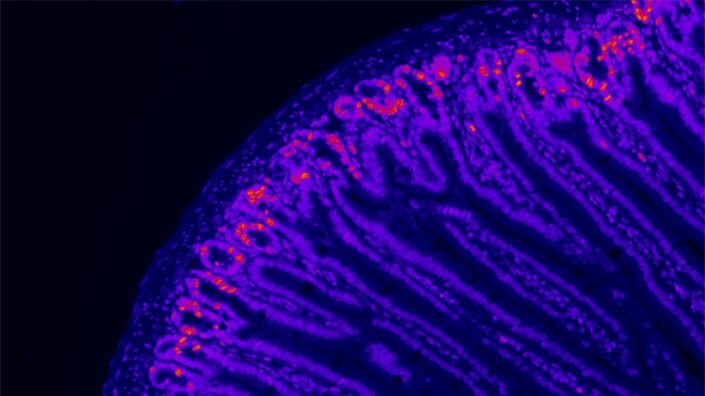
Not all plant viruses elicit immune responses in humans. In fact, the researchers tested several other viruses and found cowpea mosaic virus particles (CMVPs) to be exceptional in this regard. The team then tested CMVPs on a mouse model of colon cancer. Intraperitoneal cancers are among the deadliest and tend to metastasize quickly. Mice were injected with a dose of CMVPs and, one week later, challenged with an injection of colon cancer cells.

The treated mice demonstrated dramatically improved survival over untreated controls. 56% of the animals in the study group survived, while all mice in the control group succumbed to the disease within 25 days. On average, the treatment slowed tumor growth almost to a halt.

To see whether the treatment provided long-term protection, 40 days into the experiment, the surviving mice were re-challenged with a new dose of cancer cells. A subset of those mice was also injected with T cell-neutralizing antibodies to investigate the importance of T cells for any possible anti-tumor effect. The mice with their T cells intact showed robust survival, unlike those that had their T cells neutralized.

When another group of survivors were injected with cancer cells away from the original injection site, the protection remained robust, suggesting a systemic and tissue-agnostic nature. “Overall, the re-challenge experiments demonstrate that CPMV prophylaxis generates potent immune memory after initial challenge that is tumor-specific, but tissue-agnostic”, the paper says.

Effective in other cancers too

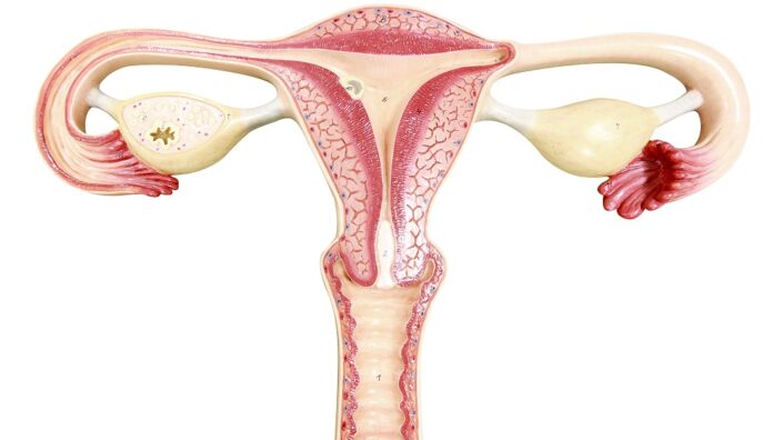
In cimilar experiments, the treatment proved effective against ovarian and breast cancer. The researchers also specifically analyzed lung metastases in a melanoma model, as lung metastases appear in many cancers, resulting in poor prognosis and low survival rates. The treatment effectively prevented melanoma cells from metastasizing into the lungs.

All these experiments were designed to mimic an adjuvant treatment aimed at preventing the recurrence and metastasis of cancer. However, people with intraperitoneal cancer often already have metastases when they are first diagnosed. To address this situation, the researchers reversed the order of things, first challenging mice with a small dose of cancer cells and waiting a week to administer this treatment, which was found to still be effective. In another experiment, they let the cancer progress and then surgically removed the tumors before administering CMVP particles. The results were still impressive, with the number of long-term survivors increasing fourfold.

“Even if you perform surgery to remove the tumors, no surgery is perfect and there is outgrowth of metastasis if no additional treatment is provided,” said Nicole Steinmetz, a professor of nanoengineering at UCSD and the study’s leading author. “Here, we use our plant virus nanoparticles after surgery to boost the immune system to reject any residual disease and prevent circulating tumor cells from metastatic seeding. We found that it works really, really well!”

We would like to ask you a small favor. We are a non-profit foundation, and unlike some other organizations, we have no shareholders and no products to sell you. All our news and educational content is free for everyone to read, but it does mean that we rely on the help of people like you. Every contribution, no matter if it’s big or small, supports independent journalism and sustains our future.

Literature

[1] Chung, Y. H., Zhao, Z., Jung, E., Omole, A. O., Wang, H., Sutorus, L., & Steinmetz, N. F. (2024). Systemic Administration of Cowpea Mosaic Virus Demonstrates Broad Protection Against Metastatic Cancers. Advanced Science, 2308237.

[2] Guan, X. (2015). Cancer metastases: challenges and opportunities. Acta pharmaceutica sinica B, 5(5), 402-418.

[3] Beisecker, A. E., Cook, M. R., Ashworth, J., Hayes, J., Brecheisen, M., Helmig, L., … & Selenke, D. (1997). Side effects of adjuvant chemotherapy: perceptions of node‐negative breast cancer patients. Psycho‐Oncology: Journal of the Psychological, Social and Behavioral Dimensions of Cancer, 6(2), 85-93.

[4] Shalhout, S. Z., Miller, D. M., Emerick, K. S., & Kaufman, H. L. (2023). Therapy with oncolytic viruses: progress and challenges. Nature Reviews Clinical Oncology, 20(3), 160-177.

[5] Wang, C., & Steinmetz, N. F. (2020). A combination of cowpea mosaic virus and immune checkpoint therapy synergistically improves therapeutic efficacy in three tumor models. Advanced functional materials, 30(27), 2002299.

Fast bicycling

Exercise May Remove Senescent Cells Through Inflammation

Exercise may be able to remove senescent cells only if acute inflammation is allowed to occur, according to a new study published in Aging.

Inflammation and senescence

Inflammation is known to be a critical part of aging, and its chronic accumulation has been labeled as a hallmark of aging: inflammaging. The SASP, the cocktail of compounds that senescent cells emit, is a key contributor to inflammaging and drives other cells senescent.

However, there is published evidence that some inflammation works in the other direction. Senescent cells can be removed by immune cells known as macrophages [1], and immune system-based therapies have been investigated for therapeutic use [2].

These researchers have previously reported that p16INK4a, a well-known marker of cellular senescence, decreases in young men 24 hours after high-intensity exercise [3]. Moderate exercise, however, did not have the same effect. They hypothesized that even though chronic inflammation encourages senescence, acute inflammation that results in CD11b-positive immune cells entering tissues discourages it.

Stimulating inflammation with exercise

The researchers recruited a dozen young men (average age of 22 years old) with no history of smoking or drug use to test this hypothesis. Half of the participants were given 400 milligrams of the anti-inflammatory medication ibuprofen, and half were given placebo. The participants cycled at high intensity for 20 seconds and took a 20-second rest for 15 sets. Their muscles were biopsied before exercise and then again 3 and 24 hours afterwards.

After 3 hours, mRNA expression of p16INK4a was decreased in both the ibuprofen and placebo groups, although significantly more in the placebo group, suggesting that the anti-inflammatory effect was also preventing the clearance of senescent cells. This trend continued at the 24-hour mark. However, not all participants responded in the same way, and some participants had significantly more p16INK4a at the beginning of the experiment than others.

Interestingly, CD11b was also decreased by exercise, at a similar rate to p16INK4a. These results were also attenuated by ibuprofen administration. CD11b was found to be directly related to p16INK4a expression in the tissue. There was also a trend towards greater expression of the DNA damage marker γ-H2AX 3 hours afterwards in the ibuprofen group and less expression 24 hours afterwards, but these were not statistically significant.

A limited but possibly valuable study

The researchers note that the total amount of exercise needed to get the observed effects was very low and conducted in short bursts: 10 minutes of exercise resulted in effects that diminished over the course of a full day.

However, this experiment was conducted solely on a relatively small cohort of young men, so it is unknown if these results are applicable to older people or women. There was no follow-up to determine long-term effects. The markers CD11b, p16INK4a, and γ-H2AX were the only metrics used; other senescence biomarkers were not used. A significant amount of future work will need to be conducted to determine if these results apply to other populations and if these findings can be used to develop therapeutic treatments or call for a reduction in anti-inflammatory use with exercise.

We would like to ask you a small favor. We are a non-profit foundation, and unlike some other organizations, we have no shareholders and no products to sell you. All our news and educational content is free for everyone to read, but it does mean that we rely on the help of people like you. Every contribution, no matter if it’s big or small, supports independent journalism and sustains our future.

Literature

[1] Kay, M. M. (1975). Mechanism of removal of senescent cells by human macrophages in situ. Proceedings of the National Academy of Sciences, 72(9), 3521-3525.

[2] Prata, L. G. L., Ovsyannikova, I. G., Tchkonia, T., & Kirkland, J. L. (2018, December). Senescent cell clearance by the immune system: Emerging therapeutic opportunities. In Seminars in immunology (Vol. 40, p. 101275). Academic Press.

[3] Jean, W. H., Hsieh, Y. W., Lai, L. F., Dewi, L., Liao, Y. C., Ye, M., … & Kuo, C. H. (2023). Senolytic effect of high intensity interval exercise on human skeletal muscle. Aging (Albany NY), 15(3), 765.

John Hancock

AgeLab to Drive Longevity Innovation and Research

John Hancock, along with its Toronto-based parent company Manulife (NYSE:MFC), today announced a five-year, multimillion-dollar research collaboration with the Massachusetts Institute of Technology (MIT) AgeLab, a multi-disciplinary research institute that works with business, government, and NGOs, leading innovation and research to help improve the quality of life for the aging population and their loved ones. Over the next five years, Manulife/John Hancock and the MIT AgeLab will collaborate to research the future of longevity innovation, developing research, thought leadership, and workshops with the goal of driving actionable insights for the business community, policymakers, and individuals and their families.

The World Economic Forum (WEF) reports that by 2050, the number of people aged over 60 is expected to double to 2.1 billion. Yet, one-fifth of an individual’s life, on average, is now expected to be lived with morbidity or in a state of illness. Lifespans and healthspans are not evenly distributed at the national or global level: a significant outcome of socioeconomic and environmental disparities. As such gaps expand, so does the need for immediate action to address what is already a major longevity crisis in our communities and the world at large. As the aging population grows, better understanding the intersection of health and wealth, including preventative health measures and retirement planning, will be paramount.

“The fastest-growing age cohort in the world is people over 85. As we continue to see people live longer lives, it is crucial that we gain new insight into how we can make longer lives synonymous with better and healthier lives,” said Brooks Tingle, president and CEO, John Hancock. “We fundamentally believe in helping make this a reality and know that this ambition does not start and end within the walls of our business. That’s why we’re committed to collaborating with leaders in the space, like the MIT AgeLab, to help provide others—from policymakers to industry leaders to healthcare advocates—with critical learnings that can scale our shared mission to drive better health and wealth outcomes.”

This research will explore the critical tenets of longevity in the United States and hopes to develop a first-of-its-kind longevity preparedness index, to be produced annually over an initial five-year period. As lifespans continue to increase worldwide, the index will measure the readiness of Americans from every generation to live a longer, healthier, and better life; and provide data-driven insights for maximizing financial planning, health and wellness habits, work and retirement transition planning, housing choices, end-of-life planning, and technological advances that support critical health and financial needs at each step of the aging process. Research on the first annual index will begin in the spring of 2024. The index is expected to expand to include Canada as the collaboration evolves.

MIT AgeLab founder and director Dr. Joseph Coughlin will lead this work with a team of social and data scientists along with experts outside of MIT. In addition to the index, the group will host several workshops throughout the year to engage in discussions and activations around longevity, generational dynamics, new technology, and behavioral insurance. Manulife/John Hancock has also joined the MIT AgeLab PLAN, an industry consortium examining how the business of advice might better prepare people to live 100 good years.

“My research team is excited to embark on this research collaboration with Manulife/John Hancock,” said MIT AgeLab’s Coughlin. “The spirit of MIT is to think and do. We want not only to identify the many different dimensions of what it takes to live longer, better; but also to measure the preparedness of a nation to live 100 good years. It is our shared objective that our work will educate and motivate people to do what it takes for themselves, their families, and their communities—to turn a longer life into a better life for all.”

The MIT AgeLab, based within MIT’s Center for Transportation & Logistics, is known internationally for its multi-disciplinary work exploring avenues of pragmatic, actionable innovation desired by, and useful to, aging consumers and workers, as well as their loved ones. This ongoing project spans sectors including automotive, healthcare, caregiving, housing and community design, and financial services; and inspired Coughlin’s bestselling 2017 book, The Longevity Economy.

This announcement supports Manulife’s Impact Agenda, which seeks to empower sustained health and well-being, and builds on John Hancock’s inaugural Longer. Healthier. Better. Symposium  hosted in Boston in September of 2023, where global leaders across public and private sectors convened to share the latest research and innovations driving the future of longevity. Globally, Manulife was proud to support WEF’s recently announced Longevity Economy Principles, which offer a strategic approach to addressing the global longevity crisis. Manulife also recently announced a multi-year partnership with UpLink, WEF’s open innovation platform, designed to help shape the future of, and investment in, longevity innovation.

Learn more about ManulifeJohn Hancock, and the MIT AgeLab.

About John Hancock and Manulife 

John Hancock is a unit of Manulife Financial Corporation, a leading international financial services provider that helps people make their decisions easier and lives better by providing financial advice, insurance, and wealth and asset management solutions. Manulife Financial Corporation trades as MFC on the TSX, NYSE, and PSE, and under 945 on the SEHK. Manulife can be found at manulife.com. One of the largest life insurers in the United States, John Hancock supports more than ten million Americans with a broad range of financial products, including life insurance and annuities. John Hancock also supports US investors by bringing leading investment capabilities and retirement planning and administration expertise to individuals and institutions. Additional information about John Hancock may be found at johnhancock.com.

We would like to ask you a small favor. We are a non-profit foundation, and unlike some other organizations, we have no shareholders and no products to sell you. All our news and educational content is free for everyone to read, but it does mean that we rely on the help of people like you. Every contribution, no matter if it’s big or small, supports independent journalism and sustains our future.
Ovaries

Stem Cell Transplants for Ovarian Aging

Experiments in mouse models show the efficacy and safety of mesenchymal stem cell transplantation in treating ovarian aging [1].

Mesenchymal stem cells as a therapy

Mesenchymal stem cells (MSCs) are stem cells derived from mesodermal tissue, such as the umbilical cord, umbilical cord blood, the placenta, fat tissue, and bone marrow [2]. MSCs are a promising new therapeutic approach for various diseases [3], including this paper’s focus: female infertility caused by ovarian aging.

The quantity and quality of oocytes start to decline relatively early in a female’s life. [4] Since contemporary women frequently postpone motherhood, the number of females diagnosed with infertility increases. [5]

At this moment, MSC therapy for human ovaries is in the preliminary stage of clinical application [6, 7]. Studies to date have focused on women with premature ovarian failure and ovarian hyporesponsiveness and used MSCs derived from their own bodies.

The efficacy of MSCs

The difference between this study and the previous mouse study is that in previous studies, stem cells were injected through the animals’ tail veins, which required cells to travel from the site of injection to the ovaries. In this study, the researchers used orthotopic transplantation, which allows the cells to be delivered closer to the site of their action to increase efficiency.

Among other groups, the researchers used young (4-5 months) mice and aged (10-12 months) mice, which had reduced reproductive functions, in order to evaluate the efficacy and toxicity of MSC treatment. MSCs were derived from healthy donors’ fat tissue (adipose tissue, AD) obtained during liposuction surgery and from full-term umbilical cord (UC) tissue following neonatal delivery. The researchers observed improved ovarian functioning in aging mice when AD-MSCs and UC-MSCs were used. However, AD-MSCs were shown to be more effective than UC-MSCs.

Following the injection of MSCs, the mice were monitored for eight days to track their oestrous cycle, the murine equivalent of monthly hormonal changes in human females. MSC transplantation improved this cycle compared to the control animals. For UC-MSCs, the changes in the duration of cycle phases were statistically significant, but for AD-MSCs, they were not.

After 1 and 3 weeks following the transplantation, the mice were sacrificed, and their tissues were analyzed. Analysis of the ovaries revealed that old mice that received AD-MSCs had a significantly increased proportion of proliferating cells compared to controls. UC-MSCs led to a slight increase in the proportion of proliferating cells, but it wasn’t statistically significant.

MSC ovarian transplantation didn’t increase the total number of follicles, which contain immature egg cells, in the ovaries. Still, the number of primary follicles significantly increased after UC-MSC and AD-MSC transplantation compared to controls. Also, both types of stem cells increased blood vessel proliferation in older animals’ ovaries.

An analysis of gene expression showed that UC-MSC and AD-MSC transplantation led to increased expression of MAPK cascade components, “central signaling pathways that regulate a wide variety of stimulated cellular processes, including proliferation, differentiation, apoptosis and stress response” [8]. AD-MSCs caused more changes in gene expression than UC-MSCs, including “cell-cell adhesion and positive regulation of the immune response.” The researchers also observed that these treatments had different short-term and long-term effects. While short-term effects involved different signaling pathways, “long-term effects were enriched in the activation of immune function.“

Good safety profile

Since the treatment’s efficacy showed promising results, the next step was to assess the safety of these transplants, which is indispensable in assessing theiir clinical utility.

Analyzing the mice sacrificed in this experiment indicated that they appeared healthy and that the transplanted MSCs did not generate tumors. Similarly, toxicity testing showed that “there was no significant acute toxic reaction” following MSC transplantation. Additionally, the expression of immune molecules wasn’t significantly increased, and the number of immune cells didn’t significantly increase in most mice. This data suggests that AD-MSC and UC-MSC transplantation does not significantly stimulate the immune system.

To function properly, orthotopically transplanted cells must reach their destination and not accumulate somewhere else in the body. The researchers injected a few mice with fluorescently labeled cells, and then searched the mice’s organs for these clearly visible cells after sacrifice. These cells accumulated mainly in the ovaries, with a small number of cells also observed in the uterus and spleen.

Therapy that holds potential

Based on the results of their experiments, the authors believe that MSCs hold great potential for clinical applications. Since they can be produced industrially, they can benefit many patients. However, first, the efficacy and safety of MSCs need to be shown during clinical trials.

Safety validation experiments confirmed that both AD-MSCs and UC-MSCs were not tumorigenic, with no acute toxic reactions, low immunogenicity, and a small amount of nondeterministic distribution. Furthermore, the mechanisms underlying the long-term and short-term effects after MSC transplantation differed, yet both led to an enhancement of the MAPK cascade. Collectively, orthotopic transplantation of MSCs has significant efficacy and high safety in the treatment of ovarian ageing.

We would like to ask you a small favor. We are a non-profit foundation, and unlike some other organizations, we have no shareholders and no products to sell you. All our news and educational content is free for everyone to read, but it does mean that we rely on the help of people like you. Every contribution, no matter if it’s big or small, supports independent journalism and sustains our future.

Literature

[1] Pei, W., Fu, L., Guo, W., Wang, Y., Fan, Y., Yang, R., Li, R., Qiao, J., & Yu, Y. (2024). Efficacy and safety of mesenchymal stem cell therapy for ovarian ageing in a mouse model. Stem cell research & therapy, 15(1), 96.

[2] Kouroupis, D., Sanjurjo-Rodriguez, C., Jones, E., & Correa, D. (2019). Mesenchymal Stem Cell Functionalization for Enhanced Therapeutic Applications. Tissue engineering. Part B, Reviews, 25(1), 55–77.

[3] Galipeau, J., & Sensébé, L. (2018). Mesenchymal Stromal Cells: Clinical Challenges and Therapeutic Opportunities. Cell stem cell, 22(6), 824–833.

[4] Secomandi, L., Borghesan, M., Velarde, M., & Demaria, M. (2022). The role of cellular senescence in female reproductive aging and the potential for senotherapeutic interventions. Human reproduction update, 28(2), 172–189.

[5] Carson, S. A., & Kallen, A. N. (2021). Diagnosis and Management of Infertility: A Review. JAMA, 326(1), 65–76.

[6] Herraiz, S., Romeu, M., Buigues, A., Martínez, S., Díaz-García, C., Gómez-Seguí, I., Martínez, J., Pellicer, N., & Pellicer, A. (2018). Autologous stem cell ovarian transplantation to increase reproductive potential in patients who are poor responders. Fertility and sterility, 110(3), 496–505.e1.

[7] Yan, L., Wu, Y., Li, L., Wu, J., Zhao, F., Gao, Z., Liu, W., Li, T., Fan, Y., Hao, J., Liu, J., & Wang, H. (2020). Clinical analysis of human umbilical cord mesenchymal stem cell allotransplantation in patients with premature ovarian insufficiency. Cell proliferation, 53(12), e12938.

[8] Plotnikov, A., Zehorai, E., Procaccia, S., & Seger, R. (2011). The MAPK cascades: signaling components, nuclear roles and mechanisms of nuclear translocation. Biochimica et biophysica acta, 1813(9), 1619–1633.

Evening exercise

Evening Aerobic Exercise Associated with Least Mortality

A new study suggests that aerobic medium-to-vigorous physical activity benefits obese and diabetic people the most when conducted in the evening hours [1].

Do you really need morning runs?

Physical activity is an essential part of the longevity-conscious lifestyle and one of the best life-extending interventions currently available. However, questions remain as to what types of physical activity are the healthiest, in what quantities, and so on. Exercise is a heterogeneous universe, and finding an individual mix involves such factors as age, sex, and medical conditions.

Recently, the timing of physical activity has become a widely explored issue. There is still a widespread public misconception that to lead a healthy lifestyle means to get up at six in the morning and go for a run. Many people find this regimen too hard to maintain. Thankfully, the reality seems to be more lenient. Numerous studies have suggested that at least some types of exercise bring greater health benefits when performed in the afternoon and evening hours [2].

30,000 accelerometer-wearing participants

In this new study, Australian scientists utilized data from UK Biobank, a huge repository of longitudinal health data on about half a million British citizens. A subset of UKB participants wore sophisticated accelerometers for a week to register their physical activity patterns. The resulting data has spawned numerous enlightening studies.

These researchers wanted to determine the best timing of aerobic medium-to-vigorous physical activity (MVPI) in obese and diabetic people. They assessed such outcomes as mortality, cardiovascular disease, and microvascular disease.

While the researchers used their own sophisticated way to categorize physical activity, generally, medium physical activit is between 3 to 6 metabolic equivalents (METs), a scale in which 1 is energy consumption at rest and vigorous is everything north of 6. For instance, brisk walking falls into the medium category, while jogging is considered vigorous.

Aerobic MVPI was defined in this study as at least three continuous minutes of MVPI. The participants were divided into three groups according to when in the day most of their MVPI occurred: 6 AM to 12 PM for the morning group, 12 AM to 6 PM for the afternoon group, and 6 PM to 12 AM for the evening group.

Of about 30 thousand participants, all with body mass indices (BMIs) of more than 30, 2,995 had type 2 diabetes at baseline. The median age was 62, and gender distribution was fairly even. All results were controlled for potentially confounding factors, including non-health-related deaths, cholesterol levels, blood pressure, ethnicity, socioeconomic status, and the season of accelerometer wear time.

Go for a walk after work

The reference group included people who averaged less than one bout of aerobic MVPI per day. Compared to that group, all those who were more active showed less all-cause mortality. However, the evening exercisers stood out with a hazard ratio of 0.39: that is, they were almost three times less likely to die than non-exercisers. The reduction in mortality was less pronounced in the morning and afternoon groups (HRs of 0.67 and 0.60, respectively).

Exercise HRs

People diagnosed with type 2 diabetes seemed to benefit from evening exercise even more (HR of 0.24, which translates to more than a four-fold reduction in all-cause mortality). Afternoon MVPI also showed strong results in this subgroup (HR of 0.44). Interestingly, no statistically significant association was found for morning exercisers in this subgroup (HR of 0.86).

The results were largely similar for cardiovascular disease, with modest and almost similar reductions for the morning and afternoon groups (HR of 0.83 and 0.84 respectively), and a much larger one for the evening group (HR of 0.64). In the subset of diabetic participants, only evening exercise showed a statistically significant effect: HR of 0.54.

Different results were recorded for microvascular disease, which included nephropathy, neuropathy, and retinopathy. Here, all groups showed rather small effect sizes, although evening exercise still came out on top.

The frequency of aerobic bouts seemed to be more important for their association with health outcomes than the overall duration of daily activity. Concordantly, recent research suggests that short bouts of physical activity can have a strong health-promoting effect [3].

Populational studies like this one can only establish correlation and not causation, and their results can be affected by hard-to-eliminate confounding factors and arbitrary design decisions.

Building upon previous clinical studies, our analyses underscore the consistent association of evening MVPA with the lowest risk in mortality, as well as strong associations with the incidence of CVD and MVD, when compared with not undertaking aerobic MVPA bouts. These findings are robust and extend to the subset of participants with T2D, in whom evening MVPA exhibited even more pronounced associations with mortality and cardiovascular morbidity. Sensitivity analyses demonstrated that when controlling for more even temporal distributions of aerobic MVPA, evening MVPA was associated with the greatest reduction in mortality, whereas more evenly spread MVPA was associated with the greatest reduction in MVD incidence.

We would like to ask you a small favor. We are a non-profit foundation, and unlike some other organizations, we have no shareholders and no products to sell you. All our news and educational content is free for everyone to read, but it does mean that we rely on the help of people like you. Every contribution, no matter if it’s big or small, supports independent journalism and sustains our future.

Literature

[1] Sabag, A., Ahmadi, M. N., Francois, M. E., Postnova, S., Cistulli, P. A., Fontana, L., & Stamatakis, E. (2024). Timing of Moderate to Vigorous Physical Activity, Mortality, Cardiovascular Disease, and Microvascular Disease in Adults With Obesity. Diabetes care, 47(5), 890-897.

[2] Moholdt, T., Parr, E. B., Devlin, B. L., Debik, J., Giskeødegård, G., & Hawley, J. A. (2021). The effect of morning vs evening exercise training on glycaemic control and serum metabolites in overweight/obese men: a randomised trial. Diabetologia, 64(9), 2061-2076.

[3] Stamatakis, E., Ahmadi, M. N., Gill, J. M., Thøgersen-Ntoumani, C., Gibala, M. J., Doherty, A., & Hamer, M. (2022). Association of wearable device-measured vigorous intermittent lifestyle physical activity with mortality. Nature Medicine, 1-9.

Hungry mouse

Aged Bacteria Increase Gut Inflammation in Younger Animals

Researchers have discovered negative ways in which intestinal bacteria change over time, spurring gut inflammation, and introduced older bacteria into a population of younger mice to determine their effects.

A well-known link

It has been thoroughly documented that gut bacteria have an influence on aging [1], and age-related changes to gut inflammation can lead to systemic inflammation [2, 3]. This has been proposed as a cause of inflammaging, the age-related increase in inflammation that leads to a host of order disorders.

These researchers attempted to shed more light on this subject by directly analyzing the guts of younger and older mice and then determining what effects that their respective bacterial populations were having.

Some bacteria are better than others

First, the researchers compared the colonic contents of 3- to 4-month-old and 19- to 20-month-old Black 6 mice, comparing diversity and taxonomic diversity. Their findings concurred with those of previous work involving human beings, showing that such taxonomic groups as Bacteroides were more common in the young than the aged, being replaced by such groups as Clostridium. Also as in previous work, gut diversity significantly decreased with age.

Inflammatory compounds were also more strongly expressed in the colons of older mice, including chemokine ligand 2 (Ccl2), which attracts macrophages. Calprotectin, which is found in immune cells and is a marker of inflammatory bowel disease (IBD) in people, was far more prevalent in the guts of the old mice. These changes in gene expression revealed that bacteria had become embedded into the tissues of the gut and that immune cells were present to fight them.

Specific bacterial changes were associated with these inflammatory markers. In particular, an increase of Erysipelatoclostridium and a decrease of Lachnospiraceae were found to be associated with more calprotectin and an increase in inflammatory signaling.

The researchers focused on one particular inflammatory signal, toll-like receptor 4 (TLR4). They found that bacterial samples taken from aged mice had more effect than those of young mice in activating TLR4 in immune cells, regardless of what area of the gut the samples were taken from. A related compound, TLR5, was found to be statistically unaffected.

Aged bacteria cause some inflammation in young animals

In another experiment, these researchers transplanted bacteria from the guts of old or young mice into those of young mice that had been raised without any gut bacteria at all. Four weeks later, the young mice whose intestines were colonized by bacteria from younger mice fared much better than those that had received bacteria from older mice. The aged-bacteria recipients had considerably greater inflammation in the colon and more signs of toxicity.

However, these younger animals were able to negate some of the effects. TLR4 was not found to be elevated after four weeks, and Erysipelatoclostridium levels did not differ between the recipients at that time point either. This demonstrates that the intestines of the mice themselves play a role in regulating bacterial contents.

Young and old mice respond differently to antibiotics

To test the resilience of bacterial populations, the researchers gave young and old animals broad-spectrum antibiotics in their drinking water for a week. The responses of young and old animals were slightly different: for example, aged mice had their bacterial diversity further decreased, while young animals had an increase in Erysipelatoclostridium. Both age groups had decreases in Lachnospiraceae, which produce the vital compound butyrate and are associated with less inflammation. Antibiotic treatment, like aging, was also found to cause an increase in reactive oxygen species (ROS).

These findings highlight the potential dangers of broad-spectrum antibiotic use while opening up questions relating to the relationship between bacteria and aging. It is largely explained how harmful bacteria that accumulate with aging can cause colonic inflammation; however, it is still unexplained exactly how younger animals maintain relatively healthier gut populations even after being exposed to aged bacteria.

We would like to ask you a small favor. We are a non-profit foundation, and unlike some other organizations, we have no shareholders and no products to sell you. All our news and educational content is free for everyone to read, but it does mean that we rely on the help of people like you. Every contribution, no matter if it’s big or small, supports independent journalism and sustains our future.

Literature

[1] DeJong, E. N., Surette, M. G., & Bowdish, D. M. (2020). The gut microbiota and unhealthy aging: disentangling cause from consequence. Cell Host & Microbe, 28(2), 180-189.

[2] Malik, J. A., Zafar, M. A., Lamba, T., Nanda, S., Khan, M. A., & Agrewala, J. N. (2023). The impact of aging-induced gut microbiome dysbiosis on dendritic cells and lung diseases. Gut Microbes, 15(2), 2290643.

[3] Thevaranjan, N., Puchta, A., Schulz, C., Naidoo, A., Szamosi, J. C., Verschoor, C. P., … & Bowdish, D. M. (2017). Age-associated microbial dysbiosis promotes intestinal permeability, systemic inflammation, and macrophage dysfunction. Cell host & microbe, 21(4), 455-466.

Cynomolgus Monkey

iPSC-derived Cardiomyocytes Rescue Heart Damage in Monkeys

Scientists have successfully used heart muscle cells created from induced pluripotent stem cells to counter the deleterious effects of heart attack in non-human primates.

Reprogrammed cells for heart regeneration

Since Dr. Yamanaka’s discovery, there has been a massive effort to turn this discovery into working therapies, with some of those experimental treatments currently nearing fruition. For instances, induced pluripotent stem cells (iPSCs) can be differentiated again into various types of youthful functioning cells.

Scientists have tried for a while to utilize iPSC-derived heart muscle cells (cardiomyocytes), to treat the consequences of myocardial infarction (heart attack) [1]. Healing after a heart attack is imperfect: stiff fibrotic scar tissue forms where healthy muscle was, leading to impaired heart function. However, attempts to infuse the healing muscle with iPSC-derived cardiomyocytes have been hampered by poor engraftment and posttransplant arrythmias, the latter caused by significant differences between the electrical qualities of adult cardiomyocytes and iPSC-derived ones.

Furthermore, before such problems can arise, most injected cardiomyocytes are thought to be simply washed away by circulating blood. Even those that remain are subject to a special type of cellular death, anoikis, caused by poor attachment to the extracellular matrix.

Spheroids instead of single cells

The Japanese researchers who authored this new study had learned about all these problems the hard way, from the results of their previous studies. This time, they decided to do things differently. In this new study performed on non-human primates (cynomolgus monkeys), which are far better models than rodents, instead of inoculating the monkeys with “free-floating” iPSC-derived cardiomyocytes, the researchers used spheroids: tiny balls of cells that form after several divisions. The researchers hoped that this would lead to better survival, engraftment, and integration of the cells into the existing electrical network of the heart muscle.

After receiving frozen human iPSCs from Kyoto University, the researchers differentiated them into cardiomyocytes. The cells were then frozen again for transportation to the animal lab and thawed without hurting their viability. The freezing-thawing part, according to the authors, is important because it shows suitability for future clinical use. Having grown the spheroids, the scientists injected them into 10 monkeys who had undergone heart attacks.

Partial rescue of heart damage

At first, the dose used was 20 million cells. Four weeks after transplantation, the heart contractile function of the treated monkeys was significantly improved versus controls, but the difference fell below the level of statistical significance at the 12-week mark, despite the difference between the 4-week and the 12-week readings being minuscule.

Since the resulting grafts were small, the researchers decided to increase the dose. Another ten monkeys were inoculated with 60 million cells each. This led to much better engraftment and to sustained (albeit only slightly bigger) benefits in cardiac function. Moreover, the improvement at the 12-week mark was more significant than at the 4-week mark. On the other hand, comparing cardiac function of the treated monkeys to that of healthy ones suggested that the treatment had only resulted in partial restoration.

iPSC Cardiomyocytes

Despite the greater number of transplanted cells, none of them were detected in other tissues such as lungs, suggesting highly targeted engraftment. The graft cells’ maturation levels were close to those of the host cells, and the graft was adequately supplied by blood vessels. Unfortunately, the larger dose also caused more arrythmias, but those symptoms were mild and short-lived. The researchers did not detect “any apparent immune response”, which is an encouraging result even though the monkeys were immunosuppressed.

According to the researchers, cardiac spheroids “have several advantages and may be an ideal form of a cardiomyocyte product for cardiac regenerative therapy”. For instance, they proved to be more viable than single cells, surviving for at least four hours at 4°C (39°F), which significantly improves transportability.

We would like to ask you a small favor. We are a non-profit foundation, and unlike some other organizations, we have no shareholders and no products to sell you. All our news and educational content is free for everyone to read, but it does mean that we rely on the help of people like you. Every contribution, no matter if it’s big or small, supports independent journalism and sustains our future.

Literature

[1] Wu, P., Deng, G., Sai, X., Guo, H., Huang, H., & Zhu, P. (2021). Maturation strategies and limitations of induced pluripotent stem cell-derived cardiomyocytes. Bioscience Reports, 41(6), BSR20200833.

Upside and downside

Clearing Senescent Cells Can Be a Double-Edged Sword

Researchers have found that removing senescent cells makes it easier for mice to fight initial infections but harder for them to develop immune memory.

Senescence and immunity

Introducing their paper, the researchers discuss the SASP and the relationship of senescent cells to the immune system, noting that “the aged microenvironment is one of the key drivers of dysfunction in CD4 and CD8 T-cell responses” and that CD8 T cells are responsible for clearing viruses. Additionally, with aging, CD4 cells fail to differentiate into the memory cells needed for long-term immunity [1].

These researchers had previously found that senolytics may have beneficial effects on the immune system, including rescuing CD4 T-cell differentiation [2], and others found that senolytics improve survival in mice against a coronavirus [3].

In an effort to identify causal relationships, these researchers employed aged, genetically engineered mice provided by Dr. Judith Campisi. These mice’s p16-expressing senescent cells can be removed by the administration of ganciclovir (GCV), which, itself, has no effect on the flu that was used to challenge their immune systems. However, some of the results showed a significant downside.

The mostly expected results

Shortly after having their senescent cells cleared, the mice also had a slight, non-significant decrease in the senescence marker SA-β-gal. Interestingly, they also had a similarly slight increase in the DNA damage marker γ-H2AX.

12 to 30 days after being exposed to influenza, the GCV-exposed model mice had cleared significantly more of the virus from their systems than the unexposed control group. This was traced to a decrease in CD127, a receptor for IL-7. T cells with more CD127 are more likely to survive after a virus and become memory cells. However, T cells with less CD127 are more likely to be involved in clearing the virus [4].

In other words, the upside and downside are closely connected: clearing p16-expressing cells improves short-term immune efficiency at the cost of long-term immune memory. This finding was heavily corroborated by further experiments with a different flu virus: the number of flu-specific memory T cells was significantly less than in the control group, and rechallenging those mice with the same flu again showed that their memory response was less effective.

However, these mixed results may be confined to p16-expressing CD8 cells. These researchers have done previous work on CD4 cells, targeting them with the senolytic combination of dasatinib and quercetin with positive results [2]. This serves as strong evidence that senescent cells, and their effects, are heterogenous and cannot be treated as a single factor.

We would like to ask you a small favor. We are a non-profit foundation, and unlike some other organizations, we have no shareholders and no products to sell you. All our news and educational content is free for everyone to read, but it does mean that we rely on the help of people like you. Every contribution, no matter if it’s big or small, supports independent journalism and sustains our future.

Literature

[1] Haynes, L., Eaton, S. M., Burns, E. M., Randall, T. D., & Swain, S. L. (2003). CD4 T cell memory derived from young naive cells functions well into old age, but memory generated from aged naive cells functions poorly. Proceedings of the National Academy of Sciences, 100(25), 15053-15058.

[2] Lorenzo, E. C., Torrance, B. L., Keilich, S. R., Al‐Naggar, I., Harrison, A., Xu, M., … & Haynes, L. (2022). Senescence‐induced changes in CD4 T cell differentiation can be alleviated by treatment with senolytics. Aging cell, 21(1), e13525.

[3] Camell, C. D., Yousefzadeh, M. J., Zhu, Y., Prata, L. G. L., Huggins, M. A., Pierson, M., … & Robbins, P. D. (2021). Senolytics reduce coronavirus-related mortality in old mice. Science, 373(6552), eabe4832.

[4] Obar, J. J., & Lefrançois, L. (2010). Memory CD8+ T cell differentiation. Annals of the New York Academy of Sciences, 1183(1), 251-266.

Longevity Desci Recap April 2024

Longevity and DeSci Recap – April 2024

In this latest edition of the Longevity and DeSci Recap, we’ll be taking a look back over a month full of events, research, and investments. As we head into spring, renewed life is on the horizon with continued investments in the longevity sector and the announcement of a permanent longevity/DeSci hub being launched on the sandy shores of the Caribbean.

Upcoming conferences and events

Global Synthetic Biology Conference

Spanning four days this May, the Global Synthetic Biology Conference will take place in San Jose, California, hosting 180+ content experiences, 20+ networking opportunities, and 200+ sponsors and exhibitors. This year, topics are set to include human health, planetary health, tools and technology, and the business of biology and society. Tickets for the event are available here.

SALT iConnections is coming to New York

This May 20-21, SALT, the global investment platform, will host an event centered on the impact of disruptive technologies on global finance, geopolitics and economics. Expected to be attended by prominent figures from New York’s financial ecosystem, the event offers a variety of sessions covering growth, innovation, and opportunities. Tickets are still available online.

Annual therapeutics summit returns

This June 11-13, the Age-Related Disease Therapeutics Summit is back for its sixth edition. Featuring 20+ speakers, works, and more, the event will host industry names such as Eli Lilly, Takeda, Turn Bio, BioAge, and Rubedo Life Sciences.

Off to Dublin for the Longevity Summit 2024

From June 13-16, the capital city of the Emerald Isle will play host to one of the biggest longevity events of the season. This year’s conference will host talks on rejuvenation biotechnology and longevity, with speakers to include Dr. Irina Conboy and Matthew “Oki” O’Connor. Tickets are still available here.

11th Aging Research and Drug Discovery Meeting

ARDD is back in 2024 this August. This year’s event is set to delve into such topics as senolytics, stem cell research, healthy longevity, and epigenetic reprogramming with prominent industry speakers, such as Nir Barzilai, Vera Gorbunova, and Aubrey de Grey. Tickets and event details are available here.

Tech breakthroughs & new research

Gero combines wearable tech and longevity research in new study

Aimed at uncovering the digital biomarkers of aging through the use of wearable technology, Gero has announced the launch of a new study conducted through its GeroSense app. The company has launched a non-profit initiative that draws upon specific data, such as daily step patterns and heart rate, to help improve predictive accuracy for aging. The study seeks to uncover the relationship between such things as exercise, diet, mental health, and medication on aging in the context of human behavior.

$1 billion launch for AI drug discovery company Xaira

The season of biotech investment has arrived with the formation of Xaira, an AI drug discovery company. Backed by $1 billion in initial funding and investors such as ARCH Venture Partners and Foresite Labs, Xaira is set to emerge from stealth mode and tackle data scarcity within drug development studies with its AI platform.

Gordian Biotechnology reveals new platform for drug development

Launching with a $60 million investment, Gordian Biotechnology has announced a proprietary in vivo therapeutic screening platform. Designed to target age-related diseases by streamlining the process for drug development through technology, the company boasts up to 80% predictive power for illnesses such as heart failure and osteoarthritis. With similar approaches leading to drug discovery already yielding promising results, the team behind Gordian Biotechnology seek to accelerate the discoveries with enhanced efficiency.

DAOs and communities

Vitalia pop-up city event draws to a close — what’s next?

Community living with a longevity twist, Vitalia was the second of its kind, a pop-up city event focused on growing the longevity community and its governance, following the success of Zuzalu. Spanning the first four months of 2024, the pop-up city located on the Roatán island in Honduras hosted longevity enthusiasts, experts, and curious health seekers alike from all over the world. Now, due to its success, the organizers have announced that Vitalia now has a permanent hub on Roatan within the special economic zone of Próspera, an area that facilitates legal autonomy suitable for pioneering longevity research and start-ups. lifespan.io’s Arkadi Mazin visited the city and tells of its potential now and in the future.

For people thinking of visiting over the next few months, here’s a taste of what’s on the schedule:

  • May 24-26 will see a focus on designing and building infrastructure for the DeSci community. With a move to the new Beta district planned, construction technologists, biomedical visionaries, and healthcare infrastructure builders are welcomed to attend.
  • Next up from June 18-22, medicine and biotech acceleration will come to the forefront with a deep dive collaboration into regenerative medicine and accelerated clinical trials.
  • July 26-28 will see biohacking take center stage as interested parties question “What does it mean to upgrade humanity?”

More events are planned till the end of the year, with the schedule constantly evolving. Stay tuned here and on the Vitalia website for the latest details.

DAOs for funding and collaborative development

As the concept of DAOs and their potential within the physical sciences grows, increasing research is being conducted and their impact recognized as a tangible solution. This latest article, published in Nature, highlights the potential of DAOs, challenges ahead, and their future role in life sciences, including VitaDAO as a case study.

Social media pages to follow this month

Research Hub Foundation offers the latest collaborations and crypto uses within the research sphere.

Longevity Summit Dublin provides the newest updates on one of the year’s biggest longevity conferences.

ValleyDAO delves into the wider ecosystem of longevity.

We would like to ask you a small favor. We are a non-profit foundation, and unlike some other organizations, we have no shareholders and no products to sell you. All our news and educational content is free for everyone to read, but it does mean that we rely on the help of people like you. Every contribution, no matter if it’s big or small, supports independent journalism and sustains our future.
Mother and baby rat

NMN and NR Reverse Ovarian Aging in Rats

In a recent Pharmaceutical Research paper, the researchers explored the molecular processes through which the NAD+ precursors NMN and NR reversed ovarian aging in middle-aged rats [1].

Use it or lose it

Female fertility decreases rather quickly in life. Aging leads to a decrease in the number and quality of oocytes, and fertilization success declines in a woman’s 30s [2]. Therefore, multiple research groups have been aiming to attack the aging ovary problem from many angles.

The researchers of this paper focused specifically on how aging affects mitochondrial fragmentation (fission) and mitochondrial merging (fusion) mechanisms in ovaries. These processes are essential for proper mitochondrial functioning and mitochondria-dependent biological processes [3].

Previous research has shown that increased NAD+ levels can improve mitochondrial function and reverse ovarian aging [4]. Since NAD+ precursors, namely nicotinamide mononucleotide (NMN) and nicotinamide riboside (NR), are commonly consumed as supplements and have good safety profiles, these researchers determined that it was worth testing whether supplementing rats with NMN or NR can improve ovarian aging.

Better weight and better looks

The researchers compared four groups of rats, with each group containing six animals: young, middle-aged, middle-aged + NMN, and middle-aged + NR. The treated animals received MNM and NR for 17 days. The next day, researchers compared the animals’ biomarkers.

First, the researchers compared the body weight versus the ovarian weight of the animals to calculate the ovarian index, which is used as an indicator of female fertility. A high ovarian index indicates better fertility [5]. The results showed a slight increase in the ovarian index following NMN and NR treatments.

Researchers also looked into the morphology of the organ. They observed increased amounts of corpus luteum in the middle-aged rats treated with NMN and NR. The corpus luteum is a structure that forms in the ovary following ovulation. It secrete progesterone, a hormone essential for implantation and pregnancy [6]. Aging leads to a diminishment of the corpus luteum [7].

Another indication of the state of ovarian aging in the middle-aged rats that showed improvements upon NMN and NR treatments was the increased number of antral follicles and decreased number of atretic follicles. Follicles in the ovary are sacs that contain immature eggs. Antral follicles are large follicles that are preparing for ovulation, while atretic follicles are characterized by apoptotic bodies, a degenerating oocyte, and fragmentation of the oocytic nucleus [8].

Hormones are another essential component necessary for proper ovarian functioning and reproduction and are impacted by ovarian aging. Based on the rats’ luteinizing hormone/follicle stimulating hormone (LH/FSH) ratio, the researchers learned “that ovarian aging disrupted the LH/FSH balance” and increased ovarian follicular atresia, a process of follicular degeneration or resorption accompanied by apoptosis. However, here again, NMN and NR applications improved those parameters: they helped to rebalance the LH/FSH ratio and decreased follicular atresia.

Mitochondria and sirtuins in service of better ovarian health

The previously described phenotypes are the usual suspects in ovarian aging. However, in this paper, the researchers also decided to investigate mitochondrial phenotypes as a marker of ovarian health since mitochondrial fission and fusion proteins are essential in oogenesis, embryogenesis, implantation, and protection of the ovarian follicular reserve [9, 10].

Compared to young rats, middle-aged rats had significantly reduced levels of gene transcripts of mitochondrial fusion-associated genes. NMN and NR treatment helped to increase the expression of those genes in middle-aged rats close to the levels seen in young rats.

The mitochondrial fission-associated gene transcript levels in middle-aged rats were increased compared to those in younger rats. NMN and NR treatment significantly reduced those gene levels in the rats’ ovaries. The protein analysis confirmed the positive impact of NMN and NR.

Seeing the connection between NMN, NR, and mitochondria prompted the researchers to test the levels of sirtuins. Sirtuins were previously reported to help delay ovarian aging and balance mitochondrial dynamics, and they are regulated by NAD+ [11]. Therefore, measuring their levels was essential in this experimental setup.

The researchers observed decreased levels of Sirt1 transcripts in the middle-aged group compared to the young rats, probably due to a decrease in NAD+ caused by aging. Treatment with the NAD+ precursors NMN and NR increased Sirt1 levels in ovaries. Those results were confirmed by measuring SIRT1 protein levels.

Bringing it all together and moving forward

Based on the current results and previous research, they hypothesized that NAD+ released from NMN and NR supplementation led to SIRT1 activation. Activated SIRT1 led to a decrease in DRP1, one of the fission-related proteins, which decreased the frequency of mitochondrial fission.

The authors point out that previous research on model animals and humans shows that NMN and NR supplementation is safe even at high doses. This is good news for future testing of NMN and NR supplementation in humans for delaying ovarian aging.

This study displays that […] the administration of a NAD+ precursor (NMN or NR) restores LH/FSH balance and mitochondrial dynamics, increases SIRT1 activity and alleviates folliculogenesis problems in middle-aged rats. Therefore, we consider that NMN and NR may be used as drug or supplement for reduction of aging-induced folliculogenesis or ovulation problems.

We would like to ask you a small favor. We are a non-profit foundation, and unlike some other organizations, we have no shareholders and no products to sell you. All our news and educational content is free for everyone to read, but it does mean that we rely on the help of people like you. Every contribution, no matter if it’s big or small, supports independent journalism and sustains our future.

Literature

[1] Arslan, N. P., Taskin, M., & Keles, O. N. (2024). Nicotinamide Mononucleotide and Nicotinamide Riboside Reverse Ovarian Aging in Rats Via Rebalancing Mitochondrial Fission and Fusion Mechanisms. Pharmaceutical research, 10.1007/s11095-024-03704-3. Advance online publication.

[2] Amanvermez, R., & Tosun, M. (2016). An Update on Ovarian Aging and Ovarian Reserve Tests. International journal of fertility & sterility, 9(4), 411–415.

[3] Tilokani, L., Nagashima, S., Paupe, V., & Prudent, J. (2018). Mitochondrial dynamics: overview of molecular mechanisms. Essays in biochemistry, 62(3), 341–360.

[4] Yang, L., Lin, X., Tang, H., Fan, Y., Zeng, S., Jia, L., Li, Y., Shi, Y., He, S., Wang, H., Hu, Z., Gong, X., Liang, X., Yang, Y., & Liu, X. (2020). Mitochondrial DNA mutation exacerbates female reproductive aging via impairment of the NADH/NAD+ redox. Aging cell, 19(9), e13206.

[5] Li, S., Liu, M., Ma, H., Jin, Q., Ma, Y., Wang, C., Ren, J., Liu, G., & Dai, Y. (2021). Ameliorative effect of recombinant human lactoferrin on the premature ovarian failure in rats after cyclophosphamide treatments. Journal of ovarian research, 14(1), 17.

[6] Taketa Y. (2022). Luteal toxicity evaluation in rats. Journal of toxicologic pathology, 35(1), 7–17.

[7] Acuña, E., Fornes, R., Fernandois, D., Garrido, M. P., Greiner, M., Lara, H. E., & Paredes, A. H. (2009). Increases in norepinephrine release and ovarian cyst formation during ageing in the rat. Reproductive biology and endocrinology : RB&E, 7, 64.

[8] Saatcioglu, H. D., Cuevas, I., & Castrillon, D. H. (2016). Control of Oocyte Reawakening by Kit. PLoS genetics, 12(8), e1006215.

[9] Liu, X. M., Zhang, Y. P., Ji, S. Y., Li, B. T., Tian, X., Li, D., Tong, C., & Fan, H. Y. (2016). Mitoguardin-1 and -2 promote maturation and the developmental potential of mouse oocytes by maintaining mitochondrial dynamics and functions. Oncotarget, 7(2), 1155–1167.

[10] Zhang, M., Bener, M. B., Jiang, Z., Wang, T., Esencan, E., Scott, R., Horvath, T., & Seli, E. (2019). Mitofusin 2 plays a role in oocyte and follicle development, and is required to maintain ovarian follicular reserve during reproductive aging. Aging, 11(12), 3919–3938.

[11] Iljas, J. D., Wei, Z., & Homer, H. A. (2020). Sirt1 sustains female fertility by slowing age-related decline in oocyte quality required for post-fertilization embryo development. Aging cell, 19(9), e13204.

Chromosomes

Exploring How Caloric Restriction Impacts Telomere Length

A recent study from Penn State University may shed more light on the link between telomeres, aging, and why caloric restriction appears to influence them.

Telomeres are linked to aging

Telomeres are small protective caps on the ends of our chromosomes that guard our DNA. Telomere attrition, in which these caps slowly erode, is a hallmark of aging. The shortening of telomeres limits the amount of cell divisions, ultimately causing a decline in cell numbers in organs and tissues.

Shortened telomeres are linked to multiple age-related diseases and idiopathic pulmonary fibrosis. For that reason, telomeres, and their length and rate of loss, have long been a biomarker of aging.

They are also a potential target for therapies that restore lost telomere length, and a few companies are working on this approach. If successful, it may be possible to reduce the risks of several age-related diseases associated with short telomeres.

Investigating how caloric restriction impacts telomeres

Today, we want to highlight further data coming from the CALERIE clinical trial that could help us understand why caloric restriction may increase lifespan [1].

Last year, we wrote about how caloric restriction may slow down human aging with results from the CALERIE clinical trial. Those results suggested that caloric restriction does have a beneficial effect on longevity. However, the study also raised a number of questions and had limitations that we discussed in that article.

This time, the scientists examined the telomere lengths of 175 individuals involved in the CALERIE study. Measurements were taken at the beginning, one year in, and at the end of the 24-month study period, during which two-thirds of participants followed a caloric restriction regimen and one-third served as a control group.

The researchers noted that during the course of the study, the rate of telomere loss changed. In the first year, the participants on caloric restriction lost weight and saw increased telomere loss. The following year saw weight loss stabilized for those on caloric restriction. In the second year, however, the researchers found that telomere loss slowed down compared to the control group.

After the two year mark, both the test and control groups’ rate of loss was similar. There was no statistical difference between the two groups at this point.

The results are unclear

The researchers had originally hypothesized that there would be a reduction in telomere loss in the calorically restricted group; however, this was not the case. In fact, the rate of telomere attrition did the opposite and increased in the first year before stabilizing.

This study raises more questions than it answers. What would happen if the data for a third year was checked; would the telomere loss rate change again? Would that data change again in a fourth year of caloric restriction? Finally, why did the rate of loss initially speed up when the expectation was for it to slow down?

This is another demonstration that the dynamics of telomere loss are complex and not well understood. The study participants are due for another check-in at the 10-year mark, so it will be quite a while yet before we will know more.

That said, some data suggests that caloric restriction may help reduce cholesterol and blood pressure levels, so it could still be worthwhile to do it. If you want to find out more, check out our topic on fasting and time-restricted feeding.

We would like to ask you a small favor. We are a non-profit foundation, and unlike some other organizations, we have no shareholders and no products to sell you. All our news and educational content is free for everyone to read, but it does mean that we rely on the help of people like you. Every contribution, no matter if it’s big or small, supports independent journalism and sustains our future.

Literature

[1] Hastings, W. J., Ye, Q., Wolf, S. E., Ryan, C. P., Das, S. K., Huffman, K. M., … & Shalev, I. (2024). Effect of long‐term caloric restriction on telomere length in healthy adults: CALERIE™ 2 trial analysis. Aging cell, e14149.

LongX Logo

LongX Launches the Xplore Program for Students

LongX, an initiative dedicated to providing early-career avenues into longevity, announced a call for applications for its 2024 Xplore Program. The program lasts from June 2024 until September 2024 and features a one-month longevity primer course followed by the opportunity to gain educational experience with a company operating in the longevity biotechnology sector.

LongX ApplicationsThe program is fully remote and open to high school and undergraduate students of any background. Fellows in the program will have the opportunity to learn about aging biology, product management, longevity tools and roadmaps, the ethics of aging interventions, and more. They will be guided throughout the program by university graduates and gain additional insights through fireside chats with experts.

2024 industry partners include Vincere Bio, Senexell, AgeRate, and several more.

To apply or learn more about the program, please visit LongX.

About LongX

LongX was launched in 2023 as a platform for youth interested in longevity. We prioritize fostering innovation and interdisciplinary collaboration, aiming for both short-term impact and long-term progress. We encourage exploration beyond traditional roles and aim to equip future experts with the skills to drive progress. Our Substack provides regular articles on our thoughts, experiences, and interviews.

Contact

Denisa Lepădatu, Co-Founder | Outreach

longevityxplorer@gmail.com

We would like to ask you a small favor. We are a non-profit foundation, and unlike some other organizations, we have no shareholders and no products to sell you. All our news and educational content is free for everyone to read, but it does mean that we rely on the help of people like you. Every contribution, no matter if it’s big or small, supports independent journalism and sustains our future.
Kdineys

A New Potential Path to Treating Kidney Fibrosis

Researchers have found a new protein target for senescence-related kidney diseases and published their findings in Aging Cell.

Kidney disease and fatty acid use

Chronic kidney disease (CKD) is all too common in older people, as a full third of people over 70 have moderate to severe forms of it [1]. The kidneys need surprising amounts of blood to function, as they consume a fifth of the heart’s output [2]. They are also unusually good at metabolizing fat, but problems with this system can lead to kidney fibrosis and further problems [3]. This stems from malfunctions in fatty acid oxidation (FAO), and a decrease in the related enzymes stops fats from being used for energy [4] and is directly linked to kidney disease [5].

Previous work has found that protease-activating receptor 2 (PAR2), which promotes inflammation [6], hinders fat metabolism and encourages the development of fatty liver disease [7]. These researchers, therefore, decided to investigate the relationship between PAR2, fatty acid oxidation, kidney fibrosis, and cellular senescence in the kidneys.

Rodents’ kidneys act much like humans’

The researchers first investigated kidney disease in a standard breed of rats, comparing rats aged 6 and 20 months. Interestingly, the aging of the kidneys was found to be very sex-dependent in these animals: male rats had significant elevations in damage-related gene and protein expressions along with related physical changes, including fibrosis. Female rats had far fewer statistically significant differences in the kidneys between young and old.

These changes were found to be directly related to cellular senescence. The senescence markers p16, p21, and p53 dramatically increased, as did compounds related to the SASP. However, like the physical changes, these were only predominant in male rats.

The researchers continued their work from rats to mice. Feeding mice an adenine-rich diet harms their kidneys, but like with rats, the damage was found to be more significant in males. Also just like with the aged rats, markers of cellular senescence in the kidney tubes coincided with CKD and inflammation.

In both rats and mice, expression of PAR2 was found to be signiicantly associated with senescence and CKD, specifically in the kidney tube cells analyzed previously. In aged male rat kidneys, these were found to physically coincide in the same region. Further confirming these findings, the researchers induced kidney disease in mice by using cisplatin, finding a similar relationship between PAR2, senescence, and kidney fibrosis.

An examination at the cellular level gave some clues as to why this is the case. As expected, PAR2 was found to decrease the ability of the kidney tube cells to properly process fats. This led to accumulation of these fats and an increase in lactate brought on by glycolysis, an alternate method of energy production. There was also a reduced expression of Cpt1a, a protein that decreases with senescence.

Does taking away PAR2 fix the problem?

The researchers then performed their final steps of the experiment by acquiring a mouse model that does not express PAR2. Against adenine and cisplatin both, these PAR2-knockout mice were more resistant to induced kidney disorders, showing less senescence and less lipid accumulation in the tube cells. Inflammation and fibrosis were also decreased.

While this research showed only benefits for knocking out PAR2, it is rare that an aspect of biology can be simply taken away without side effects. Furthermore, it was not tested in naturally aged animals. Further research will need to be done to determine if anti-PAR2 therapies could potentially be used in people.

Literature

[1] Kovesdy, C. P. (2022). Epidemiology of chronic kidney disease: an update 2022. Kidney international supplements, 12(1), 7-11.

[2] Duann, P., & Lin, P. H. (2017). Mitochondria damage and kidney disease. Mitochondrial Dynamics in Cardiovascular Medicine, 529-551.

[3] Kang, H. M., Ahn, S. H., Choi, P., Ko, Y. A., Han, S. H., Chinga, F., … & Susztak, K. (2015). Defective fatty acid oxidation in renal tubular epithelial cells has a key role in kidney fibrosis development. Nature medicine, 21(1), 37-46.

[4] Miguel, V., Tituaña, J., Herrero, J. I., Herrero, L., Serra, D., Cuevas, P., … & Lamas, S. (2021). Renal tubule Cpt1a overexpression protects from kidney fibrosis by restoring mitochondrial homeostasis. The Journal of clinical investigation, 131(5).

[5] Kang, H. M., Ahn, S. H., Choi, P., Ko, Y. A., Han, S. H., Chinga, F., … & Susztak, K. (2015). Defective fatty acid oxidation in renal tubular epithelial cells has a key role in kidney fibrosis development. Nature medicine, 21(1), 37-46.

[6] Heuberger, D. M., & Schuepbach, R. A. (2019). Protease-activated receptors (PARs): mechanisms of action and potential therapeutic modulators in PAR-driven inflammatory diseases. Thrombosis journal, 17(1), 4.

[7] Rana, R., Shearer, A. M., Fletcher, E. K., Nguyen, N., Guha, S., Cox, D. H., … & Kuliopulos, A. (2019). PAR2 controls cholesterol homeostasis and lipid metabolism in nonalcoholic fatty liver disease. Molecular Metabolism, 29, 99-113.

Mitochondria up close

Transplanting Mitochondria Improves Muscle Function

A recent study saw researchers associated with Cellvie demonstrate significant improvements to mitochondria and muscle function in aged mice by injecting additional mitochondria [1].

Mitochondrial dysfunction is a reason we age

Mitochondria are cellular powerhouses that convert nutrients into adenosine triphosphate (ATP), a form of energy that powers cells. It is not an exaggeration to say that without mitochondria, complex life, such as people, would not be possible.

With aging, the mitochondria can become damaged and less efficient over time. This is because producing energy is a dirty business that creates harmful byproducts called free radicals.

These free radicals bounce around inside of cells, damaging things that they strike, particularly mitochondrial DNA. This damage causes mutations, which make energy production less efficient and cause mitochondria to behave in harmful ways.

Mitochondria do not have an efficient DNA repair system like cells’ nuclear DNA. As time passes, the number of mutations in mitochondria increases. This leads to a downward spiral in which mitochondria become increasingly unable to produce energy and function properly.

A number of researchers are now developing therapies to combat this. If they are successful, it may be possible to slow down or even reverse aging in our cells.

Rejuvenating old cells with donated mitochondria

It has been known for some time that mitochondria can transfer between cells and can be absorbed into cells from their environment. This suggests transferring healthy mitochondria to an aged animal or person may be a viable approach for rejuvenation.

This is exactly what the researchers in this new study did. It is interesting that the mitochondria were harvested from mice of the same age and not younger donors.

The mitochondria were separated from the donor tissue and given directly to the test group mice. The animals were then injected directly into the hindleg muscle tissue, meaning delivery was relatively simple.

Compared to the control group, the test group saw improvements to mitochondrial function and muscle function. There was also an improvement in endurance.

Results: The results indicated significant increases (ranging between ~36% and ~65%) in basal cytochrome c oxidase and citrate synthase activity as well as ATP levels in mice receiving mitochondrial transplantation relative to the placebo. Moreover, there were significant increases (approx. two-fold) in protein expression of mitochondrial markers in both glycolytic and oxidative muscles. These enhancements in the muscle translated to significant improvements in exercise tolerance.

A follow-up study to better understand these results

As mentioned, the notable thing about this particular study was that even though the mitochondria were delivered from animals of the same age, the results demonstrate some level of improvement.

Perhaps there is some form of dilution of damaged mitochondria happening here that explains the results. The influx of additional mitochondria may be somewhat offsetting the burden of mutated mitochondria in the recipient.

That said, it is reasonable to assume that delivering mitochondria from a younger donor would be more advantageous. It would make for an ideal follow up experiment using mitochondria harvested from younger animals.

There is also the consideration of if mitochondrial haplotypes need to match between donor and recipient or not. Mitochondrial haplotypes refer to groups of closely linked genetic markers present on mitochondrial DNA. These haplotypes are used to trace maternal lineages and genetic variations in populations.

Understanding the role of haplotypes in a follow-up study would also be beneficial. If they do not need to match to be effective, that would also simplify production at scale.

An inroad for rejuvenation research

Mitochondrial dysfunction is a hallmark of aging, but it’s unlikely that “aging” will be the target of this research. Companies working on mitochondrial transfer will probably initially target frailty and muscle loss in older adults for clinical trials. Once approved, the approach could be used off label for other related conditions and aspects of aging.

The great news is that growing mitochondria in culture is likely a scalable technology. This is very important and directly relates to the ability of people to access and afford therapies.

Many rejuvenation biotechnology supporters are worried about being able to access it in the future. After all, it’s no use if a therapy works but is unaffordable for most people. Hopefully, companies such as Cellvie and Mitrix will use bioreactor-grown mitochondria to cost-effectively produce them en masse.

It’s likely that medical tourism in countries with less regulations will offer mitochondrial transplants to those with the means. Obviously, those first adopters need to consider the risks of doing so. Meanwhile, the rest of us will be waiting for therapies to get through the clinic via the FDA, EMA, etc.

This avenue of research looks promising if the challenges of manufacturing at scale and access can be overcome. We will continue to follow research progress closely and hopefully will have more to report in the near future.

We would like to ask you a small favor. We are a non-profit foundation, and unlike some other organizations, we have no shareholders and no products to sell you. All our news and educational content is free for everyone to read, but it does mean that we rely on the help of people like you. Every contribution, no matter if it’s big or small, supports independent journalism and sustains our future.

Literature

[1] Arroum, T., Hish, G. A., Burghardt, K. J., McCully, J. D., Hüttemann, M., & Malek, M. H. (2024). Mitochondrial Transplantation’s Role in Rodent Skeletal Muscle Bioenergetics: Recharging the Engine of Aging. Biomolecules, 14(4), 493.

Elderly clinical trial

Stem Cells Alleviate Frailty in Clinical Trial

A placebo-controlled Phase 1/2 trial conducted in East Shanghai has found that administering umbilical cord-derived mesenchymal stem cells reduces frailty in older people.

Stem cells against frailty

These researchers begin by defining frailty as “a state of heightened vulnerability to potential stressors as a consequence of reduction in physiological reserves across multiple systems” [1]. This vulnerability destroys the strength and endurance of older people, exhausting their stamina and greatly increasing their risks of death and disability, and a metric has been determined to measure this [2]. However, while vitamin supplements may help people with nutritional deficiencies, there are no medically approved drugs to treat frailty [3].

As stem cell exhaustion has been pinpointed as a cause of frailty [4], replacement stem cells have been investigated as a possible treatment. In particular, mesenchymal stem cells (MSCs), which are naturally attracted to injury sites [5], appear to be the most promising. MSCs have multiple potential sources for derivation [6], and previous trials have been conducted to treat frailty by using MSCs derived from bone marrow (BM-MSCs), with positive results [7, 8].

This study, however, was conducted on stem cells that were originally derived from the human umbilical cord (HUC-MSCs). These cells are easy to mass produce [9], have been successfully clinically tested against other diseases such as heart failure [10] and arthritis [11], and fight inflammation [12]. This, however, is the first trial of HUC-MSCs for frailty.

Frail adults only

All participants had to meet three criteria: to be between the ages of 60 and 80, to score between 1 and 4 on the Fried frailty scale [2], and to be expected to live another year. A large variety of co-morbidities were screened out, such as uncontrolled diabetes, serious cardiovascular problems, infections, and viral diseases. This was a double-blinded trial from which 80 potential candidates were excluded. 15 patients received placebo, and 15 received MSCs, for 6 months.

This study measured physical performance by testing grip strength, the timed up-and-go test, walking speed, and the ability to stand up and sit back down. Inflammatory cytokines such as interleukins were also measured, and sleep quality, quality of life, and mental health were also assessed.

Only good significant effects

There were no significant adverse effects. Three participants had suffered from ailments during the trial, two of which were in the placebo group and the third of which had dizziness not related to the MSCs.

Physical function, the primary endpoint of the study, was strongly affected by the MSCs. Even with only 30 total participants, not all of which participated in every assessment, the researchers were able to obtain, against baseline, a p-value of .003 after only one week of treatment and p-values under .001 for 1 and 6 months. Against placebo, the p-value at the end of the 6-month study was .042.

There were possible effects on mental health and sleep quality but those could be statistically attributed to the placebo effect. However, the treatment improved total quality of life with a p-value of 0.002 against placebo at the end of the study.

MSC Trial

Cytokines had less clear effects; the placebo group spiked in TNF-α and IL-17 at 6 months while the MSC group did not.

While not all of the endpoints were hit, this study was against frailty, and it is clear from these results that MSCs have beneficial impacts on frailty in human beings. However, this study was conducted in one country among 30 people. Further work, with a larger sample size and more testing sites, will need to be conducted to determine if these results hold up under further scrutiny.

We would like to ask you a small favor. We are a non-profit foundation, and unlike some other organizations, we have no shareholders and no products to sell you. All our news and educational content is free for everyone to read, but it does mean that we rely on the help of people like you. Every contribution, no matter if it’s big or small, supports independent journalism and sustains our future.

Literature

[1] Clegg, A., Young, J., Iliffe, S., Rikkert, M. O., & Rockwood, K. (2013). Frailty in elderly people. The lancet, 381(9868), 752-762.

[2] Fried, L. P., Tangen, C. M., Walston, J., Newman, A. B., Hirsch, C., Gottdiener, J., … & McBurnie, M. A. (2001). Frailty in older adults: evidence for a phenotype. The Journals of Gerontology Series A: Biological Sciences and Medical Sciences, 56(3), M146-M157.

[3] Dent, E., Morley, J. E., Cruz-Jentoft, A. J., Woodhouse, L., Rodríguez-Mañas, L., Fried, L. P., … & Vellas, B. (2019). Physical frailty: ICFSR international clinical practice guidelines for identification and management. The Journal of nutrition, health and aging, 23(9), 771-787.

[4] Schulman, I. H., Balkan, W., & Hare, J. M. (2018). Mesenchymal stem cell therapy for aging frailty. Frontiers in Nutrition, 5, 108.

[5] Golpanian, S., Wolf, A., Hatzistergos, K. E., & Hare, J. M. (2016). Rebuilding the damaged heart: mesenchymal stem cells, cell-based therapy, and engineered heart tissue. Physiological reviews, 96(3), 1127-1168.

[6] Zhang, J., Huang, X., Wang, H., Liu, X., Zhang, T., Wang, Y., & Hu, D. (2015). The challenges and promises of allogeneic mesenchymal stem cells for use as a cell-based therapy. Stem cell research & therapy, 6, 1-7.

[7] Golpanian, S., DiFede, D. L., Khan, A., Schulman, I. H., Landin, A. M., Tompkins, B. A., … & Hare, J. M. (2017). Allogeneic human mesenchymal stem cell infusions for aging frailty. Journals of Gerontology Series A: Biomedical Sciences and Medical Sciences, 72(11), 1505-1512.

[8] Tompkins, B. A., DiFede, D. L., Khan, A., Landin, A. M., Schulman, I. H., Pujol, M. V., … & Hare, J. M. (2017). Allogeneic mesenchymal stem cells ameliorate aging frailty: a phase II randomized, double-blind, placebo-controlled clinical trial. Journals of Gerontology Series A: Biomedical Sciences and Medical Sciences, 72(11), 1513-1522.

[9] Sarugaser, R., Lickorish, D., Baksh, D., Hosseini, M. M., & Davies, J. E. (2005). Human umbilical cord perivascular (HUCPV) cells: a source of mesenchymal progenitors. Stem cells, 23(2), 220-229.

[10] Bartolucci, J., Verdugo, F. J., González, P. L., Larrea, R. E., Abarzua, E., Goset, C., … & Khoury, M. (2017). Safety and efficacy of the intravenous infusion of umbilical cord mesenchymal stem cells in patients with heart failure: a phase 1/2 randomized controlled trial (RIMECARD trial [randomized clinical trial of intravenous infusion umbilical cord mesenchymal stem cells on cardiopathy]). Circulation research, 121(10), 1192-1204.

[11] Wang, L., Huang, S., Li, S., Li, M., Shi, J., Bai, W., … & Liu, Y. (2019). Efficacy and safety of umbilical cord mesenchymal stem cell therapy for rheumatoid arthritis patients: a prospective phase I/II study. Drug design, development and therapy, 4331-4340.

[12] Uccelli, A., Pistoia, V., & Moretta, L. (2007). Mesenchymal stem cells: a new strategy for immunosuppression?. Trends in immunology, 28(5), 219-226.