The Blog

Building a Future Free of Age-Related Disease

Food clock

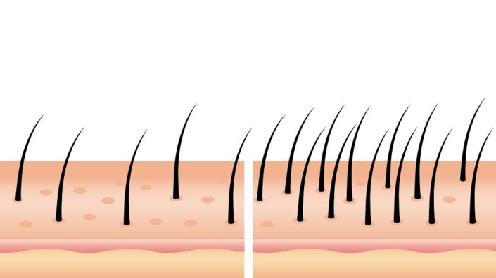
What Time-Restricted Eating Does and Doesn’t Affect

In Cell Reports Medicine, researchers have published a study demonstrating that, while it has no impact on many aspects of intestinal function, time-restricted eating (TRE) reduces markers of blood glucose.

A matter of when rather than what

People who conduct TRE, choosing to consume food only at certain times of the day, often report improvements to health due to the ensuing weight loss; they consume less food per day in general [1]. However, human clinical trials that have controlled the amount of food consumed, so that everyone involved is eating the same amount, have found that it improves glucose use and reduces biomarkers of aging [2]. More studies have found that it improves muscle performance in healthy men [3].

Other research has found that the gastrointestinal system changes throughout the day, following the circadian rhythms of other bodily systems [4]. However, the researchers suggest that this is more due to TRE being associated with overeating; someone who only eats during certain times of the day often eats a large meal, and doing this can slow down the gastric system [5].

Better glucose metabolism confirmed

To test their hypothesis, the researchers conducted a six-day test in which 16 healthy adults followed personalized eating and drinking recommendations between either 8:00 AM to 2:00 PM (TRE) or 8:00 AM to 8:00 PM (control). The diets provided were balanced, with moderate amounts of eggs, fowl, and dairy products; no red meat was present. The TRE group and control group had similar eating schedules, with the TRE group’s being compressed into half the time.

All participants were studied on a large number of gastrointestinal and metabolic metrics, including stool examinations, subjective hunger, resting energy expenditure, gene expression, and metrics related to glucose and insulin use.

Time-restricted eating

The results were largely in accordance with previous studies, showing that glucose metabolism was, indeed, positively affected by TRE. Total glucose was diminished, and glycemic variability, the fluctuations in glucose over a day, was also significantly reduced by TRE. However, this study’s key findings are largely of the things that didn’t happen.

No effects on nutrients, but effects on hunger

Analyzing the stool of the participants, the researchers found that energy use was not affected by TRE. Digestibility of fats, carbohydrates, and proteins was unaffected, as was fiber and consistency of stool. Urine was similarly unaffected by TRE, both in amount and in nitrogen content. Resting energy expenditure was also unaffected, as were the metabolites in blood plasma and the bacterial contents of stool.

The researchers also used indigestible dye capsules to determine how long it takes for stool to pass through the digestive system. This metric was similar between both the study and the control groups. Hydrogen production due to fermentation in the gut was also not affected. This, the researchers note, affects the circadian rhythm by increasing satiety during daylight hours.

However, the TRE group was considerably less hungry during the day than the control group. Participants were asked about their hunger, their capacity to eat, and their desire to eat, all of which were not affected before breakfast but significantly reduced in the TRE group at noon and at 5:00 PM. On the other hand, they became significantly (and expectedly) more hungry at 9:00 PM.

While many of these results show that nothing significant has happened in many respects, this makes them of interest to nutritionists and anyone considering a meal plan that involves TRE. This approach seems to have beneficial effects on glucose metabolism but does not affect how the gut absorbs nutrients. This was a small study, but it appears to be the case that a nutritionist creating a defined meal plan could compress it into an early-morning meal plan without adverse effects.

We would like to ask you a small favor. We are a non-profit foundation, and unlike some other organizations, we have no shareholders and no products to sell you. All our news and educational content is free for everyone to read, but it does mean that we rely on the help of people like you. Every contribution, no matter if it’s big or small, supports independent journalism and sustains our future.

Literature

[1] Pellegrini, M., Cioffi, I., Evangelista, A., Ponzo, V., Goitre, I., Ciccone, G., … & Bo, S. (2020). Effects of time-restricted feeding on body weight and metabolism. A systematic review and meta-analysis. Reviews in endocrine and metabolic disorders, 21, 17-33.

[2] Jamshed, H., Beyl, R. A., Della Manna, D. L., Yang, E. S., Ravussin, E., & Peterson, C. M. (2019). Early time-restricted feeding improves 24-hour glucose levels and affects markers of the circadian clock, aging, and autophagy in humans. Nutrients, 11(6), 1234.

[3] Jones, R., Pabla, P., Mallinson, J., Nixon, A., Taylor, T., Bennett, A., & Tsintzas, K. (2020). Two weeks of early time-restricted feeding (eTRF) improves skeletal muscle insulin and anabolic sensitivity in healthy men. The American journal of clinical nutrition, 112(4), 1015-1028.

[4] Segers, A., & Depoortere, I. (2021). Circadian clocks in the digestive system. Nature Reviews Gastroenterology & Hepatology, 18(4), 239-251.

[5] Basolo, A., Hohenadel, M., Ang, Q. Y., Piaggi, P., Heinitz, S., Walter, M., … & Krakoff, J. (2020). Effects of underfeeding and oral vancomycin on gut microbiome and nutrient absorption in humans. Nature Medicine, 26(4), 589-598.

Stressed intestines

Stress Impairs Gut Health via the Microbiome

A new study done in mice sheds light on how mental stress contributes to intestinal problems by altering microbiome composition [1].

Mind over matter

Some experiences are not called “gut-wrenching” just metaphorically, as the brain can indeed influence gastrointestinal function in a myriad of ways. While most of them are adaptive, brain signals can also cause intestinal problems. For instance, mental stress is thought to contribute to IBS (irritable bowel syndrome), which affects hundreds of millions of people worldwide, along with inflammatory bowel disease (IBD), an umbrella term that includes Crohn’s disease, ulcerative colitis, and other conditions [2]. However, the exact mechanisms behind this brain-gut interplay are not sufficiently understood.

Impaired mitochondrial function and differentiation

In this new study published in Cell Metabolism, the researchers subjected mice to two weeks of chronic stress to study their intestinal responses. This caused notable morphological changes in the mice’s guts consistent with a loss of intestinal function, which were still present up to two weeks after the mice were no longer subjected to stress. Moreover, the researchers observed a substantial decrease of proliferative cells along the intestinal villi (tiny folds) following stress. “These data,” the researchers suggest, “indicate that chronic psychological stress in mice triggers intestinal epithelia disturbance that is not easily repairable.”

Intestinal epithelial cells have an especially high turnover rate, the highest in the body [3]. Their whole population is renewed every few days. This means that intestinal stem cells (ISC) must work hard and proliferate profusely to be able to replenish all the cells that are constantly being shed.

Stress was shown to only mildly affect the proliferation of ISCs. However, it had a more profound dampening effect on their differentiation. ISCs receive cues from another type of cell, Panet cells, but the researchers were able to prove that stress affected ISCs specifically.

RNA sequencing showed that stress altered numerous molecular pathways in ISCs, especially those related to mitochondrial function. As a result, oxygen consumption rate in affected cells was much lower than in controls. Electronic microscopy confirmed changes in mitochondrial morphology in ISCs from stressed mice, and single-cell RNA analysis helped establish the link between this mitochondrial dysfunction and the impaired ability of ISCs to differentiate.

The microbial connection

Clearly, stress affects ISCs, but the mechanisms were unclear. Several options, such as variations in caloric intake and signaling by stress hormones, were ruled out. What if an intermediary were involved? Signals from the sympathetic nervous system are known to remodel the microbiome [4], and the researchers discovered that this is exactly what had happened to stressed mice.

The researchers conducted a series of clever experiments, such as transplanting the microbiota of stressed mice to healthy controls, which then started showing similar intestinal symptoms. They then tried treating mice with various narrow-action antibiotics to see which bacteria strains were responsible. Their attention turned to bacteria of Lactobacillus genus. One of the strains, L. murinus, turned to be highly enriched in the stressed mice. Colonizing the guts of germ-free mice with L. murinus recapitulated many of the effects of stress.

Looking for the specific metabolite responsible, the researchers noticed that the levels of indoleacetic acid (IAA) were increased several-fold in the guts of the stressed mice as well as in germ-free mice that received L. murinus transplants.

Finally, intestinal specimens were directly treated with IAA in concentration that mimicked stress condition. The treatment impaired intestinal regeneration and largely mimicked the effects of stress on ISCs’ differentiation and mitochondrial homeostasis. The researchers then established that α-ketoglutarate (AKG), a molecule that recently emerged as a possible geroprotector, was effective in abrogating the effects of stress on ISCs and intestinal function.

To initially assess whether their findings were relevant to humans, the researchers analyzed a microbiome composition dataset on people with major depressive disorder (MDD). Both Lactobacillus content and IAA levels were markedly increased in people with MDD. Finally, the researchers recruited their own cohort of patients with several mood disorders and received similar results. “Overall,” the researchers conclude, “these findings reveal a microbially driven brain-to-gut metabolic axis that could be therapeutically targeted for gut-brain comorbidities driven by chronic stress.”

Psychological stress exerts a significant yet poorly understood impact on the gut-brain axis. In the clinic, stressful lifestyle is critically involved in the exacerbation of brain-gut comorbidities such as IBD and IBS. Here we reveal a pathway by which the gut microbiome relays stress signals from the brain to shape the susceptibility to intestinal epithelial injury in mice. Specifically, this brain-to-gut circuit entails microbial metabolite IAA as a niche signal that hampers mitochondrial respiration to skew ISC fate decision and epithelial renewal. Importantly, IAA is consistently elevated in the gut of patients with mental distress, and targeting IAA signals proves effective to protect mice from the detrimental effects of stress.

We would like to ask you a small favor. We are a non-profit foundation, and unlike some other organizations, we have no shareholders and no products to sell you. All our news and educational content is free for everyone to read, but it does mean that we rely on the help of people like you. Every contribution, no matter if it’s big or small, supports independent journalism and sustains our future.

Literature

[1] Wei, W., Liu, Y., Hou, Y., Cao, S., Chen, Z., Zhang, Y., … & Hao, H. (2024). Psychological stress-induced microbial metabolite indole-3-acetate disrupts intestinal cell lineage commitment. Cell Metabolism.

[2] Bisgaard, T. H., Allin, K. H., Keefer, L., Ananthakrishnan, A. N., & Jess, T. (2022). Depression and anxiety in inflammatory bowel disease: epidemiology, mechanisms and treatment. Nature Reviews Gastroenterology & Hepatology, 19(11), 717-726.

[3] Williams, J. M., Duckworth, C. A., Burkitt, M. D., Watson, A. J. M., Campbell, B. J., & Pritchard, D. M. (2015). Epithelial cell shedding and barrier function: a matter of life and death at the small intestinal villus tip. Veterinary pathology, 52(3), 445-455.

[4] Gershon, M. D., & Margolis, K. G. (2021). The gut, its microbiome, and the brain: connections and communications. The Journal of clinical investigation, 131(18).

Desci January

Longevity and DeSci Recap – January 2024

Welcome back to the latest edition of the Longevity and DeSci Recap and the first of 2024. The past year has been packed with new discoveries and releases, including Exo’s Iris, a hand-held AI ultrasound device; large-scale deals, such as Fauna Bio x Lilly at $494 million and ExcepGEN $14 million in seed funding; and innovations such as AthenaDAO and Zuzalu, the first pop-up city.

It seems that 2024 is shaping up to be an eventful year already. January has started with a bang with the launch of the latest pop-up city event, Vitalia, in Honduras, the news of a unique piece of technology, and an AI tool that could reinvent healthcare consultations. Here are the insights, interviews, and research in the longevity technology sphere over the past month.

Upcoming conferences

Here are some of the first quarter’s upcoming conferences and events for all longevity enthusiasts.

The City of Life is live. Join Vitalia in Roatan, Honduras: The redefinition of the longevity landscape is already underway in Roatan, Honduras, where up to 200 residents and 500+ visitors interested in longevity are joining forces to rejuvenate the world. Inspired by the first pop-up city, Zuzalu, Vitalia’s schedule is packed with talks, co-working, events, and other attractions for longevity enthusiasts. Broken into four separate bi-weekly thematic areas, culminating in a summit, Vitalia’s participants will have a chance to explore:

  • Longevity & Human Improvement (Jan 15 – 21): How longevity is an integral part of human development, business models for longevity start-ups, human improvement, biohacking and more, with an overall focus on life-enhancing solutions
  • Start-up Societies (Jan 29 – Feb 4): The impact of crypto on governance and longevity societies, network states and new governance, real-life examples of crypto communities and their challenges
  • AI & Technological Progress (Feb 5 – 11): The journey of AI as an accelerant in the technology world, the history of progress and stagnation in technology, how AI can play a role in advancing biotech, and the wider ecosystem of AI and its potentials
  • Pathways to Life Extension (Feb 19 – 25): The potential for new drug development, innovative design solutions, clinical advancements, and healthcare, and presenting projects and concrete solutions that have been developed during the pop-up city event

Longevity and Biohacking Convention in Cancun: The Longevity and Biohacking convention in Cancun, Mexico is set to add to the fervor. For two days from March 2, 2024 to March 3, 2024, the event’s attendees will gather together to explore the topics of longevity and achieving peak performance, mental and physical. The schedule is packed with 34 keynotes, 60 exhibits showing the latest in longevity technology, workshops, and networking events. It will culminate with a final dinner party to round up the exciting two-day event.

Head back in time with the Turn Around Aging Conference: To be held in Munich in March, the Turn Around Aging Conference is dedicated to healthy longevity and solutions that can improve human health and lifespan. Supplements, lifestyle factors, and hormone replacement are set to be hot topics of discussion at the event, with ticket prices starting from €129. This conference will be held in the German language.

Over to DC for the Longevity Science Fly-In: From March 21-22, 2024, the Longevity Science Fly-In will be the host of this exclusive longevity networking-centric event. Notable names such as Newt Gingrich and experts in biotechnology will be present at the luxury Hay-Adams hotel, close to the White House. Registration for the top-level event is set at $2,000.

Rejuvenation Start-up Summit 2024 is back in Berlin: Set to be the world’s largest in-person gathering that brings together longevity start-ups, investors, and enthusiasts, this year’s conference will facilitate a variety of world-renowned speakers, such as Matthew Rosen of CoRegan, Alexander Leutner of cell blocks, Lifespan’s Stephanie Dainow, and others.

Longevity investment and development news

Health biotech company Timeline raises $66 million in funding: Longevity is set to take the consumer market with Timeline’s latest Series D funding round. The company raised a massive $66 million in an oversubscribed round led by BOLD, L’Oréal Groupe’s venture capital fund to further Timeline’s proprietary technology, Mitopure. Mitopure, backed by 15 years of research, is claimed to enhance cellular longevity and improve mitochondrial function, a key factor in skin, strength and cognition. This latest funding will allow the company to continue its venture into the food, beauty and health sectors, bringing its longevity solution to a larger market.

New funding for early heart disease detection: CardioSignal, the tech company dedicated to improving remote cardiac care, just received $10 million in Series A funding to advance its heart disease detection technology. The smartphone-based solution aims to detect the early signs of heart disease using gyroscopic and accelerometer sensors to measure heart movements aiming to reduce deaths from cardiac disease.

Tech breakthroughs & new research

Neuralink has been implanted for the first time: Much anticipated with both fear and awe, the wait is over for those speculating whether or not Elon Musk’s Neuralink would ever get off the ground. The start of 2024 saw the first successful implant of the proprietary technology into a human, using its unique product, Telepathy. The technology is designed to “generalized brain interface to restore autonomy to those with unmet medical needs today and unlock human potential tomorrow” and is aimed at those with paralysis and degenerative disease.

After much consideration, and initial safety concerns, the chip (Telepathy) was approved by the FDA for human trials and works by capturing brain signals and transmitting them via Bluetooth technology.

Can a chatbot do better medical consultations than a human?: This is the question asked by lifespan.io’s Arkadi Mazin when reviewing the latest research into a Google-made AI chatbot designed to conduct medical consultations. The technology, Articulated Medical Intelligence Explorer (AMIE) was trained using media resources and refined via iterations. It was then put to the test against primary care physicians, outperforming them in 24 out of 26 categories, including differential diagnosis, empathy and communication.

However, this study evaluates only chatting ability, which is not an area that physicians are typically trained in. Yet, this technology does present the potential to democratize healthcare and deliver a solution for resource-poor communities.

DAOs and communities

AthenaDAO announces its first Entrepreneur-in-Residence: Competition was fierce as five finalists competed to be the first-ever EIR at AthenaDAO. The final event was held as a pitch session and evaluated by three judges: Dr.Lynae Brayboy, MD, Chief Medical Officer & Co-Founder of Ovom Care; Alok Tayi, PhD, Founder and CEO of Vibe Biotechnology; and James Brodie, Chief Investment Officer at ID Theory. After a long debate, the judges selected Maria Such, PhD for the honor.

World longevity news

Manulife partners with UpLink in multi-year longevity initiative: The global investment management group Manulife has just announced a partnership with UpLink, the World Economic Forum’s open innovation platform. UpLink, launched in 2020, supports start-ups that come up with solutions for the World’s Sustainable Development Goals and has supported over 350 entrepreneurs since its inception. The multi-year initiative led by Manulife and UpLink seeks to address the demographic and financial challenges of aging, promote longevity, and build upon the previous partnership focused on nature-based climate solutions.

Social media pages to follow this month

Life Noggin YouTube: If you haven’t already subscribed, now’s the time to learn more about longevity and other topics in quick, easy-to-understand videos.

lifespan.io News: Hosted by Emmett Short, lifespan.io News covers the latest in longevity in video form.

Christian Angermayer’s Instagram: Christian Angermayer is an entrepreneur, biotech founder and longevity enthusiast. Follow to hear the latest insights and investment news.

We would like to ask you a small favor. We are a non-profit foundation, and unlike some other organizations, we have no shareholders and no products to sell you. All our news and educational content is free for everyone to read, but it does mean that we rely on the help of people like you. Every contribution, no matter if it’s big or small, supports independent journalism and sustains our future.
Hair restoration

Rapamycin Rescues Hair Growth in Mice

A new study has found that autophagy plays an important role in activating hair follicle stem cells and keeping the hair growth cycle going. By boosting autophagy, rapamycin improved hair growth in mice and in a human hair organ culture [1].

Round and round it goes

The hair leads a cyclical life. First, a hair grows from the follicle in the anagen stage. It lasts for several years in humans, which and defines maximum hair length. Next comes a short period called catagen, when the follicle shrinks and stops getting nutrients from the skin. Finally, there’s telogen, when the old hair is replaced with a new one. The new one starts to grow, and the cycle repeats itself.

Hair follicle stem cells (HFSC) are a crucial element of this process. They are located in the bulge zone of hair follicles and are usually quiescent. Upon receiving certain molecular signals, they get activated and differentiate into various types of hair-related cells, ensuring that a new anagen phase will ensue. In hair growth disorders, such as androgenetic alopecia and stress-induced alopecia, HFSC dysfunction can cause extended telogen [2].

Autophagy, the process of recycling intracellular junk, is an important modulator of cellular metabolism. Autophagy levels decrease with age, including in stem cells, which is thought to contribute to their dysfunction [3]. In this new study published in Cell & Bioscience, the researchers wanted to investigate how autophagy levels in HFSCs affect hair regeneration.

Autophagy drives hair growth

In an initial mouse experiment, the researchers first confirmed that autophagy levels in HFSCs fluctuate across the hair growth cycle. During the shift from telogen to anagen, autophagy levels were elevated and then diminished once the anagen phase was reached. This pattern of autophagy variation in HFSCs aligned with the hair follicle cycle phases, indicating a potentially significant role of autophagy in a hair follicle’s progression from the telogen to the anagen stage.

The researchers then treated the mice with rapamycin, a drug that hardly needs introduction in the longevity community. Rapamycin, which upregulates autophagy by inhibiting the protein mTOR, is currently the most potent small molecule in terms of lifespan extension in mice [4]. Another group was treated with the autophagy inhibitor 3-MA. Inhibiting autophagy produced an effect opposite to that of rapamycin.

Modifying autophagy had an obvious impact on hair growth. Autophagy inhibition delayed the telogen-anagen transition in hair follicles, while boosting autophagy with rapamycin expedited it. “Surprisingly, on day 11, mice in the rapamycin group regained almost intact hair, whereas most hair follicles in the 3-MA group remained in the telogen”, the researchers report. The scientists also discovered that this was synchronized with HFSC activation. The cells’ proliferation rates were higher in the rapamycin group and much lower in the 3-MA group than in the control group.

Not just mice

Similar results were obtained in an organ culture with human hairs, showing that the findings were not specific to mouse hair. The researchers detected morphological differences between the groups: 3-MA hampered hair shaft growth, while rapamycin boosted it. As a result, one week into the experiment, the control group had 30% hair follicles in anagen, the 3-MA group had less than 20% follicles in anagen, and in the rapamycin group, more than half of the follicles remained in anagen: that is, they continued to generate hair growth.

In another in vitro experiment performed in HFSC cultures, the percentage of live cells was similar across the groups, but 3-MA inhibited cell proliferation, while rapamycin promoted it in a dose-dependent manner. This also corresponded to higher or lower amounts of autophagy. The researchers however note that since autophagy is a common phenomenon across cell types, it is possible that part of the effect observed is driven by cells other than HFSC, which should be a topic of further research.

Cellular autophagy maintains the metabolic homeostasis of cells and regulates stem cell activation by degrading soluble macromolecules and denatured organelles in the cytoplasm. Autophagy in mouse dorsal skin remains low during the telogen and increases upon entering the anagen. The Inhibition of autophagy causes hair follicles to enter the catagen earlier, highlighting the importance of autophagy in initiating the normal hair follicle cycle and hair regeneration under physiological conditions. In a mouse model of alopecia areata, researchers found that autophagy within hair follicles was suppressed, and the induction of autophagy alleviated alopecia symptoms, whereas its inhibition advanced disease development.

We would like to ask you a small favor. We are a non-profit foundation, and unlike some other organizations, we have no shareholders and no products to sell you. All our news and educational content is free for everyone to read, but it does mean that we rely on the help of people like you. Every contribution, no matter if it’s big or small, supports independent journalism and sustains our future.

Literature

[1] Sun, P., Wang, Z., Li, S., Yin, J., Gan, Y., Liu, S., … & Miao, Y. (2024). Autophagy induces hair follicle stem cell activation and hair follicle regeneration by regulating glycolysis. Cell & Bioscience, 14(1), 1-18.

[2] Shin, W., Rosin, N. L., Sparks, H., Sinha, S., Rahmani, W., Sharma, N., … & Biernaskie, J. (2020). Dysfunction of hair follicle mesenchymal progenitors contributes to age-associated hair loss. Developmental cell, 53(2), 185-198.

[3] Revuelta, M., & Matheu, A. (2017). Autophagy in stem cell aging. Aging cell, 16(5), 912-915.

[4] Harrison, D. E., Strong, R., Sharp, Z. D., Nelson, J. F., Astle, C. M., Flurkey, K., … & Miller, R. A. (2009). Rapamycin fed late in life extends lifespan in genetically heterogeneous mice. nature, 460(7253), 392-395.

Cardiovascular exercise

How Cardiovascular Mortality Goes Down with Exercise

In the International Journal of Cardiology, researchers have described five styles of exercise and compared them to mortality risk.

How much is the right amount?

It is well known that physical activity is associated with a significantly diminished risk of death by cardiovascular disease, the leading cause of death around the world. However, it has remained unclear just how much exercise is good at protecting against this cause of death, and previous studies have relied on self-reported examinations [1], which are subjective and therefore unreliable.

Previous work has determined that there is a relationship between disruptions in natural circadian rhythms, which govern sleep and activity cycles, and cardiovascular mortality [2]. However, how these rhythms in physical activity affect cardiovascular mortality is very difficult to research due to inaccuracies in self-reporting and problems with data collection. A part of the UK Biobank gave these researchers the data they needed to fill this gap.

Enter the accelerometer

While there were some exclusions from certain parts of this study due to specific health issues, a full 102,323 participants in the UK Biobank, which took data between 2006 and 2010, had wearable accelerometer data. Each of these participants wore a wrist accelerometer for a week, recording their normal amounts of activity.

An algorithm was used to cluster these participants into five distinct groups. In order, they were a persistently low group, a moderate-and-stable group, a single-increase group, a double-increase group with morning and evening peaks, and a vigorous group that performed more physical activity than anyone else.

Accelerometer Groups

As expected, the lowest activity group had the strongest correlations with cardiovascular disease and diabetes, being tied with the second-lowest group for cancer. They also had considerably higher BMIs.

In nearly 13 years following the data collection, the deaths of the UK Biobank participants were recorded. Even after adjusting for BMI, age, and other confounders in Model 3, the low-activity group was far more likely to die of any cause and of cardiovascular disease than any of the other groups, with even moderate activity associated with a lower risk. Greater amounts of activity, largely regardless of when it was conducted, was correlated with decreasing risk.

Mortality Impact Physical Activity

Inflammation is a factor

Three metrics were used to estimate the effects of biological aging: phenotypic age, systemic immune inflammation, and systemic inflammation response. As expected, phenotypic age was associated with all-cause mortality, and in a non-linear way: people with the highest phenotypic age were far more likely to die than anyone else. The inflammation indices were somewhat more linear, with the response index’s associations leveling out near its median.

Here, too, physical activity was found to have a strong association. As before, the three most vigorous clusters differed little, moderate exercisers had a slight advantage, and the people who exercised the least were found to have the greatest phenotypic ages and the most inflammation. The authors found that these anti-inflammatory effects played a mediating role in cardiovascular risk.

The authors also note that the double increase group seemed to be protected from ischemic heart disease as well as the vigorous exercise group, despite the latter group conducting much more physical activity during the day. This led them to conclude that circadian effects may be an important part of physical activity. However, it is also clear from the data that simply getting a sufficient amount of exercise on a regular basis is very strongly correlated with reduced mortality, from all causes and from cardiovascular disease.

We would like to ask you a small favor. We are a non-profit foundation, and unlike some other organizations, we have no shareholders and no products to sell you. All our news and educational content is free for everyone to read, but it does mean that we rely on the help of people like you. Every contribution, no matter if it’s big or small, supports independent journalism and sustains our future.

Literature

[1] Castetbon, K., Vernay, M., Malon, A., Salanave, B., Deschamps, V., Roudier, C., … & Hercberg, S. (2009). Dietary intake, physical activity and nutritional status in adults: the French nutrition and health survey (ENNS, 2006–2007). British Journal of Nutrition, 102(5), 733-743.

[2] Man, A. W., Li, H., & Xia, N. (2021). Circadian rhythm: potential therapeutic target for atherosclerosis and thrombosis. International journal of molecular sciences, 22(2), 676.

Rejuvenation Roundup January 2024

Rejuvenation Roundup January 2024

As work on epigenetic treatments continues, we continue to showcase all the novel research and possible therapies that might make several age-related diseases much more treatable. Here’s what happened in January.

LEAF News

Interviews

Andrew Steele InterviewAndrew Steele: “A Mindset Shift Is Required”: Andrew Steele is a physicist who became interested in longevity several years ago and began a career as a bioinformatician before becoming a longevity advocate, quickly making a name for himself with numerous media appearances.

Mikhail Batin’s Crusade Against Aging: There’s no other way to put it: Mikhail (Misha) Batin is an oddball in the longevity space. In this field, which is populated mostly by scientists, entrepreneurs, and enthusiasts, Batin belongs to a different breed: activists.

Advocacy and Analysis

Elderly womanFemale-Specific Aging Trajectories Remain Understudied: In a recent Perspective published in Nature Aging, researchers noted a lack of studies on female-specific physiological factors on aging trajectories. They proposed solutions, including model systems, to address the issue.

Research Roundup

Study Suggests Trade-Offs Between Reproduction and Lifespan: Scientists might have found support for the antagonistic pleiotropy theory of aging, which suggests that evolution selects some genetic traits that are associated with shorter lifespans because they also help us reproduce.

Rabbit eating carrotA Carrot Extract Improves the Metabolism of Old Mice: In Nature Communications, researchers have published their discovery of a compound found in carrots that promotes mitochondrial health and improves the physical abilities of older mice.

TLR5 Activation Improves Health and Lifespan in Aged Mice: By activating toll-like receptor 5, an important element of the innate immune system, scientists have increased lifespan and improved multiple health measurements in old mice, despite having started the treatment late in life.

Knee jointTreating Arthritis with Rapamycin-Releasing Nanoparticles: Researchers have described how specialized nanoparticles, which deliver rapamycin and the antioxidant astaxanthin, restore macrophage balance and reduce inflammation in a mouse model of osteoarthritis.

David Sinclair on Human Trials of Anti-Aging Compounds: A new review authored by three acclaimed geroscientists paints a promising picture of past and ongoing human clinical trials of prospective anti-aging drugs.

Stress in pregnancyStress in Pregnancy Linked to Shorter Infant Telomeres: A new study published in BMC Psychiatry reviewed literature that links perceived stress during pregnancy with newborns’ telomere length. Telomeres are protective caps at the end of chromosomes. The shortening of the telomeres, which happens with each DNA replication cycle, is one of the Hallmarks of Aging.

An Exercise-Related Protein May Protect the Heart: Researchers publishing in Aging have pinpointed a protein that increases after exercise and is associated with a reduced risk of coronary artery disease. Coronary artery disease directly kills more people than anything else in the world.

Fat tissue communicationImproved Brain-Fat Communication Extends Lifespan in Mice: A new study suggests that the dysregulation of the crosstalk between hypothalamus and white adipose tissue affects aging. Rescuing this “communication channel” led to significant lifespan extension.

How Parkinson’s Disease Perpetuates Itself: A new paper elaborates on how and why microglia fail to clean up the α-synuclein protein of Parkinson’s disease, gradually making the disease worse.

Shielded cellsEnhanced CAR T Cells Eradicate Multiple Cancers: Scientists have created improved CAR T cells that also express the cytokine IL-10. The new cells proved highly effective in mice and in a pilot human trial. Chimeric antigen receptor (CAR) T cells are among the most promising treatments in oncology.

Combining Common Anti-Aging Interventions and Exercise: A review recently published in BMC Biology suggests that taking gerotherapeutics while exercising doesn’t have advantages over separate treatments. Exercise is a well-established intervention that ameliorates several aspects of aging.

Metabolic syndrome diagnosisA Marker of Insulin Resistance Predicts Kidney Disease: A recent study published in GeroScience has investigated whether or not a common index of metabolic syndrome can be applied to chronic kidney disease (CKD).

Google’s Chatbot Does Medical Interviews Better than Humans: Scientists at Google have created an AI chatbot for conducting medical interviews. It matched or surpassed human primary care practitioners on most criteria, including accuracy, politeness, and empathy.

Enriched environmentA Better Environment May Ameliorate Early Aging: Publishing in Aging, researchers have reversed the early aging of prenatally harmed young mice by exposing them to a more enriched environment. Newborn rodents from sleep-deprived mothers have conditions that are biologically similar to the processes of aging.

Physical Activity May Decrease Disability-Related Mortality: In GeroScience, researchers have published an analysis of a cohort study, concluding that adherence to physical activity recommendations can mitigate the increased mortality associated with disability.

Plant proteinPlant Protein Is Associated with Lifelong Health: A new large cohort study suggests that protein might be healthier than some other nutrients and that plant protein is especially beneficial for lifelong health. The ideal amount of dietary protein has always been a subject of significant scientific debate.

Different Risks for Men and Women’s Cardiovascular Health: In a recent paper published in Nutrients, the authors investigated longitudinal data regarding dietary and lifestyle factors that impact cardiovascular risk in males and females

IbuprofenFasting and NSAIDs May Work the Same Way: In a new paper in Cell Reports, researchers have named a little-known lipid as a core reason why inflammation decreases with fasting and may also shed light on how non-steroidal anti-inflammatory drugs (NSAIDs) work.

CAR T Therapy Lowers Senescence, Improves Health in Mice: Scientists have created CAR T cells that target senescent cells. This approach alleviated metabolic dysfunction in mice.

EpigeneticsEpigenetic Reprogramming to Repair Genetic Damage: Steve Horvath, Vera Gorbunova, Alejandro Ocampo, and their team have used partial reprogramming to repair DNA damage in a mouse model. They published their findings in Frontiers in Aging.

Epicatechin Protects Rats from Myocardial Infarction: A new study suggests that the flavonoid epicatechin, found in chocolate, tea, and berries, provides robust protection against reperfusion injury in myocardial infarction. When blood rushes back to a previously oxygen-deprived tissue, it creates reperfusion injury, which accounts for about half of the total damage.

Brain scansBrain Cancer and Therapy May Lead to Brain Aging: In a recent study published in Aging Cell, researchers found similarities between glioblastoma and Alzheimer’s disease patients in their gene expression and protein profiles. The majority of patients with brain tumors develop irreversible, progressive neurocognitive dysfunction, sometimes long after their treatment.

The combination of physical activity with fruit and vegetable intake associated with life satisfaction among middle-aged and older adults: Adopting multiple healthy behaviors in daily life is a safe and effective approach to promote life satisfaction among older adults.

OXR1 maintains the retromer to delay brain aging under dietary restriction: Multi-omic analyses in flies and humans showed that decreased Mtd/OXR1 is associated with aging and neurological diseases.

Impact of Intermittent Fasting and/or Caloric Restriction on Aging-Related Outcomes in Adults: A Scoping Review of Randomized Controlled Trials: These findings suggest that intermittent fasting has health benefits in a variety of conditions and may be better accepted and tolerated than caloric restriction, but more comparative research is required.

Effect of multivitamin-mineral supplementation versus placebo on cognitive function: results from a meta-analysis of 3 cognitive studies: This meta-analysis indicates that daily multivitamin-mineral supplements significantly benefit both global cognition and episodic memory.

Association of sleep duration, chronotype, social jetlag, and sleep disturbance with phenotypic age acceleration: This study aimed to examine sleep characteristics with biological age.

Epigenetic drift underlies epigenetic clock signals, but displays distinct responses to lifespan interventions, development, and cellular dedifferentiation: The authors identify key linkages between epigenetic disorder and epigenetic clocks and demonstrate the multifaceted nature of epigenetic aging.

Epigenetic Reprogramming as a Key to Reverse Ageing and Increase Longevity: The reviewers highlight the major epigenetic changes during ageing and the state-of-art of the current emerging epigenetic reprogramming strategies leveraging on transcription factors.

Longevity interventions modulate mechanotransduction and extracellular matrix homeostasis in C. elegans: These results provide in-vivo evidence that coordinated ECM remodeling through mechanotransduction is required and sufficient to promote longevity, offering potential avenues for interventions targeting ECM dynamics.

Growing old while staying young: The unique mechanisms that defy aging in plants: Senescence in plants does not work the same way as it does in animals.

Seven knowledge gaps in modern biogerontology: Implications of these knowledge gaps, especially in the context of ongoing attempts to develop effective interventions in ageing and longevity, are also discussed.

Chitosan alleviates ovarian aging by enhancing macrophage phagocyte-mediated tissue homeostasis: The low molecular weight chitosan used in this study is a potential therapeutic strategy for age-related ovarian dysfunction.

Researching New Drug Combinations with Senolytic Activity Using Senescent Human Lung Fibroblasts MRC-5 Cell Line: The authors found two effective senolytic combinations with dasatinib in addition to dasatinib and quercetin by using resveratrol and ellagic acid.

Autophagy induces hair follicle stem cell activation and hair follicle regeneration by regulating glycolysis: Autophagy activated hair follicles by promoting the transition from regular metabolism to glycolysis, ultimately initiating the hair follicle cycle and promoting hair growth.

News Nuggets

Kizoo FoundersKizoo Leads Financing Round in Reservoir Neuroscience: Reservoir Neuroscience announced today the completion of a $4M financing round to support its development of new therapies for neurodegenerative diseases. Reservoir will use the funds to develop its novel class of drug compounds designed to restore health to the brain’s blood vessels in order to rejuvenate the aging brain.

Coming Up

Join Leading VCs at the Rejuvenation Startup Summit 2024: The Rejuvenation Startup Summit (Berlin, May 10-11, 2024) is a vibrant, in-person networking event that brings together startups and members of the longevity venture capital/investor ecosystem – all aiming to create therapies to vastly extend the healthy human lifespan.

We would like to ask you a small favor. We are a non-profit foundation, and unlike some other organizations, we have no shareholders and no products to sell you. All our news and educational content is free for everyone to read, but it does mean that we rely on the help of people like you. Every contribution, no matter if it’s big or small, supports independent journalism and sustains our future.

Brain scans

Brain Cancer and Therapy May Lead to Brain Aging

In a recent study published in Aging Cell, researchers found similarities between glioblastoma and Alzheimer’s disease patients in their gene expression and protein profiles [1].

Brain tumors and neurocognitive dysfunction

The majority of patients with brain tumors develop irreversible, progressive neurocognitive dysfunction, sometimes long after their treatment [2-4]. It reduces their quality of life and, in some cases, can lead to dementia [5, 6].

Radiotherapy and chemotherapy play a major role in the development of this neurocognitive dysfunction [7, 8]. Therefore, the authors of this paper investigated the brains of patients with glioblastoma, a brain cancer, on a molecular level and noticed interesting similarities to brains with Alzheimer’s disease.

Gene expression similarities

The authors obtained postmortem brain samples from glioblastoma patients. They compared normal-appearing non-tumor brain tissue from glioblastoma patients who received cancer treatment to the tissue from the same brain regions of Alzheimer’s disease patients who didn’t have brain tumors.

The researchers performed a transcriptomic analysis, determining which genes in these tissues were active and at what levels. The three samples from glioblastoma patients included two samples from regions close to the tumor and one further away. The control samples from people without cancer came from the same regions and people of similar age.

The researchers identified over 1200 differentially expressed genes in each region closer to the tumor and 301 differentially expressed genes in the further region. Reduction in gene expression changes in the regions further from the tumor suggests that these changes are associated with the tumor environment or are induced by radiation therapy.

When the researchers compared all (near and far) normal-appearing brain tissue samples from glioblastoma patients to control samples, they found the gene expression differed between samples. The analysis found 601 upregulated genes and 596 downregulated genes when patients were compared with controls.

Next, the authors performed gene ontology analysis, in which genes are grouped based on the biological processes in which they are involved, their molecular functions, and where in the cell they are located. They noted that the most common terms in the upregulated genes group were related to inflammation and regulation of inflammatory response. The most common terms in the downregulated genes were related to oxidative phosphorylation and proton transmembrane transport.

The researchers aimed to understand whether changes in the glioblastoma patients’ brain tissues resemble any other disease, comparing their results with previously published data. They found that the 140 upregulated and 156 downregulated genes identified in the glioblastoma patients overlap with genes identified in an Alzheimer’s study [9] but not with other analyzed neurodegenerative diseases.

Protein quantity similarities

The analysis of the gene expression data also pointed to mitochondrial dysfunction in glioblastoma patients’ brains. To further investigate this, the researchers measured mitochondrial protein levels. They didn’t observe changes in total levels, but one protein, called OPA1, was reduced in normal-appearing brain tissues from glioblastoma patients.

OPA1 is responsible for, among other processes, mitochondria fusion, mitochondrial stability, and energy output. Those results align with previous gene expression analyses that suggested reduced oxidative phosphorylation.

The authors also analyzed levels of several other proteins and compared them to those of the Alzheimer’s patients. This analysis showed increased levels of oxidative stress and DNA damage in normal-appearing glioblastoma patients’ brain tissues, similar to that of Alzheimer’s patients.

Glioblastoma patients’ normal-appearing brain tissue also had increased levels of lysosomal lipofuscin compared to controls. The accumulation of this protein is a hallmark of aging, age-related neurodegeneration, and Alzheimer’s disease [10].

Similarly, levels of tau protein, which is hyperphosphorylated and forms aggregates in Alzheimer’s disease [11], were significantly higher in normal-appearing brain tissue from glioblastoma patients. However, the researchers didn’t observe a significant increase in amyloid-β42, a known hallmark of Alzheimer’s disease.

The authors summarized that “these data suggest that the brain of GBM patients contains hallmarks of accelerated aging and AD-like neuropathological features.”

The need for better treatment

The authors admit they cannot exclude the possibility that these glioblastoma patients had pre-existing Alzheimer’s disease in their brains. However, the gradient in gene expression changes between near and far brain regions suggests that these changes are tumor or tumor-therapy dependent.

The researchers suggest that radiotherapy and chemotherapy play a role in those changes, as previous work in rodents has shown irradiation leading to “behavioral and cognitive changes, and neuroinflammation” [4, 12, 13].

The authors believe that research into how the human brain responds to cancer and the associated treatment is essential to develop interventions that can help patients and improve their quality of life.

We would like to ask you a small favor. We are a non-profit foundation, and unlike some other organizations, we have no shareholders and no products to sell you. All our news and educational content is free for everyone to read, but it does mean that we rely on the help of people like you. Every contribution, no matter if it’s big or small, supports independent journalism and sustains our future.

Literature

[1] Ainslie, A. P., Klaver, M., Voshart, D. C., Gerrits, E., den Dunnen, W. F. A., Eggen, B. J. L., Bergink, S., & Barazzuol, L. (2024). Glioblastoma and its treatment are associated with extensive accelerated brain aging. Aging cell, e14066. Advance online publication.

[2] Al Dahhan, N. Z., Cox, E., Nieman, B. J., & Mabbott, D. J. (2022). Cross-translational models of late-onset cognitive sequelae and their treatment in pediatric brain tumor survivors. Neuron, 110(14), 2215–2241.

[3] Lustberg, M. B., Kuderer, N. M., Desai, A., Bergerot, C., & Lyman, G. H. (2023). Mitigating long-term and delayed adverse events associated with cancer treatment: implications for survivorship. Nature reviews. Clinical oncology, 20(8), 527–542.

[4] Makale, M. T., McDonald, C. R., Hattangadi-Gluth, J. A., & Kesari, S. (2017). Mechanisms of radiotherapy-associated cognitive disability in patients with brain tumours. Nature reviews. Neurology, 13(1), 52–64.

[5] Liu, R., Page, M., Solheim, K., Fox, S., & Chang, S. M. (2009). Quality of life in adults with brain tumors: current knowledge and future directions. Neuro-oncology, 11(3), 330–339.

[6] DeAngelis, L. M., Delattre, J. Y., & Posner, J. B. (1989). Radiation-induced dementia in patients cured of brain metastases. Neurology, 39(6), 789–796.

[7] Dietrich J. (2010). Chemotherapy associated central nervous system damage. Advances in experimental medicine and biology, 678, 77–85.

[8] Hoffmann, C., Distel, L., Knippen, S., Gryc, T., Schmidt, M. A., Fietkau, R., & Putz, F. (2018). Brain volume reduction after whole-brain radiotherapy: quantification and prognostic relevance. Neuro-oncology, 20(2), 268–278.

[9] Blalock, E. M., Geddes, J. W., Chen, K. C., Porter, N. M., Markesbery, W. R., & Landfield, P. W. (2004). Incipient Alzheimer’s disease: microarray correlation analyses reveal major transcriptional and tumor suppressor responses. Proceedings of the National Academy of Sciences of the United States of America, 101(7), 2173–2178.

[10] Moreno-García, A., Kun, A., Calero, O., Medina, M., & Calero, M. (2018). An Overview of the Role of Lipofuscin in Age-Related Neurodegeneration. Frontiers in neuroscience, 12, 464.

[11] Iqbal, K., Liu, F., Gong, C. X., & Grundke-Iqbal, I. (2010). Tau in Alzheimer disease and related tauopathies. Current Alzheimer research, 7(8), 656–664.

[12] Gibson, E. M., & Monje, M. (2021). Microglia in Cancer Therapy-Related Cognitive Impairment. Trends in neurosciences, 44(6), 441–451.

[13] Simmons, D. A., Lartey, F. M., Schüler, E., Rafat, M., King, G., Kim, A., Ko, R., Semaan, S., Gonzalez, S., Jenkins, M., Pradhan, P., Shih, Z., Wang, J., von Eyben, R., Graves, E. E., Maxim, P. G., Longo, F. M., & Loo, B. W., Jr (2019). Reduced cognitive deficits after FLASH irradiation of whole mouse brain are associated with less hippocampal dendritic spine loss and neuroinflammation. Radiotherapy and oncology : journal of the European Society for Therapeutic Radiology and Oncology, 139, 4–10.

Epicatechin Protects Rats from Myocardial Infarction

A new study suggests that the flavonoid epicatechin, found in chocolate, tea, and berries, provides robust protection against reperfusion injury in myocardial infarction [1].

Reperfusion injury: friendly fire

Myocardial infarction (heart attack) is the injury caused by complete or partial cessation of blood flow to a portion of the myocardium, the heart’s muscle tissue. This, in turn, usually happens when arteries become blocked by atherosclerotic plaques due to aging and various lifestyle and environmental factors. Heart attacks are responsible for almost 700,000 deaths each year in the US, or one in five.

While the best course of action in the case of a heart attack is to immediately restore the blood flow, there is a catch: when blood rushes back to a previously oxygen-deprived tissue, it creates reperfusion injury, which accounts for about half of the total damage [2]. Hence, making the myocardium resistant to reperfusion injury can save many lives.

Strong protection

Myocardial infarction / reperfusion injury, or MI/RI, has been linked to increased inflammation. In this new study published in Aging, the researchers investigated the effects of priming rats with epicatechin, a flavonoid found in tea, cocoa, herbs, and other foods, on MI/RI. Flavonoids have been shown to have cardioprotective effects, explained at least in part by their potent anti-inflammatory and antioxidant activity [3].

Rats were given either 1 mg/kg or 2 mg/kg daily of epicatechin for 15 days prior to the myocardial injury. Diltiazem, a potent antihypertensive prescription drug, was used as a positive control, in a much higher concentration of 20 mg/kg/day.

Pre-treatment with epicatechin, especially with the bigger dose, provided robust protection against MI/RI. There was much less injury-associated arrythmia and inflammatory cell infiltration (white areas on the B image below). While in the untreated group, infarction affected about half of the myocardium, this number was much lower in the 2 mg/kg epicatechin group and basically on par with the positive control group.

Reperfusion injury

The treatment also greatly improved antioxidant activity in the injured tissues, driving down the levels of destructive reactive oxygen species (ROS). Here, too, the higher dose was found to be considerably more effective.

When downregulating autophagy is a good thing

Injured myocardium is characterized by high levels of autophagy and ferroptosis, a type of iron-dependent cellular death. Autophagy is a beneficial process that clears out intracellular junk [4]. Increasing autophagy is thought to be a mechanism behind many longevity-promoting treatments. As a matter of fact, autophagy protects the heart from stress induced by factors such as energy deprivation.

However, in the specific context of MI/RI, excessive autophagy seems to be detrimental [5]. The 2mg/kg epicatechin treatment was effective in curtailing autophagy and ferroptosis – again on par with the positive control treatment.

To elucidate additional details of epicatechin’s protective effect, the researchers performed a number of experiments in vitro on H9C2 cells (a cell line derived from a rat embryo’s heart). Using high-resolution imaging, they captured the process of autophagic vesicles fusing with lysosomes for degradation. Treating cells with epicatechin induced dose-dependent autophagy downregulation.

Investigating how exactly catechin protects cells from reperfusion injury, the researchers found that the levels of several autophagy-related proteins were altered by the MI/RI-imitating treatment. In particular, it suppressed USP-14, an autophagy inhibitor. Treating the cells with the USP-14 inhibitor IU-1 abrogated the effects of epicatechin.

Epicatechin is a safe and widely available phytochemical, with benefits probably going far beyond the narrow context of myocardial infarction. Notably, the doses used in this study were relatively low (2 mg/kg translates to less than 200 mg for an average human, which can be obtained from supplements).

In the present study, a rat myocardial ischemia-reperfusion model treated with ligation and reperfusion of the left anterior descending coronary artery (LAD) surgery was established in vivo, and a hypoxic repletion treated H9C2 cell model was established in vitro to assess the protective effect of EPI on MI/RI. By analyzing the differences in the expressions of key proteins related to autophagy and ferroptosis, as well as changes in oxidative stress levels, the protective effect of EPI on MI/RI were revealed, and the mechanism by which EPI alleviates MI/RI was clarified, intending to explore a potential drug for the treatment of MI/RI.

We would like to ask you a small favor. We are a non-profit foundation, and unlike some other organizations, we have no shareholders and no products to sell you. All our news and educational content is free for everyone to read, but it does mean that we rely on the help of people like you. Every contribution, no matter if it’s big or small, supports independent journalism and sustains our future.

Literature

[1] Junhong, K., Yun, T., Guangxing, S., Yuhan, D., Qian, X., & Haowen, Z. (2024). (-)-Epicatechin protects against myocardial ischemia/reperfusion injury via autophagy-dependent ferroptosis. Aging, 15.

[2] Frank, A., Bonney, M., Bonney, S., Weitzel, L., Koeppen, M., & Eckle, T. (2012, September). Myocardial ischemia reperfusion injury: from basic science to clinical bedside. In Seminars in cardiothoracic and vascular anesthesia (Vol. 16, No. 3, pp. 123-132). Sage CA: Los Angeles, CA: SAGE Publications.

[3] Khan, J., Deb, P. K., Priya, S., Medina, K. D., Devi, R., Walode, S. G., & Rudrapal, M. (2021). Dietary flavonoids: Cardioprotective potential with antioxidant effects and their pharmacokinetic, toxicological and therapeutic concerns. Molecules, 26(13), 4021.

[4] Barbosa, M. C., Grosso, R. A., & Fader, C. M. (2019). Hallmarks of aging: an autophagic perspective. Frontiers in endocrinology, 9, 790.

[5] Ma, S., Wang, Y., Chen, Y., & Cao, F. (2015). The role of the autophagy in myocardial ischemia/reperfusion injury. Biochimica et Biophysica Acta (BBA)-Molecular Basis of Disease, 1852(2), 271-276.

Epigenetics

Epigenetic Reprogramming to Repair Genetic Damage

Steve Horvath, Vera Gorbunova, Alejandro Ocampo, and their team have used partial reprogramming to repair DNA damage in a mouse model. They published their findings in Frontiers in Aging.

Building a path to rejuvenation

The researchers begin their paper by discussing these four well-known factors along with the concept of partial cellular reprogramming, which resets the cells’ epigenetics, sending them back into a functionally younger state. Substantial prior work has demonstrated that partial reprogramming mitigates multiple hallmarks of aging in living animals [1]. Both protein machinery and RNA transcription has been affected in animal models [2, 3].

However, only limited work has focused on epigenetic reprogramming’s effects on base genomics and the ability of cells to repair their own DNA, particularly when the cells have already sustained significant damage. To that end, the researchers used a mouse model of progeric, rapid aging that lacks the vital repair factor Ercc1.

A greater response

Fibroblast cells from the tail tips of the Ercc1-lacking mice, as well from mice that had normal DNA damage repair abilities, were tested for the DNA damage marker γH2AX. As expected, the Ercc1-less mice had increased amounts of this marker, and their cells were enlarged as well.

All of these animals were genetically engineered to express the OSKM factors in the presence of doxycycline. Some animals were only administered this compound, while others also received Vitamin C, which lowers natural barriers to epigenetic reprogramming, along with an activator of the Wnt pathway, which affects cellular proliferation and development.

Samples from both Ercc1-depleted and more ordinary animals had positive responses to OSKM treatment, particularly alongside the other two compounds. The epigenetic age of one sample was reduced by more than half after four days. Heterochromatin, which is increased in Ercc-less cells as a response to damage, was restored to the levels of ordinary cells, as was γH2AX and nuclear size. This coincided with the epigenetic reset, leading these researchers to concur with previous work [1] that there is a causative relationship involved.

Most interestingly, the Ercc1-depleted cells showed a stronger epigenetic response after reprogramming than the cells from more ordinary mice. Vitamin C and the Wnt promoter (VC) were found to have a very significant effect on DNA repair, strongly upregulating various genes responsible for rejoining the ends of damaged DNA, along with fixing mismatches and missing DNA bases.

Reprogramming DNA repair

A potential shortcut to DNA repair

This study’s findings coincide with previous work showing that reprogramming inhibits the TGFb pathway, which is associated with aging phenotypes and whose inhibition has been found to extend lifespan in worms [4]. The researchers also examined the related bone morphogenic pathway (BMP).

In Ercc1-depleted fibroblasts, most of the inhibitors that affected either TGFb or BMP were found to also decrease γH2AX. The effective inhibitors also reduced nuclear size. Three inhibitors were also found to lead to significant decreases in epigenetic age. showing a strong link between TGFb inhibition, reprogramming, and genetic repair.

RNA sequencing confirmed these findings. Many of the gene sequences that were found to be upregulated and downregulated with TGFb inhibition were also affected in the same way at day 4 of reprogramming. The affected gene sequences spanned the range of cellular functions, including the extracellular matrix, other structural proteins, protein metabolism, and alcohol metabolism. However, some DNA repair processes that were enhanced with reprogramming were not enhanced with TGFb inhibition in Ercc1-depleted fibroblasts.

While performed primarily on cells derived from a heavily modified mouse model and certainly does not provide definitive evidence, this study provides guidelines for future research. If TGFb inhibition is found to be less dangerous than reprogramming, and can be conducted on living organisms without severe side effects, it may be possible to use this technique to reduce aging phenotypes and so ameliorate age-related diseases.

We would like to ask you a small favor. We are a non-profit foundation, and unlike some other organizations, we have no shareholders and no products to sell you. All our news and educational content is free for everyone to read, but it does mean that we rely on the help of people like you. Every contribution, no matter if it’s big or small, supports independent journalism and sustains our future.

Literature

[1] Ocampo, A., Reddy, P., Martinez-Redondo, P., Platero-Luengo, A., Hatanaka, F., Hishida, T., … & Belmonte, J. C. I. (2016). In vivo amelioration of age-associated hallmarks by partial reprogramming. Cell, 167(7), 1719-1733.

[2] Benevento, M., Tonge, P. D., Puri, M. C., Hussein, S. M., Cloonan, N., Wood, D. L., … & Heck, A. J. (2014). Proteome adaptation in cell reprogramming proceeds via distinct transcriptional networks. Nature communications, 5(1), 5613.

[3] Plath, K., & Lowry, W. E. (2011). Progress in understanding reprogramming to the induced pluripotent state. Nature reviews genetics, 12(4), 253-265.

[4] Schoenfeldt, L., Paine, P. T., Kamaludeen M, N. H., Phelps, G. B., Mrabti, C., Perez, K., & Ocampo, A. (2022). Chemical reprogramming ameliorates cellular hallmarks of aging and extends lifespan. bioRxiv, 2022-08.

CAR T Cells

CAR T Therapy Lowers Senescence, Improves Health in Mice

Scientists have created CAR T cells that target senescent cells. This approach alleviated metabolic dysfunction in mice [1].

Immunotherapy is not just for cancer

Cellular senescence plays a complex and context-dependent role. It is important in organismal development and wound healing, but as the number of senescent cells increases with age, their effects become harmful [2].

Despite senescence being incompletely understood and highly heterogeneous, eliminating senescent cells or rendering them benign is among the most widely explored directions in geroscience. Most attempts to do so have involved small molecules. However, small molecule therapy can have a lot of off-target effects and is transient, requiring regular consumption of a drug. Recently, scientists began exploring a different path: immunotherapy.

T cells versus senescent cells

In this new study, published in Nature Aging, the researchers report creating genetically engineered chimeric antigen receptor (CAR) T cells that target senescent cells via the protein urokinase plasminogen activator receptor (uPAR). CAR T cells have often been used for anti-cancer immunotherapy, but there is no reason why they cannot be engineered to search and destroy any type of cells.

Previously, this group showed that uPAR is present on the surface of senescent cells across a variety of cell types and triggers of senescence and that CAR T cells engineered to target uPAR can selectively eliminate senescent cells in young mice without harming healthy cells [3]. This time, the researchers explored the effects of their therapy in old mice.

First, they confirmed that Plaur, the gene that encodes uPAR, as well as the levels of uPAR itself, were significantly upregulated in several organs in aged mice compared to young ones. This increase correlated with that of the most popular senescence marker, beta-galactosidase (β-gal). Further analysis confirmed that uPAR and β-gal are mostly expressed by the same cells. Using a different dataset, the researchers found a similar upregulation of uPAR in aged human tissues.

18- to 20-month-old mice infused with uPAR-CAR T cells showed downregulation of uPAR and β-gal and a decrease in plasma levels of SASP factors. Despite uPAR being present in some healthy cells as well, the researchers report that the treatment was well tolerated.

Metabolic benefits

Aging in mice, just like in humans, drives metabolic problems such as impaired glucose tolerance [4]. The researchers observed that aged mice that received CAR T therapy had much lower fasting glucose levels and better pancreatic function than age-matched controls. The treated mice also experienced a noted improvement in exercise capacity at 2.5 months after treatment.

While small molecule senolytics must be administered regularly, CAR T cells linger in the body. Scientists confirmed this by infusing young mice with uPAR CAR T cells and monitoring them over a long period of time. CAR T cells remained active in these mice, protecting them from age-related metabolic decline. Just as with old animals that received therapeutic treatment, prophylactic uPAR CAR T administration led to significantly lower fasting glucose levels, better glucose tolerance, enhanced pancreatic function, and higher exercise capacity later in life.

Finally, the researchers modeled metabolic syndrome in mice by putting them on a high-fat diet. This led to accelerated accumulation of senescent cells, similar to what happens during aging. After two months on this unhealthy diet, the mice were given uPAR CAR T cell treatment. 20 days later, the treated mice had much lower body weight, better glucose control, and lower senescent cell burden. The treatment was also effective when given before starting the diet, as a preventative measure. Its protective effect was still evident 5.5 months later, despite the mice remaining on a high-fat diet.

CAR T Senescence Glucose

The unasked questions

“If we give it to aged mice, they rejuvenate. If we give it to young mice, they age slower. No other therapy right now can do this”, said Corina Amor Vegas, assistant professor at Cold Spring Harbor Laboratory and the lead author of the study. Professor David Sinclair of Harvard seemed to share her enthusiasm, sharing the paper on X. However, he also pointed at two important questions the study did not ask: “I’d like to see the effects of the (currently irreversible) treatment on wound healing (might be defective, which would be bad news) & on lifespan (might be extended, which would be good news).” Hopefully, we will get these answers down the line.

We would like to ask you a small favor. We are a non-profit foundation, and unlike some other organizations, we have no shareholders and no products to sell you. All our news and educational content is free for everyone to read, but it does mean that we rely on the help of people like you. Every contribution, no matter if it’s big or small, supports independent journalism and sustains our future.

Literature

[1] Amor, C., Fernández-Maestre, I., Chowdhury, S., Ho, Y. J., Nadella, S., Graham, C., … & Lowe, S. W. (2023). Prophylactic and long-lasting efficacy of senolytic CAR T cells against age-related metabolic dysfunction. Research Square.

[2] Campisi, J. (2013). Aging, cellular senescence, and cancer. Annual review of physiology, 75, 685-705.

[3] Amor, C., Feucht, J., Leibold, J., Ho, Y. J., Zhu, C., Alonso-Curbelo, D., … & Lowe, S. W. (2020). Senolytic CAR T cells reverse senescence-associated pathologies. Nature, 583(7814), 127-132.

[4] Jackson, R. A. (1990). Mechanisms of age-related glucose intolerance. Diabetes Care, 13(Supplement_2), 9-19.

Ibuprofen

Fasting and NSAIDs May Work the Same Way

In a new paper in Cell Reports, researchers have named a little-known lipid as a core reason why inflammation decreases with fasting and may also shed light on how non-steroidal anti-inflammatory drugs (NSAIDs) work.

Why high-calorie diets increase inflammation

Previous research has identified the core reason why a high-calorie diet leads to inflammation. Excessive amounts of oxidized LDL cholesterol overwhelm macrophages, causing them to produce the protein NLRP3, which is also stimulated by saturated fats [1]. This protein allows for the activation of well-known inflammatory factors, such as IL-1β and TNF-α [2].

Fasting has been found to suppress NLRP3 [3], but the causative mechanisms were not fully explained. Lipid metabolism has been identified as a factor in NLRP3-related inflammation, although it has been unclear whether it inhibits or increases it [4]. NSAIDs, which affect this pathway, are also not fully understood despite their widespread clinical use.

One of the key lipids involved is arachidonic acid (AA), which is oxidated to form eicosanoids that play multiple regulatory roles in inflammation [5]. This paper focuses on this lipid and its role in cells.

A lipid that plays a crucial role

In this study, 21 volunteers were fed a 500-calorie meal, did not consume calories for 24 hours, and were then fed again. As expected, IL-1β levels, which were low after a fast, substantially rose in most participants three hours after refeeding. AA, as expected, was elevated after a fast and decreased after refeeding.

This study was followed by a close look at macrophages derived from bone marrow. Ordinary macrophages, upon stimulation with nigericin, produce NLRP3 and IL-1β. They also produce greater amounts of eicosanoids, including their precursor, AA. However, macrophages that were genetically altered not to produce NLRP3 did not respond to this stimulation in the same way, despite having similar lipid profiles before the stimulation.

The researchers hypothesized that the AA produced by NLRP3 stimulation is an inhibitor that naturally limits the extent of inflammation. They then stimulated macrophages with nigericin in its presence, finding that they produced nearly none of the IL-1β that they normally would. AA did not, however, prevent cell death caused by nigericin.

Further experiments found that the harmful effects of palmitic acid, which is often elevated in metabolic syndrome and triggers NLRP3, were blunted by AA, both in terms of cell death and inflammation.

Tested individually, some eicosanoids affected inflammation on a curve, with low concentrations doing nothing against nigericin, moderate concentrations somewhat reducing NLRP3, and higher concentrations once again having no benefit. The researchers believe that this is because these molecules form a regulatory network, with AA playing the role of a master switch.

A contradictory effect

NSAIDs inhibit the COX pathway, which is responsible for converting AA into eicosanoids. However, instead of stimulating IL-1β inflammation, COX inhibitors suppress it. Both of these facts were confirmed by these researchers through tests involving macrophages and nigericin.

Therefore, the researchers proposed that a constant amount of AA in the cells was responsible for the anti-inflammatory effect of both AA treatment and COX inhibitors. They found that PLC, another key component of the NLRP3 pathway system, was inhibited by both. They concluded that AA, itself, drives a negative feedback loop that discourages the production of inflammatory compounds.

These results are from cells, not animals or people, but they are highly illuminating and may spur the development of new anti-inflammatory drugs that capitalize on this effect. Whether such drugs may be more effective than existing NSAIDs is still unclear, and there is definitely no pill on the near horizon that completely suppresses the effects of metabolic syndrome.

We would like to ask you a small favor. We are a non-profit foundation, and unlike some other organizations, we have no shareholders and no products to sell you. All our news and educational content is free for everyone to read, but it does mean that we rely on the help of people like you. Every contribution, no matter if it’s big or small, supports independent journalism and sustains our future.

Literature

[1] Christ, A., Lauterbach, M., & Latz, E. (2019). Western diet and the immune system: an inflammatory connection. Immunity, 51(5), 794-811.

[2] Xu, J., & Núñez, G. (2023). The NLRP3 inflammasome: activation and regulation. Trends in Biochemical Sciences, 48(4), 331-344.

[3] Traba, J., Kwarteng-Siaw, M., Okoli, T. C., Li, J., Huffstutler, R. D., Bray, A., … & Sack, M. N. (2015). Fasting and refeeding differentially regulate NLRP3 inflammasome activation in human subjects. The Journal of clinical investigation, 125(12), 4592-4600.

[4] Sokolowska, M., Chen, L. Y., Liu, Y., Martinez-Anton, A., Qi, H. Y., Logun, C., … & Shelhamer, J. H. (2015). Prostaglandin E2 inhibits NLRP3 inflammasome activation through EP4 receptor and intracellular cyclic AMP in human macrophages. The Journal of Immunology, 194(11), 5472-5487.

[5] Eisenstein, A., Hilliard, B. K., Pope, S. D., Zhang, C., Taskar, P., Waizman, D. A., … & Wang, A. (2022). Activation of the transcription factor NRF2 mediates the anti-inflammatory properties of a subset of over-the-counter and prescription NSAIDs. Immunity, 55(6), 1082-1095.

Cardiovascular system

Different Risks for Men and Women’s Cardiovascular Health

In a recent paper published in Nutrients, the authors investigated longitudinal data regarding dietary and lifestyle factors that impact cardiovascular risk in males and females [1].

Cardiovascular disease risk factors

Aging, particularly vascular aging, is a major risk factor for cardiovascular diseases (CVD) [2]. Previous research has identified several factors that contribute to early vascular aging, including hypertension, psychological stress, and diet [3-6].

Therefore, the authors of the paper chose to analyze how such variables as consumption of sucrose, fructose, sodium, and potassium along with psychological stress in young adult males and females influence vascular aging and CVD risk later in life. They also analyzed sex as a variable, since previous research has suggested differences in CVD risk factors, CVD outcomes, and vascular aging are dependent on sex [7].

Shared risk factors

The authors of this retrospective observational study obtained the data of 2,656 racially diverse participants in the Coronary Artery Risk Development in Young Adults Study conducted in the United States. As young adults, the study participants underwent carotid artery ultrasound, which was used to calculate a vascular aging index. Additionally, researchers analyzed demographics, dietary data, and depression scores, both from when participants were young and after a 20-year follow-up.

Analysis of the results showed that CVD, stroke, and death were significantly predicted by BMI in both males and females. Males and females also showed similarities regarding the association between high blood pressure at baseline and stroke risk. Specifically, the authors found that blood pressure higher than “130/80 mmHg in adolescence doubles the risk of having a stroke 35 years later.”

Fructose consumption at the 20-year follow-up was also found to be a risk factor for CVD in both sexes. The authors mention that “this is the first study to demonstrate in a longitudinal cohort that fructose, not sucrose, predicts CVD risk.” It is interesting that the authors found fructose, but not sucrose (dietary sugar), to be a risk factor for CVD, as sucrose is composed of equal proportions of glucose and fructose.

The authors elaborate on the role of fructose in vascular aging and CVD. Previous preclinical research has shown that “dietary fructose contributes to vascular stiffness both in adolescence and adulthood” [8-10]. It also reduces plasma insulin and leptin but increases ghrelin concentrations [11]. The authors speculate that such hormonal changes might contribute to obesity. Another analysis of six human cohort studies linked sugar-sweetened beverages to an increased risk of hypertension [12].

This data is specifically important since the composition of high fructose corn syrup, which is commonly used in the food industry in North America, is dominated by fructose [13-15].

Sex-specific risk factors

The authors emphasized the importance of looking into sex differences while studying CVD, as previous research already pointed to some differences. For example, males were shown to more frequently suffer from heart disease, coronary heart disease, hypertension, and stroke than age-matched females [16].

It’s hypothesized that sex hormones play the main role in such differences. The hypothesis is supported by the fact that in post-menopausal women, researchers observed increased occurrence of CVD and heart failure [17].

In the analyzed data, the researchers also observed sex-specific differences in vascular aging and CVD risk factors. The first difference pertained to psychological stress. In females, but not males, depression scores at baseline were associated with vascular aging.

They note that since depression scores in males and females didn’t statistically differ, this underscores the effect that depression has on vascular aging in females but not males. The authors also found it important to follow up on this observation when females transition to menopause.

On the other hand, in males, the vascular aging index was predicted by sodium intake at year 20 of follow-up. However, for potassium intake, there was an inverse correlation. The researchers also observed an inverse correlation for males who had at least one hour per month of aerobic exercise over the past year.

Sex-specific alterations in lifestyle

The authors conclude that based on their research, males and females might need different changes in dietary and lifestyle behaviors to benefit their cardiovascular health. However, both sexes also share some factors, such as BMI, that impact their health.

Male Female CVD Risk

Literature

[1] Osborne, M., Bernard, A., Falkowski, E., Peterson, D., Vavilikolanu, A., & Komnenov, D. (2023). Longitudinal Associations of Dietary Fructose, Sodium, and Potassium and Psychological Stress with Vascular Aging Index and Incident Cardiovascular Disease in the CARDIA Cohort. Nutrients, 16(1), 127.

[2] Mikael, L. R., Paiva, A. M. G., Gomes, M. M., Sousa, A. L. L., Jardim, P. C. B. V., Vitorino, P. V. O., Euzébio, M. B., Sousa, W. M., & Barroso, W. K. S. (2017). Vascular Aging and Arterial Stiffness. Arquivos brasileiros de cardiologia, 109(3), 253–258.

[3] Harvey, A., Montezano, A. C., Lopes, R. A., Rios, F., & Touyz, R. M. (2016). Vascular Fibrosis in Aging and Hypertension: Molecular Mechanisms and Clinical Implications. The Canadian journal of cardiology, 32(5), 659–668.

[4] LaRocca, T. J., Martens, C. R., & Seals, D. R. (2017). Nutrition and other lifestyle influences on arterial aging. Ageing research reviews, 39, 106–119.

[5] Merz, A. A., & Cheng, S. (2016). Sex differences in cardiovascular ageing. Heart (British Cardiac Society), 102(11), 825–831.

[6] Sara, J. D. S., Toya, T., Ahmad, A., Clark, M. M., Gilliam, W. P., Lerman, L. O., & Lerman, A. (2022). Mental Stress and Its Effects on Vascular Health. Mayo Clinic proceedings, 97(5), 951–990.

[7] Connelly, P. J., Azizi, Z., Alipour, P., Delles, C., Pilote, L., & Raparelli, V. (2021). The Importance of Gender to Understand Sex Differences in Cardiovascular Disease. The Canadian journal of cardiology, 37(5), 699–710.

[8] Komnenov, D., Levanovich, P. E., Perecki, N., Chung, C. S., & Rossi, N. F. (2020). Aortic Stiffness and Diastolic Dysfunction in Sprague Dawley Rats Consuming Short-Term Fructose Plus High Salt Diet. Integrated blood pressure control, 13, 111–124.

[9] Komnenov, D., & Rossi, N. F. (2023). Fructose-induced salt-sensitive blood pressure differentially affects sympathetically mediated aortic stiffness in male and female Sprague-Dawley rats. Physiological reports, 11(9), e15687.

[10] Levanovich, P. E., Chung, C. S., Komnenov, D., & Rossi, N. F. (2021). Fructose plus High-Salt Diet in Early Life Results in Salt-Sensitive Cardiovascular Changes in Mature Male Sprague Dawley Rats. Nutrients, 13(9), 3129.

[11] Stanhope, K. L., Schwarz, J. M., Keim, N. L., Griffen, S. C., Bremer, A. A., Graham, J. L., Hatcher, B., Cox, C. L., Dyachenko, A., Zhang, W., McGahan, J. P., Seibert, A., Krauss, R. M., Chiu, S., Schaefer, E. J., Ai, M., Otokozawa, S., Nakajima, K., Nakano, T., Beysen, C., … Havel, P. J. (2009). Consuming fructose-sweetened, not glucose-sweetened, beverages increases visceral adiposity and lipids and decreases insulin sensitivity in overweight/obese humans. The Journal of clinical investigation, 119(5), 1322–1334.

[12] Jayalath, V. H., de Souza, R. J., Ha, V., Mirrahimi, A., Blanco-Mejia, S., Di Buono, M., Jenkins, A. L., Leiter, L. A., Wolever, T. M., Beyene, J., Kendall, C. W., Jenkins, D. J., & Sievenpiper, J. L. (2015). Sugar-sweetened beverage consumption and incident hypertension: a systematic review and meta-analysis of prospective cohorts. The American journal of clinical nutrition, 102(4), 914–921.

[13] Tappy, L., & Lê, K. A. (2010). Metabolic effects of fructose and the worldwide increase in obesity. Physiological reviews, 90(1), 23–46.

[14] Ventura, E. E., Davis, J. N., & Goran, M. I. (2011). Sugar content of popular sweetened beverages based on objective laboratory analysis: focus on fructose content. Obesity (Silver Spring, Md.), 19(4), 868–874.

[15] Walker, R. W., Dumke, K. A., & Goran, M. I. (2014). Fructose content in popular beverages made with and without high-fructose corn syrup. Nutrition (Burbank, Los Angeles County, Calif.), 30(7-8), 928–935.

[16] Tsao, C. W., Aday, A. W., Almarzooq, Z. I., Alonso, A., Beaton, A. Z., Bittencourt, M. S., Boehme, A. K., Buxton, A. E., Carson, A. P., Commodore-Mensah, Y., Elkind, M. S. V., Evenson, K. R., Eze-Nliam, C., Ferguson, J. F., Generoso, G., Ho, J. E., Kalani, R., Khan, S. S., Kissela, B. M., Knutson, K. L., … Martin, S. S. (2022). Heart Disease and Stroke Statistics-2022 Update: A Report From the American Heart Association. Circulation, 145(8), e153–e639.

[17] Sabbatini, A. R., & Kararigas, G. (2020). Menopause-Related Estrogen Decrease and the Pathogenesis of HFpEF: JACC Review Topic of the Week. Journal of the American College of Cardiology, 75(9), 1074–1082.

Plant protein

Plant Protein Is Associated with Lifelong Health

A new large cohort study suggests that protein might be healthier than some other nutrients and that plant protein is especially beneficial for lifelong health [1].

How much and from what source?

The ideal amount of dietary protein has always been a subject of significant scientific debate. Some scientists claim that the recommended protein intake guidelines are way too high, while others contend that they are way too low. Numerous studies have shown that protein restriction (as well as limiting the intake of particular amino acids such as methionine and isoleucine) improves healthspan and lifespan in animal models [2]. On the other hand, older people might need more protein to maintain their waning muscle mass [3]. Yet another dimension to the debate is the source of protein, with plant protein slowly gaining recognition as the healthier kind [4].

Plant protein scores big

This new study adds some more data to the pool. It was based on the prospective Nurses’ Health Study cohort and included 48,762 NHS participants whose age back in 1984 was less than 60. The researchers looked for correlations of the sources and total intake of protein with the odds of “healthy aging”, which was defined as “being free from 11 major chronic diseases, having good mental health, and not having impairments in either cognitive or physical function” at the time of follow-up, which mostly happened in 2014-2016. The results were adjusted for various confounding factors, including baseline age, race, education, smoking status, alcohol intake, physical activity, baseline history of high blood pressure and cholesterol, and total energy intake.

The researchers then tweaked the model some more, adding body mass index (BMI) and a couple of other variables. In all models, the total amount of protein was only mildly associated with healthy aging. Results for animal protein intake were inconclusive, with one model showing up to 20% less and the other showing 13% more chances of healthy aging for people with the highest levels of animal protein intake versus those with the lowest levels. Dairy protein intake was generally positively correlated with healthy aging, but the effect sizes were not large. Dairy protein was seen as a subset of animal protein.

Plant protein intake, on the other hand, showed consistent and strong association with healthy aging across all models. Each 3% increase in the amount of energy derived from plant protein was associated with 38% higher odds of healthy aging. In the same model, the difference in odds ratio between the quintiles with the lowest and the highest plant protein intake was 41%. Among the particular features of healthy aging, as defined by the researchers, plant protein intake was most strongly associated with the absence of physical function limitations and with mental health.

A potential for substitutions

The researchers also analyzed the effects of incremental substitution of one nutrient with another. The analysis suggests there are benefits in substituting calories from fat, especially from monounsaturated fatty acids (MUFAs), with calories from any kind of protein. Calories from carbohydrates were also found to be inferior to those from protein, to a certain extent. Here, too, plant protein reigned supreme, with much higher effect sizes than animal-based protein:

Protein hazard ratios

These results are at odds with previous research suggesting that MUFAs are a healthy type of fat [5]. MUFAs are abundant in olive oil, nuts, and some other plant-based foods.

This study has several important limitations. First, it included mostly females of European descent, and there are substantial and not fully understood differences between male and female aging. However, the results are largely consistent with other, more sexually diverse studies [6].

Second, populational studies like this one can only establish correlation and not causation, and their results are hard to interpret due to the abundance of variables. For instance, it is hypothesized that people who consume plant-based diets are generally more health-conscious, so their other behaviors might explain part of the effect. While the researchers attempted to correct for this and other factors, those corrections are always imperfect.

In conclusion, the findings from this large prospective study suggest that dietary protein intake, and especially the consumption of plant protein, in middle-aged female nurses, may be related to higher odds of healthy aging. Plant protein intake was favorably associated with several domains of health status of older adults, including good physical function and good mental health status. Our study contributes evidence to the role of protein in the etiology of healthy aging and adds specific insights to the importance of protein sources and relevant etiologic windows in midlife, which may contribute to providing recommendations regarding the amount of protein intake to promote healthy aging.

We would like to ask you a small favor. We are a non-profit foundation, and unlike some other organizations, we have no shareholders and no products to sell you. All our news and educational content is free for everyone to read, but it does mean that we rely on the help of people like you. Every contribution, no matter if it’s big or small, supports independent journalism and sustains our future.

Literature

[1] Korat, A. V. A., Shea, M. K., Jacques, P. F., Sebastiani, P., Wang, M., Eliassen, A. H., … & Sun, Q. (2024). Dietary protein intake in midlife in relation to healthy aging–results from the prospective Nurses’ Health Study cohort. The American Journal of Clinical Nutrition.

[2] Mirzaei, H., Raynes, R., & Longo, V. D. (2016). The Conserved Role for Protein Restriction During Aging and Disease. Current opinion in clinical nutrition and metabolic care, 19(1), 74.

[3] Deer, R. R., & Volpi, E. (2015). Protein intake and muscle function in older adults. Current opinion in clinical nutrition and metabolic care, 18(3), 248.

[4] Huang, J., Liao, L. M., Weinstein, S. J., Sinha, R., Graubard, B. I., & Albanes, D. (2020). Association between plant and animal protein intake and overall and cause-specific mortality. JAMA internal medicine, 180(9), 1173-1184.

[5] Schwingshackl, L., & Hoffmann, G. (2014). Monounsaturated fatty acids, olive oil and health status: a systematic review and meta-analysis of cohort studies. Lipids in health and disease, 13, 1-15.

[6] Ortolá, R., Struijk, E. A., García-Esquinas, E., Rodríguez-Artalejo, F., & Lopez-Garcia, E. (2020). Changes in dietary intake of animal and vegetable protein and unhealthy aging. The American Journal of Medicine, 133(2), 231-239.

Wheelchair exercise

Physical Activity May Decrease Disability-Related Mortality

Today in GeroScience, researchers have published an analysis of a cohort study, concluding that adherence to physical activity recommendations can mitigate the increased mortality associated with disability.

Simply being able to live

In this paper, disability refers to impediments to the basic activities of daily living (ADLs), such as moving, dressing, showering, and eating. Disability is strongly linked to mortality, as has been shown by longitudinal studies around the world [1, 2, 3]. Older people who are unable to take care of themselves are statistically much more likely to die than their more able-bodied, but same-aged, counterparts.

Some previous work has found that regular physical activity, as expected, mitigates some of the mortality associated with disability [3] as well as mortality among older people as a whole [4]. This has led to the institution of physical activity guidelines by governments and medical establishments [5]. With a very large cohort study as its backing, these researchers endeavored to determine the extent to which adherence to these guidelines is associated with reduced mortality.

An enormous cohort

After exclusions for incomplete data, this study used data from 177,360 people in the United States who were at least 60 years old and were examined between 1997 to 2018. This data was gathered from the National Health Information Survey. Respondents who needed help to do very basic tasks were listed as disabled in ADLs, and respondents who needed help to do more advanced tasks, such as shopping and chores, were listed as being disabled in instrumental activities of daily living (IADLs).

Physical activity was measured as being both moderate or vigorous, and these researchers counted vigorous physical activity to count for twice as much. A total of 150 minutes per week of physical activity is recommended by the World Health Organization (WHO).

An extensive amount of other supporting data was included, including demographic characteristics, educational attainment, smoking, alcohol consumption, and body mass. Physician-diagnosed conditions were recorded, and respondents were also asked for their functional limitations.

Expected connections

5.5% of the respondents had disabiliies in ADLs, and 11.7% of the respondents had disabilities in IADLs. As expected, most demographic factors that are associated with mortality, along with functional limitations and chronic disorders, were associated with disability. However, there were some exceptions; for example, women, who live longer than men on average, reported being more commonly disabled.

The average follow-up of this cohort was 8 years, and during that time, 66,694 of the participants had died. 22,673 had died of cardiovascular diseases, and 13,845 had died of cancer. Disability in ADLs or IADLs was associated with a 47%-49% increased risk of all-cause mortality, a 33%-41% increased risk of death from cardiovascular diseases, and an 18%-33% increased risk of fatal cancer.

Physical activity appeared to substantially help with the increased all-cause mortality risk for disability in both ADLs and IADLs, bottoming out at approximately 300 to 400 minutes per week of physical activity.

Physical activity mortality

While the cancer-related results were less consistent, cardiovascular mortality dropped linearly with increased physical activity, and that particular relationship was slightly stronger for people with disability than without it, suggesting that such a regimen may partially make up for the inability to do regular daily tasks that require motion. Even with physical activity, disabled people still had higher mortality risks than people without it.

Limitations and conclusions

This is an association study, not a clinical trial, and it relies on self-reported measurements. Disabled people are, as expected, less likely to follow WHO physical activity guidelines than able-bodied people. There was also no measurement of sedentary behavior.

Ultimately, these researchers believe that interventions on behalf of these older people are required to both improve their survival and their quality of life. Finding environmental and social ways to encourage older people to be more mobile, whether they are disabled or not, is highly likely to be beneficial.

We would like to ask you a small favor. We are a non-profit foundation, and unlike some other organizations, we have no shareholders and no products to sell you. All our news and educational content is free for everyone to read, but it does mean that we rely on the help of people like you. Every contribution, no matter if it’s big or small, supports independent journalism and sustains our future.

Literature

[1] Yang, Y., Du, Z., Liu, Y., Lao, J., Sun, X., & Tang, F. (2021). Disability and the risk of subsequent mortality in elderly: a 12-year longitudinal population-based study. BMC geriatrics, 21(1), 1-9.

[2] Wu, L. W., Chen, W. L., Peng, T. C., Chiang, S. T., Yang, H. F., Sun, Y. S., … & Kao, T. W. (2016). All-cause mortality risk in elderly individuals with disabilities: a retrospective observational study. BMJ open, 6(9).

[3] Martinez-Gomez, D., Guallar-Castillon, P., Higueras-Fresnillo, S., Garcia-Esquinas, E., Lopez-Garcia, E., Bandinelli, S., & Rodríguez-Artalejo, F. (2018). Physical activity attenuates total and cardiovascular mortality associated with physical disability: a national cohort of older adults. The Journals of Gerontology: Series A, 73(2), 240-247.

[4] Ekelund, U., Tarp, J., Fagerland, M. W., Johannessen, J. S., Hansen, B. H., Jefferis, B. J., … & Lee, I. M. (2020). Joint associations of accelerometer-measured physical activity and sedentary time with all-cause mortality: a harmonised meta-analysis in more than 44 000 middle-aged and older individuals. British journal of sports medicine, 54(24), 1499-1506.

[5] Piercy, K. L., Troiano, R. P., Ballard, R. M., Carlson, S. A., Fulton, J. E., Galuska, D. A., … & Olson, R. D. (2018). The physical activity guidelines for Americans. Jama, 320(19), 2020-2028.

Enriched environment

A Better Environment May Ameliorate Early Aging

Publishing in Aging, researchers have reversed the early aging of prenatally harmed young mice by exposing them to a more enriched environment.

Young organisms born old

Newborn rodents from sleep-deprived mothers have conditions that are biologically similar to the processes of aging. One rat study found that maternal sleep deprivation causes these animals to be born with a lack of neuroplasticity [1], the fundamental ability of the brain to learn new things, and a substantial amount of other animal research has corroborated this [2].

These researchers’ previous work has found that this initial damage, which accumulates through the lifespan of animals, can be ameliorated with exposure to a better environment [3]. However, that work did not explain the biochemical reasons why that exposure was effective.

Therefore, this research team has delved deeper into the brain and its workings, attempting to understand how and why environmental exposure can affect the same biological pathways that prenatal stress does.

Improvements across the board

For this experiment, the researchers bred mice and deprived some of their mothers of sleep; other mothers were allowed to sleep as part of a control group. At 21 days, some of the mice born from sleep-deprived mothers were exposed to an enriched environment, which resembles a home for pet mice in which play and socialization are encouraged, rather than the featureless cages used for most mouse experiments. These mice then lived in these environments until they were tested at 18 months old.

In the well-known Morris water maze test, a cognitive test focusing on the ability to seek underwater platforms, the mice born from sleep-deprived mothers performed expectedly poorly compared to unmodified controls. The mice that were given enriched environments, however, performed as well as the mice born from the control group. Both male and female mice seemed to be equally affected by both the maternal sleep deprivation and the environmental exposure.

The researchers then turned their attentions to brain chemistry. First, they focused on oxidative stress, analyzing the levels of reactive oxygen species (ROS) and superoxide dismutase, an antioxidant enzyme. The level of this enzyme was substantially reduced in the mice that were born to sleep-deprived mothers and kept in ordinary cages, and ROS was similarly increased. However, lifelong exposure to a better environment restored those mice to the control group’s level.

ROS Environment

The same was also found to be true for known inflammatory factors, including inflammatory cytokines and tumor necrosis factor alpha (TNF-a). As with the oxidative stress, beneficial effects were found across brain regions.

Likewise, multiple proteins that are involved in brain function and metabolism, including neurotransmitter proteins and the well-known sirtuin Sirt1, were reduced with maternal sleep deprivation and restored with lifelong exposure to a better environment, as measured by RNA analysis. The researchers hold that the generation of mitochondria, as reflected by Sirt1, was primarily responsible for the effects on oxidative stress.

A mouse study, but informative

One group that this study lacks is that of mice that were not born to sleep-deprived mothers and were given an enriched environment. This would allow for confirmation that such an environment is, or is not, beneficial to mice as a whole rather than just to mice born under unfavorable circumstances.

Of course, this is also still just a mouse experiment, and it is unethical to perform this sort of experiment on human beings, and it is unknown to what degree various environmental influences have on people. However, it is likely that the lifestyle factors that have been long mentioned as influencing longevity have measurable and substantial effects on the brain and other tissues. It may be feasible to conduct a cohort study to determine such a relationship and determine if a more enriched lifestyle or environment can help people live longer.

We would like to ask you a small favor. We are a non-profit foundation, and unlike some other organizations, we have no shareholders and no products to sell you. All our news and educational content is free for everyone to read, but it does mean that we rely on the help of people like you. Every contribution, no matter if it’s big or small, supports independent journalism and sustains our future.

Literature

[1] Yu, Y., Huang, Z., Dai, C., Du, Y., Han, H., Wang, Y. T., & Dong, Z. (2018). Facilitated AMPAR endocytosis causally contributes to the maternal sleep deprivation-induced impairments of synaptic plasticity and cognition in the offspring rats. Neuropharmacology, 133, 155-162.

[2] Pires, G. N., Benedetto, L., Cortese, R., Gozal, D., Gulia, K. K., Kumar, V. M., … & Andersen, M. L. (2021). Effects of sleep modulation during pregnancy in the mother and offspring: Evidences from preclinical research. Journal of Sleep Research, 30(3), e13135.

[3] Zhang, Y. M., Wei, R. M., Li, X. Y., Feng, Y. Z., Zhang, K. X., Ge, Y. J., … & Chen, G. H. (2023). Long-term environmental enrichment overcomes depression, learning, and memory impairment in elderly CD-1 mice with maternal sleep deprivation exposure. Frontiers in Aging Neuroscience, 15, 1177250.