The Blog

Building a Future Free of Age-Related Disease

Baton passing

Vesicles from Senescent Cells Encourage Young Stem Cells

Scientists have shown that extracellular vesicles derived from senescent stem cells can improve the proliferation, viability, and migration capacity of healthy stem cells [1].

Cell-to-cell packages

Extracellular vesicles (EVs, not to be confused with electric vehicles) are tiny membrane-bound bubbles that are emitted by cells and carry various molecular cargo such as proteins and miRNA. EVs have been gaining recognition as an important tool of intercellular communication [2] that can trigger functional changes in the receiving cell. EVs are also highly indicative of the emitting cell’s health, which has led to a recent proposal to recognize them as a new hallmark of aging.

EVs emitted by senescent cells have been implicated in inducing senescence in the receiving cells (paracrine senescence) [3]. However, as this new study suggests, such EVs can also have a positive effect.

Increased antioxidant production

The researchers experimented on stem cells taken from human dental pulp. In the body, those cells reside in a low-oxygen environment (3%-6%). Placing them under room conditions (21% oxygen) results in severe oxidative stress, which drives senescence, as this same group of researchers demonstrated in an earlier study [4].

Oxidative stress conditions led to an increase in EV production. It has been hypothesized that EVs also serve as a method of “waste disposal” excreting harmful molecules when the cell’s inner recycling machinery is overwhelmed, such as when the cell is under a lot of stress [5].

The researchers isolated EVs from the senescent cells. They then co-cultured those EVs with healthy young dental pulp stem cells kept under favorable conditions (3% oxygen) and compared mRNA levels of three major antioxidant enzymes – superoxide dismutase (SOD), catalase (CAT), and glutathione peroxidase (GPx) – in the senescent cells, healthy stem cells infused with EVs derived from senescent cells, and healthy controls.

The production of those antioxidants was increased in the senescent cells, apparently to counteract oxidative stress. Interestingly, EVs derived from those senescent cells caused a significant upregulation of those molecules in healthy cells.

Senescent EVs 1

Altered mitochondrial function

Senescence-associated EVs also caused changes in mitochondrial function in the recipient cells. This included an increase in basal respiration and a decrease in maximal respiration. The difference between those two is called the spare or reserve bioenergetic capacity and is generally indicative of mitochondrial health, although this is context-dependent.

The researchers interpreted this as the recipient cells slowing down their mitochondrial bioenergetic flow to reduce the intracellular production of reactive oxygen species (ROS). In other words, cells “thought” they were already in an oxidant environment and tried producing less ROS to not exacerbate the situation.

The researchers suggest that this hypothesis is supported by the fact that the treatment did not significantly change mitochondrial membrane potential and the levels of mitochondrial peroxides, indicating preserved mitochondrial health and function, which had declined in the senescent cells.

EVs boost healthy cells’ fitness

Interestingly, the EV treatment did seem to increase the recipient cells’ fitness. While proliferation and viability were impaired in the senescent cells, EVs caused the opposite effect in healthy cells. The treatment also highly significantly decreased apoptosis (cell death) levels even compared to healthy controls.

Senescent EVs 2

The same dynamic was observed when the researchers tested the cells’ migration capacity in a wound-healing assay. The EV-treated stem cells closed the gap faster than healthy controls, which, in turn, did this much faster than the senescent cells. The beneficial effect of senescent cells in wound healing is well known, and the researchers suggest that conditioning other cells via EV transfer might be one of the mechanisms involved.

Senescent EVs 3

The results show that extracellular vesicles from senescent stem cells induce overexpression of antioxidant genes (MnSOD, CAT, and GPx) in young stem cells, which show an increased non-mitochondrial oxygen consumption, accompanied by reduced maximal respiration and spare respiratory capacity without altering mitochondrial membrane potential. This is accompanied by improved cell proliferation, viability, and migration rates and a reduction of apoptosis. In conclusion, extracellular vesicles from senescent stem cells trigger an adaptive response in young stem cells which improves their antioxidant defenses and their proliferation, migration, and survival rates. This suggests that extracellular vesicles can modulate the cells’ microenvironment and the balance between proliferation and senescence.

Conclusion

Cellular senescence is a phenomenon that is central to aging but also has multiple other effects. This study’s intriguing results demonstrate the effects of “conditioning” healthy cells by EVs emitted by senescent cells, which might partially explain their role in wound healing and tissue remodeling. This could possibly be utilized to “supercharge” stem cells for cancer therapy and other uses.

We would like to ask you a small favor. We are a non-profit foundation, and unlike some other organizations, we have no shareholders and no products to sell you. All our news and educational content is free for everyone to read, but it does mean that we rely on the help of people like you. Every contribution, no matter if it’s big or small, supports independent journalism and sustains our future.

Literature

[1] Mas-Bargues, C., Sanz-Ros, J., Romero-García, N., Huete-Acevedo, J., Dromant, M., & Borrás, C. (2023). Small extracellular vesicles from senescent stem cells trigger adaptive mechanisms in young stem cells by increasing antioxidant enzyme expression. Redox Biology, 102668.

[2] van Niel, G., Carter, D. R., Clayton, A., Lambert, D. W., Raposo, G., & Vader, P. (2022). Challenges and directions in studying cell–cell communication by extracellular vesicles. Nature Reviews Molecular Cell Biology, 23(5), 369-382.

[3] Jeon, O. H., Wilson, D. R., Clement, C. C., Rathod, S., Cherry, C., Powell, B., … & Elisseeff, J. H. (2019). Senescence cell–associated extracellular vesicles serve as osteoarthritis disease and therapeutic markers. JCI insight, 4(7).

[4] Mas-Bargues, C., Sanz-Ros, J., Román-Domínguez, A., Gimeno-Mallench, L., Inglés, M., Viña, J., & Borrás, C. (2020). Extracellular vesicles from healthy cells improves cell function and stemness in premature senescent stem cells by miR-302b and HIF-1α activation. Biomolecules, 10(6), 957.

[5] Picca, A., Guerra, F., Calvani, R., Bucci, C., Lo Monaco, M. R., Bentivoglio, A. R., … & Marzetti, E. (2019). Mitochondrial dysfunction and aging: Insights from the analysis of extracellular vesicles. International journal of molecular sciences, 20(4), 805.

Elderly smiling

Senescent Cells and Loose Teeth

A team of researchers has recently published a study on the effects of senescence on periodontal tissues in Aging.

Connecting bones and teeth

We have previously outlined a study showing a relationship between gum disease and biomarkers of inflammation. Noting that aging itself is a risk factor for periodontitis [1], this study approaches this topic in a different way, focusing on the senescent cells that are directly responsible for the inflammatory factors that make up the SASP.

Connecting bones and teeth

Along with a discussion of the SASP, this study’s introduction includes information about the periodontal ligament (PDL), a fibrous tissue that connects teeth to the underlying bone. PDL cells produce extracellular matrix proteins for this purpose [2], and they also produce adhesion molecules and cytokines to serve as a barrier between the bone and the tooth [3].

This study focuses on the various gene expressions of these cells in the context of aging, focusing on the microRNAs (miRNAs) that modulate inflammation and senescence [4].

What happens to the PDL with aging

This study’s first experiment was a careful study of the bone and tissue surrounding murine teeth. 6-week-old mice had considerably more bone structure than 68- to 104-week-old mice. As expected, the older mice had significantly more of the inflammatory biomarker IL-6 and the senescence biomarkers p16 and SA-β-gal in their PDL cells, along with a reduction in the expression of sirtuins, which are associated with longevity [5].

The researchers then turned to cultured human PDL cells. Encouraging these cells to divide repeatedly, the researchers drove them replicatively senescent, with biomarkers to match. They then subjected these cells to a variety of other analyses, finding that they produce more reactive oxygen species, have dysregulated chromosomes, and, most importantly, express the SASP.

Significant increases in inflammation with senescence

Senescent PDL cells produce very large quantities of SASP-related factors compared to younger cells. IL-6 and IL-8 levels rose more than tenfold as the cells divided towards senescence, while MMP-1 and MMP-2 were also dramatically increased. Exposing these cells to bacteria commonly present in the mouth had no influence on this inflammation.

The paper’s focus then turned to miRNA, finding that the amounts of multiple miRNA structures are significantly changed with senescence. The researchers took particular note of miR-34a, which has been previously reported to influence sirtuin expression [6]. Here, the researchers found promising results introducing miR-34a into PDL cells caused greater expression of IL-6 and reduced sirtuins, and suppressing miR-34a did the opposite, reducing IL-6 and boosting sirtuin expression.

Resveratrol, which is known to enhance sirtuin expression, had similar beneficial effects, showing that sirtuins are likely to cause reduced IL-6 expression.

Conclusion

This research points the way towards a promising approach for dealing with increased inflammation in gum tissue. It may be possible for an intervention to directly suppress miR-34a, reducing SASP biomarkers and helping older people keep their natural teeth for longer. Further research is necessary to determine if this is feasible in human beings and whether it has benefits for other tissues.

We would like to ask you a small favor. We are a non-profit foundation, and unlike some other organizations, we have no shareholders and no products to sell you. All our news and educational content is free for everyone to read, but it does mean that we rely on the help of people like you. Every contribution, no matter if it’s big or small, supports independent journalism and sustains our future.

Literature

[1] Reynolds, M. A. (2014). Modifiable risk factors in periodontitis: at the intersection of aging and disease. Periodontology 2000, 64(1), 7-19.

[2] Yamada, S., Murakami, S., Matoba, R., Ozawa, Y., Yokokoji, T., Nakahira, Y., … & Okada, H. (2001). Expression profile of active genes in human periodontal ligament and isolation of PLAP-1, a novel SLRP family gene. Gene, 275(2), 279-286.

[3] Okada, H., & Murakami, S. (1998). Cytokine expression in periodontal health and disease. Critical Reviews in Oral Biology & Medicine, 9(3), 248-266.

[4] Olivieri, F., Rippo, M. R., Monsurrò, V., Salvioli, S., Capri, M., Procopio, A. D., & Franceschi, C. (2013). MicroRNAs linking inflamm-aging, cellular senescence and cancer. Ageing research reviews, 12(4), 1056-1068.

[5] Giblin, W., Skinner, M. E., & Lombard, D. B. (2014). Sirtuins: guardians of mammalian healthspan. Trends in Genetics, 30(7), 271-286.

[6] Yamakuchi, M., Ferlito, M., & Lowenstein, C. J. (2008). miR-34a repression of SIRT1 regulates apoptosis. Proceedings of the National Academy of Sciences, 105(36), 13421-13426.

Age Later

Dr. Nir Barzilai on How to Age Later

In “Age Later: Healthspan, Lifespan, and the New Science of Longevity,” Dr. Nir Barzilai provides an insightful and comprehensive overview of the latest research on aging and longevity. As a renowned gerontologist and the director of the Institute for Aging Research at the Albert Einstein College of Medicine, Dr. Barzilai brings a wealth of expertise and a unique perspective to the subject. Throughout the book, he thoroughly explores the biology of aging, the genetics of longevity, and the potential interventions that could help slow down aging, allowing us to live healthier and longer lives.

Accessibility and fundamentals

One of the strengths of “Age Later” lies in its accessibility. Dr. Barzilai presents complex scientific concepts in a clear and engaging manner, making it easy for readers with little or no background in biology or genetics to understand and appreciate the latest developments in aging research. By weaving together personal anecdotes, case studies, and research findings, he successfully transforms the topic of aging from a dry, scientific subject into a captivating and inspiring narrative.

The book begins by delving into the biology of aging, exploring how the aging processes affect our bodies and examining the cellular mechanisms that contribute to aging. Dr. Barzilai outlines the roles of genetics and epigenetics in aging, discussing how some individuals are predisposed to live exceptionally long lives due to specific genetic factors. He introduces the concept of longevity genes, which are genetic variations that help protect against age-related diseases and promote overall health.

Dr. Barzilai’s work with centenarians, people who have reached the age of 100, forms a significant part of the book. Through his research, he has identified common traits among centenarians, including specific genetic markers, lifestyle factors, and environmental influences that contribute to their exceptional longevity. These findings offer valuable insights into the factors that could help us live longer, healthier lives.

Interventions and pharmacology

“Age Later” offers a comprehensive examination of several interventions that hold potential in decelerating aging. Dr. Barzilai delves into the advantages of caloric restriction and intermittent fasting, strategies that involve reducing calorie intake or alternating between periods of eating and fasting. Research has demonstrated that these approaches can extend the lifespan of various organisms, including yeast, worms, flies, and rodents, while improving their overall health. The book also covers the crucial role of exercise in promoting healthy aging, highlighting the benefits of regular physical activity, which range from enhancing cognitive function to maintaining muscle mass and reducing the risk of age-related diseases.

Moreover, Dr. Barzilai discusses pharmacological interventions that have gained attention in recent years for their potential in prolonging both healthspan and lifespan. He provides an in-depth analysis of metformin, a widely-used diabetes medication that has demonstrated promising effects on aging-related biomarkers, and rapamycin, an immunosuppressant drug that has been found to significantly extend the lifespan of mice. These drugs, along with other emerging compounds, are paving the way for a new era in geroscience, where targeted therapies could revolutionize how we age and combat age-related diseases.

In combining these various interventions, “Age Later” paints a comprehensive picture of the current state of aging research and the multidimensional approaches that can be employed to promote healthy aging. By harnessing the power of lifestyle modifications, exercise, and pharmacological interventions, we inch closer to the possibility of extending human healthspan and lifespan, transforming the way we perceive and experience aging.

One of the most compelling aspects of the book is its exploration of geroscience, the interdisciplinary field that investigates the relationship between aging and age-related diseases. Dr. Barzilai contends that targeting the biological processes of aging could prevent or delay the onset of multiple chronic diseases simultaneously, transforming the way we approach healthcare. He emphasizes the potential of geroscience to revolutionize medicine, allowing us to live not only longer lives but healthier ones as well.

Conclusion

The book culminates with an optimistic outlook on the future of aging research, highlighting ongoing advances in the field and their potential impact on our lives. Dr. Barzilai discusses the potential for personalized medicine, based on individual genetics and tailored interventions, as well as the ethical considerations surrounding the pursuit of extended lifespans.

In conclusion, “Age Later” by Dr. Nir Barzilai is an enlightening and thought-provoking read for anyone interested in understanding the intricacies of aging and the potential for extending human healthspan and lifespan. By weaving together captivating stories of centenarians, cutting-edge scientific research, and the promise of geroscience, Barzilai skillfully conveys the profound impact that a deeper comprehension of aging can have on our lives and the future of medicine. “Age Later” inspires hope that, through continued research and innovation, we can unlock the secrets to a longer, healthier lives for generations to come.

We would like to ask you a small favor. We are a non-profit foundation, and unlike some other organizations, we have no shareholders and no products to sell you. All our news and educational content is free for everyone to read, but it does mean that we rely on the help of people like you. Every contribution, no matter if it’s big or small, supports independent journalism and sustains our future.
Zehnder Interview

Ashley Zehnder on Harnessing Animal Genes Against Aging

Many species have developed amazing mechanisms to cope with various drivers of aging. We have previously interviewed two prominent experts that study those evolutionary marvels: Steven Austad and Emma Teeling. However, this research is not purely academic anymore. A handful of bold entrepreneurs are trying to go commercial, and one of them is Ashley Zehnder, DVM, PhD, co-founder and CEO of Fauna Bio, a biotech startup that looks for protective genotypes in animals such as hibernating squirrels in order to weaponize them against human diseases.

I must ask: how did you muster enough courage to start such a company?

Myself and my two cofounders, who were in the same postdoc lab at Stanford, realized that we had a unique set of skills. My background is more from a clinical, translational side: I come from a veterinary background of comparative physiology, clinical disease management in different species. So, I’m always thinking about the clinical impact of the work that we do.

My cofounders were the two other legs of that stool. Linda (Goodman) came from a human genomics background and realized the power of using evolutionary genomics to try to highlight and prioritize targets that would be more impactful for human disease, simply speaking, using a hundred million years of evolution to shine the spotlight on what are the most important parts of the human genome. Katie’s (Grabek) background is in human genetics, but she also has particular expertise in this one species, the 13-lined ground squirrel, and in building an amazing databank of very precisely timed samples throughout a very dynamic hibernation time course. All this gave us a starting point.

It’s not all that dissimilar to how BioAge started with their biobank of centenarians, I think. We’re trying to look for aging protection in a particular and highly specialized biobank. It’s a similar approach in that we’re looking for those extreme phenotypes, but here, we’re looking at highly conserved genes. The platform that we built at Fauna is optimized to rapidly translate insights from genetic changes in these extreme model organisms, then map them to human diseases and rapidly identify compounds that we can move into the clinic.

The problem is that a lot of work that’s done in academia is not translationally focused. A lot of investigators will focus on one species, or even an organ system within a species, and they’ll just try to characterize it very well, but not always with an eye toward how this relates to an unmet need in human populations. That’s exactly the niche we fill, that link between the two, by trying to find the best labs and the most interesting new model organisms, and then linking that directly to disease signatures in people.

I suppose that’s easier said than done. Could you explain Fauna Bio to me? That is, the whole process, step by step?

Sure. We at Fauna start from animal biology. We start from tissues at time points and species that show natural protection. Animals that are protected from scarring or fibrosis, animals that can reverse early signs of neurodegeneration, tau phosphorylation in the brain, that can connect and disconnect neuronal dendrites in the brain.

A lot of those natural phenotypes evolved as protective mechanisms. Say, hibernating mammals. As they go down into the depths of torpor, they go to almost freezing, and they must have robust mechanisms to protect their own tissues from damage.

Every organ in the body – brain, heart, lung, kidney, liver – must be protected from periods of low oxygen, oxidative stress, inflammation, low temperatures. They must change their metabolism. One of the programs we’re working on right now is a gene that is essential for the brain to be able to switch from glucose to alternative fuel sources like beta-hydroxybutyrate and lactate. That’s how the hibernating brain stays alive even when it’s almost frozen.

Essentially, what we do at Fauna is define protective gene signatures in specific tissues at specific time points where those tissues are physiologically protected from damage. We then define that gene expression signature and map it to human diseases.

We have a knowledge graph called Centaur that brings in a large amount of genomic data, including data from UK Biobank and other repositories such as DisGeNet where people have defined what a gene expression signature looks like – for example, for a heart failure in humans. We can then say, “Okay, we have squirrel heart tissue where we know these animals are protected at these specific time points,” then we literally overlap those in space and ask, “What are the genes that go down in the protected species but go up in humans with heart failure?”

We then can statistically enrich for networks where we see this opposite regulation, where we have proof through nature that these genes are protective, if we can alter their regulation. Then, we can take those genes as genetic targets either through an AAV modulation, which we’re doing for one program, or we can map that gene expression signature directly to small molecules. We have a component of our platform called LEOTM, which maps compounds to gene expression signatures. This data has been optimized from an NIH dataset called L1000.

People will often use that dataset for repurposing by taking a disease signature from a human disease setting, computationally reversing it, and trying to find a compound that matches. We, on the other hand, just find this reverse signature in nature. We say: ok, where is nature optimized to reverse this damage? Then, we just map it directly to a small molecule.

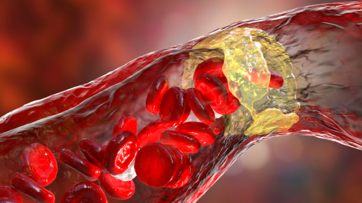
That’s how we found our lead program, Faun1003: by looking how the 13-lined ground squirrel responds to low oxygen. We found a signature that was highly protective in squirrels and mapped it to a small molecule that we are now internally optimizing for use in pulmonary fibrosis.

I understand that, at least for now, you decided to bet on small molecules. Could you explain this decision?

At the core of Fauna Bio, we’re modality-agnostic. We just try to find the best genetic target for a disease that we’re working on. Internally, we’ve optimized the platform to be able to rapidly find small molecules that hit our genes of interest, because it’s a good starting point.

But, not all targets that we find are amenable to a small molecule approach. So, the second program that I mentioned, the metabolism program, is an AAV therapy program, where we are working on a specific gene that turns on in the brain when it needs to switch to beta-hydroxybutyrate or lactate.

As other investigators have found, that gene is very relevant for diseases like retinitis pigmentosa. So, we’re looking at that as a potential gene-agnostic therapy for retinitis pigmentosa, along with some other genes. Internally, we’ve built a few more modules for small molecules development, but we’re not exclusive to that.

Can you give me an example of one such animal superpower and how you worked with that?

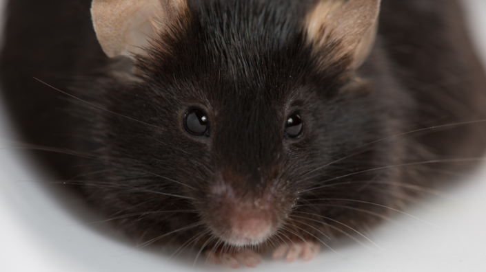
Let’s take the 13-lined ground squirrel, which is one of the best-described models of deep hibernation. Every organ in their body is adapted to survive extreme changes in oxygen levels and temperature. They can increase their metabolic rate 235-fold in an hour. That’s, by the way, one of the reasons we are partnering with Novo Nordisk on programs for obesity: they’re looking for ways to increase energy expenditure in humans, particularly in those that have lost a lot of weight on drugs like semaglutide. After losing weight, people reset their metabolism to a lower level. It’s known as “the biggest loser effect”. You lost a lot of weight, but then your body slows down metabolism, and you’re stuck there. It’s hard to change this baseline, but these animals do it every couple of weeks.

That was the basis of that exploration. We have other partnerships around other species, we’ve expanded beyond the 13-lined ground squirrel. We now work with a lab at UNLV with Frank van Breukelen. He works on a species called tenrecs. They’re amazing, they look a bit like hedgehogs, but they are actually closely related to African elephants.

What’s interesting about tenrecs is that they’re able to maintain aspects of stem-like quality in their heart cells for much longer in adulthood, so their hearts retain some ability to regenerate even later in life. We are now sequencing tenrecs to see if we can replicate that ability to maintain stemness. That should help with cardiac repair and resistance to damage.

We’re also working with a group at the University of Florida, which happens to be where I went to vet school. There’s a consortium of six or seven labs that are working with a species called the spiny mouse. It’s an African mouse that’s highly adapted to be able to repair damage to many organs without scarring. People originally started looking into them because they could repair large skin defects without any scarring. Then scientists realized that those mice can actually repair parts of their brain, including spinal cord, parts of their kidneys, all that, without laying down any scar tissue, unlike us humans.

We have an intern from the University of Montana who studies highland-adapted deer mice – that is, mice adapted to high-altitude, low-oxygen environments. This is very similar to human populations in places like Nepal. There are many implications here for mitochondrial biology, how the body uses energy and ATP.

These are just examples. We have an internal team called The Dream Team, which stands for “Discovery, Research, and Emerging Animal Models”. It’s a cross-functional team of physiologists, genomicists, and wet lab folks who go out and talk to various investigators, trying to find people who are doing the best science with the most interesting models.

There are indeed so many marvels of nature around that it must be hard to choose which ones to work on.

It’s a bit like what happens when we talk to Big Pharma. They have a strategic roadmap of disease areas, indications, and data types they want to work with. Similarly, we’re looking for species that have well-documented protection phenotypes, where the related diseases map into our indication spaces of high unmet need and commercial tractability. We have a good partnering interest, and we know we can develop molecules in a disease area that has commercial potential. We have our own internal rubric, literally a flowchart of yes or no decisions about what partnerships make the most sense for us.

It’s impressive that an early-stage startup has forged partnerships with entities like NIH and Novo Nordisk. Have you also encountered a “you people are crazy” kind of attitude?

It’s really funny: people either love what we do and are so thrilled that we exist, because they’ve always thought a company like this should exist, but they never knew there was one, or they think we’re a bit crazy.

We have people working for us who found us through webinars and other activities. We didn’t even have any job openings, but they were, like, “I have to work for you, you’re the only company that I want to work for”. And we found homes for those people because they really believe in what we do. But yes, it’s usually either one or the other.

Fauna Bio is probably a very exciting place to be.

It’s a lot of fun. I just talked to a grad student last week. She’s looking at all the labs in the field, she had interviews with Emma Teeling and a few other folks, and she was, like, “oh, I didn’t know there was a company that was doing this”.

So, yes, we get a lot of that. People are so excited when they find out that there’s a way to actually translate the work that’s coming out of these labs. I think that’s where people really get frustrated as postdocs and early-stage PIs: how do you actually start to translate these amazing findings?

It takes a lot of setting up the infrastructure, figuring out what it takes to build drug programs, what are pharma partners looking for, what does a good tractable commercial indication look like? There’s a lot of learning to do in terms of how you translate the work, but the science is in a place where genomes are good enough, the sequencing is cheap enough. It’s not an impossible hill to climb anymore, with us being able to get good molecular characterization of some of these emerging model species and look directly at a link between them and human diseases. It’s this beautiful intersection of technology and some of the work that we do that allows us to get into these new species.

Would you say that you have created a blueprint for other companies to jump in?

I’d like to think that we’ve shown it’s possible, that there’s a path to directly translating these insights into therapies that can help humans live longer and healthier, which is what we all want to do, but there are many other kinds of interesting biology that are just not necessarily a good fit for us. For instance, there are people who work on different properties of venom, which, pharmacologically, has been a rich place for discovery. That’s where we got things like ACE inhibitors and GLP-1 agonists from. So, there are many different aspects of natural biology that are under-leveraged, and there’s a huge amount of opportunity here. People don’t think to look outside humans and a few model organisms. You have to know that the data is there and what to do with it.

Have you considered working with long-lived species, such as the naked mole rat or bats?

Yes, we get this question all the time. We do. We have been thinking about the right way to ask the right questions about these species. But I think we’re getting an advantage by working with species such as hibernating mammals. If you take them as a class, they tend to live about 30% to 50% longer than their non-hibernating cousins, and there are many links between aging and hibernation in terms of DNA repair and protection and cellular regeneration.

Another nice thing about hibernation, discovery-wise, is that it’s transient. Part of the year they’re protected, and part of the year they’re not. So, you can compare those timepoints directly, looking at RNAseq profiles and organs at different time points, and the signal just jumps out in terms of what genes are responsible.

If you have an animal like the naked mole rat, they’re protected all the time. Trying to narrow it down to what’s driving that protection is pretty hard, but we have an internal team that’s thinking about how we can use data from some of those long-lived species.

That’s a long-standing question for us: “What’s the right data type, what’s the right disease area?” There’s been a lot of work characterizing those models and not so much work in the translation space, I mean high-throughput, high-quality -omics data generated from some of those species as we would need to do our thing.

We’re actually talking to one pharma partner who has an interest in some of these long-lived species in certain disease areas, but part of this is that we would need to do some robust sequencing to get the kind of data we need to feed into our platform. The currently available datasets are just not good enough. So, that’s a great question, and we think about it internally all the time, but it requires the right datasets, the right time points, the right samples, and the right disease area.

So, it has something to do with you being a company, right? You can’t just study long-lived species, you have to work towards a certain indication.

Yes, obviously, you have to rapidly translate insights into action. We must think a lot about how we can rapidly find a chemical matter, or at least genetic targets that modify human diseases. Our first layer of validation in the wet lab is in human cells and disease-relevant tissues in either 2D or 3D. Things must pass that filter before we move into more traditional preclinical animal studies. So, yes, we have to be able to rapidly find things that we can test.

A somewhat related question: have you also looked at underlying causes of aging, such as oxidative stress or DNA damage that some of these species are so good at mitigating?

Yes, sure. That’s another reason why hibernating mammals make such a good starting point for this kind of investigation. A lot of oxidative stress and inflammation is generated when they re-warm over the course of an hour every couple of weeks. This rapid cycle of rewarming and cooling is particularly damaging. Some species have developed ways to elongate telomeres, others have enhanced DNA repair and protection.

In our own data, we see upregulation of a fetal genetic program. If you look at heart cells that are damaged at the peak of one of these rewarming cycles, you see genes coming up that are usually expressed in neonatal animals at the time points where they are still growing their heart tissue. They apparently reactivate their fetal regeneration programs as part of a reparative mechanism, and that very obviously links to aging mechanisms.

What’s next for Fauna.bio and your subfield in general, if there’s even a subfield?

I guess our subfield would be comparative physiology, writ large, and studying protective mechanisms in natural animal models. Like I said, it’s a really exciting time. I think this field of investigation is where the longevity field was about ten years ago: there are many interesting datasets, people are starting to look at this space, to translate insights out of it. There are also many academic labs working on it, and there are other companies that are looking at other species. Some of those companies are still in stealth, but they do exist. I think it will be interesting to see in the next few years how many companies will be taking a similar approach.

There are companies that are looking at other aspects of extreme biology, and with some of them, we share our VC backers. I’m talking about companies like Basecamp Research, which is looking at extreme proteins. Another company, called Enveda, is working with natural compounds and plants for drug discovery. There’s also one called Wild Biotech that’s looking at microbiomes in different species.

I think people are realizing that with tools currently available, many more areas have become open for discovery, and then the question is always how you translate it and commercialize it. But people are starting to figure out what those business models look like.

What’s in your pipeline? When will we see something tangible from Fauna Bio?

Hopefully, pretty soon. Like I said, we have a small molecule that’s in lead optimization right now. We’re hoping to have it in the clinic as early as 2025. It’s for some types of pulmonary fibrosis that are particularly related to the mechanism we’re working on.

We could also have a gene therapy coming along right behind that. Fauna Bio could become a clinical company not too long from now, which is amazing considering that we started from generating our own datasets not that many years ago. It’s a great time, and we’re finally seeing the fruits of all of our labors and getting some very good results.

We would like to ask you a small favor. We are a non-profit foundation, and unlike some other organizations, we have no shareholders and no products to sell you. All our news and educational content is free for everyone to read, but it does mean that we rely on the help of people like you. Every contribution, no matter if it’s big or small, supports independent journalism and sustains our future.
Muscle factor

Promoting Muscle Regeneration With an Immune Factor

A paper published today in Nature Aging has shown that a macrophage-regulating factor has a significant impact on muscle regeneration.

Healing slows with aging

The researchers note two key reasons for the decline of regenerative abilities with aging. The first is stem cell exhaustion, the gradual depletion of stem cells and ensuing lack of functional cells. The second, which this paper focuses on, is changes to the surrounding environment, the effects of which are the subject of well-known parabiosis experiments.

However, instead of experimenting with young blood as a whole, the researchers focus on just one of its factors: mesencephalic astrocyte-derived neurotrophic factor (MANF) [1], which declines with aging and has been shown to have positive effects on the retina [2] and other tissues [3].

A factor necessary for regeneration

The researchers created a population of mice that stopped producing MANF upon exposure to a hormone therapy. When the quadriceps of these mice were injured and MANF blocked, these mice were found to have substantially weaker regeneration, with fewer new muscle fibers, fewer muscle stem cells, and many more dead fibers remaining in the tissue.

This is explained by the cellular populations in the tissue. Mice without MANF2 were found to have roughly the same amounts of pro-inflammatory macrophages but far fewer macrophages associated with regeneration. By blocking MANF at different times, the researchers showed that this particular effect occurs while the animal is injured; temporarily blocking MANF before an injury did not have an effect.

Further experiments in cellular culture, using cells from wholly MANF-deficient mice, showed that MANF is essential to macrophages changing their phenotypes towards regeneration and away from inflammation. MANF was also found to be valuable for lysosomal degradation of foreign proteins, and macrophages without it were unable to properly respond to necrotic debris, which is a logical explanation for the dead fibers remaining in MANF-deficient mice.

Restoring age-related MANF decline

This study confirmed that old (22-24 months) mice have substantially fewer healing-oriented macrophages than young (2-6 months) mice. Gene expression analysis showed that their macrophages had some things in common with young, MANF-deficient macrophages, particularly the problems with lysosomal degradation, but the aged cells also had other problems with cellular movement structures.

The researchers performed their final and most crucial experiment, delivering recombinant MANF to aged mice. This treatment worked; macrophages were restored to youthful populations, necrotic fibers were more thoroughly cleared, and new muscle fibers were created in greater quantities.

MANF therapy at 4 μg i.m. resulted in a complete rescue of the repair-associated myeloid response.

Conclusion

While this is a mouse study, MANF appears to play the same basic biological role in human beings. Further work, development, and clinical trials will be necessary to determine if MANF-based treatments can restore health and function to old, damaged muscle tissue.

We would like to ask you a small favor. We are a non-profit foundation, and unlike some other organizations, we have no shareholders and no products to sell you. All our news and educational content is free for everyone to read, but it does mean that we rely on the help of people like you. Every contribution, no matter if it’s big or small, supports independent journalism and sustains our future.

Literature

[1] Sousa-Victor, P., Neves, J., Cedron-Craft, W., Ventura, P. B., Liao, C. Y., Riley, R. R., … & Jasper, H. (2019). MANF regulates metabolic and immune homeostasis in ageing and protects against liver damage. Nature metabolism, 1(2), 276-290.

[2] Neves, J., Zhu, J., Sousa-Victor, P., Konjikusic, M., Riley, R., Chew, S., … & Lamba, D. A. (2016). Immune modulation by MANF promotes tissue repair and regenerative success in the retina. Science, 353(6294), aaf3646.

[3] JOntti, M., & Harvey, B. K. (2020). Trophic activities of endoplasmic reticulum proteins CDNF and MANF. Cell and Tissue Research, 382(1), 83-101.

The Journal Club is a monthly livestream hosted by Dr. Oliver Medvedik which covers the latest aging research papers.

Journal Club March 2023 + Zoom Connection Details

The Journal Club returns on Tuesday 28th March at 12:00 PM Eastern time to the lifespan.io Facebook channel. This month Dr. Oliver Medvedik will be exploring a recent study which looked at the effects of prolonged fasting on human macrophages and how metabolites from that fasting increased median lifespan in Caenorhabditis elegans.

Abstract

Periodic prolonged fasting (PF) extends lifespan in model organisms and ameliorates multiple disease states both clinically and experimentally owing, in part, to its ability to modulate the immune system. However, the relationship between metabolic factors, immunity, and longevity during PF remains poorly characterized especially in humans.

This study aimed to observe the effects of PF in human subjects on the clinical and experimental markers of metabolic and immune health and uncover underlying plasma-borne factors that may be responsible for these effects.

Join the livestream

Lifespan Heroes get access to the livestream using the connection details below:

Join Zoom Meeting https://lifespan-io.zoom.us/j/88373018752?pwd=RC9KcEZKOU1YMDFOTlpGaEt6ZzRWZz09 Meeting ID: 883 7301 8752 Passcode: 374339

Literature

Rhodes, C. H., Zhu, C., Agus, J., Tang, X., Li, Q., Engebrecht, J., & Zivkovic, A. M. (2022). Human fasting modulates macrophage function and upregulates multiple bioactive metabolites that extend lifespan in Caenorhabditis elegans: a pilot clinical studyThe American Journal of Clinical Nutrition.

Running seniors

Long-Term Resistance Exercise Increases Autophagy

In a new systemic review published in Autophagy Reports, researchers have demonstrated that exercise plays a role in regulating autophagy, depending on its type [1].

Manipulating autophagy 

Disabled autophagy was recently acknowledged as a hallmark of aging. Indeed, boosting the ability of cells to get rid of unnecessary and damaged organelles and molecules is considered a promising anti-aging strategy.

As shown in animal and some human studies, autophagy activation can be achieved with fasting and exercise, therapeutic interventions such as rapamycin and lithium, and even ultrasound treatment.

At the same time, just like with any other molecular process, autophagy manipulation should be fine-tuned in a context-specific manner to achieve beneficial results. Hyperactivation of autophagy might be toxic in some situations, such as for DNA damage repair [2].

Exercise is believed to activate autophagy, particularly mitophagy, the removal of damaged mitochondria. This conclusion mostly comes from animal studies, whereas human data on exercise-induced autophagy is less consistent.

In this systematic review, the researchers analyzed the results of 26 studies assessing the autophagic response to exercise in humans. The studies measured various autophagy markers in skeletal muscles, peripheral blood mononuclear cells (PBMCs), or both in participants engaging in various exercise forms and intensities.

Autophagy response to exercise

First, the researchers showed that the levels of two autophagy markers, microtubule-associated proteins 1A/1B light chain 3B (LC3-II) and sequestosome 1 (SQSTM1), were consistent with reduced autophagy following an acute bout of resistance exercise and increased autophagy following long-term resistance exercise.

The researchers did not detect any change in LC3-II following either acute or long-term (over several weeks) moderate- and vigorous-intensity endurance exercise. Only long-term vigorous-intensity endurance exercise was accompanied by altered levels of SQSTM1.

Next, the researchers found a tissue-specific autophagy response to exercise. Exercise was associated with lowered LC3-II and increased SQSTM1 levels in skeletal muscle, which corresponds to attenuated autophagy. This effect was largely due to resistance exercise. The opposite effect, induced autophagy, was observed in PBMCs in response to exercise. This suggests distinct autophagy regulation mechanisms in different tissues.

Finally, the researchers showed that several other autophagic and mitophagic markers, such as BNIP3, Beclin-1, and ATG12, were increased in the analyzed studies, including those that involve endurance exercise. However, because it was not accompanied by changes in LC3-II and SQSTM1 levels, the effect of endurance training on autophagy remains unclear.

Abstract excerpt

Our findings demonstrate that physical exercise probably regulates autophagy in an exercise modality- and tissue-dependent manner in humans, although further investigation is needed. Customized exercise prescriptions should be aimed for when implementing exercise to regulate autophagy in humans.

Conclusion

This study demonstrated that long-term resistance training might be the best exercise for increasing autophagy in humans. The beneficial health effects of other exercise types, such as endurance training, might be via mechanisms other than activated autophagy.

The limitations of this systematic review include autophagy measurements limited to western blot analysis of some protein markers, analysis of only two tissues, and studies conducted mostly on young and middle-aged healthy participants.

We would like to ask you a small favor. We are a non-profit foundation, and unlike some other organizations, we have no shareholders and no products to sell you. All our news and educational content is free for everyone to read, but it does mean that we rely on the help of people like you. Every contribution, no matter if it’s big or small, supports independent journalism and sustains our future.

Literature

[1] Chen X-K, Zheng C, Siu PM-F, Sun F-H, Wong SH-S, Ma AC-H. Does Exercise Regulate Autophagy in Humans? A Systematic Review and Meta-Analysis. Autophagy Reports 2023; 2: 2190202.

[2] Guo C, Zhao Y. Autophagy and DNA damage repair. Genome Instability & Disease 2020; 1: 172–183.

Mediterranean Diet Might Lower Risk of Dementia

Mediterranean Diet Might Lower Risk of Dementia

In a large-scale observational study, British scientists have shown that high levels of adherence to the Mediterranean diet might substantially lower the risk of dementia [1].

A healthy diet

The Mediterranean diet, which is based on ingredients such as olive oil, vegetables, legumes, fish, and whole grains, is considered one of the healthiest in the world. While most of the evidence comes from observational studies that have well-known limitations, there is also a handful of randomized clinical trials [2]. The Mediterranean diet has been linked to decreased risk of cardiovascular and all-cause mortality [3], improved cognitive function [4], and other health benefits, but data on its effect on dementia has been sparce and inconclusive.

Power of the study

This study, like many other recent studies, was based on data from UK Biobank, a huge repository of health information on some half a million of British citizen. Compared to previous studies, it had several strengths, including its size. The researchers assembled a cohort of about 60 thousand older adults with 882 cases of dementia recorded during a 9-year average follow-up period, which allowed for considerable statistical power.

A known problem with such studies is that they often make do with a single questionnaire to ascertain dietary patterns. In this cohort, however, participants not only filed in a detailed 24-hour assessment of their diets online, but most did this more than once, which allowed the researchers to gauge adherence.

The researchers employed two similar scores widely used to measure adherence to the Mediterranean diet: MEDAS and PYRAMID. Moreover, they improved the existing MEDAS scoring system that awards a binary score for 14 food types (you either meet the requirement or not) by making it continuous. The authors also adjusted for numerous potential confounding variables, including age, socioeconomic status, educational attainment, BMI, physical activity, smoking, and even sleep duration, and conducted several sensitivity analyses.

More adherence is associated with more benefits

In the fully adjusted model, high adherence to the Mediterranean diet according to the continuous MEDAS score was associated with a 23% decrease in the risk of all-cause dementia. This result was highly statistically significant. High adherence according to the PYRAMID score was associated with a smaller 14% risk decrease and nearly reached the significance threshold. Medium adherence did not seem to provide a lot of protection. A sensitivity analysis showed the negative correlation between Mediterranean diet and fatal cases of dementia to be the strongest.

Med diet graph

Few other studies have considered the relationship between diet and genetic risk for dementia. Those that did mostly limited it to the APOE genotype, which is known to be strongly correlated with Alzheimer’s risk. In this study, the researchers developed a polygenic score that combined information from multiple weighted risk alleles.

Importantly, no significant interaction between adherence to the Mediterranean diet and a polygenic risk for dementia was detected. In other words, the Mediterranean diet seemed to help lower the risk of dementia even in people who were genetically predisposed to it. However, this association did not hold in some scenarios, which is why the authors of the study call for more research.

Limited demographic scope

The researchers also note an important limitation inherent to UK Biobank: it mostly contains data on people who self-reported their ethnic backgrounds as British, Irish, or otherwise European, which may limit the applicability of the results to other populations. The scientists were also unable to reliably ascertain consumption of one of the most important components of the Mediterranean diet, olive oil, as the questionnaire only asked participants whether they used olive oil for cooking. It is possible that with this data available, the correlation between the Mediterranean diet and risk of dementia would have been even more robust.

Dr Oliver Shannon of Newcastle University, one of the study’s lead authors, notes in a blog post that currently, our options for treating dementia are very limited, which makes prevention especially important. “Much more research is needed to identify the best diet that people could follow to try and reduce their risk of dementia”, Shannon admits. “However, the findings from our study contribute towards a growing body of evidence to suggest that following a more Mediterranean-like diet could be an effective way to help ‘oil your cogs’ and reduce your risk of developing dementia”.

Conclusion

This study, while observational and hence insufficient to establish causation, provides further evidence of the Mediterranean diet’s connection to lifespan and healthspan.

We would like to ask you a small favor. We are a non-profit foundation, and unlike some other organizations, we have no shareholders and no products to sell you. All our news and educational content is free for everyone to read, but it does mean that we rely on the help of people like you. Every contribution, no matter if it’s big or small, supports independent journalism and sustains our future.

Literature

[1] Shannon, O. M., Ranson, J. M., Gregory, S., Macpherson, H., Milte, C., Lentjes, M., … & Stevenson, E. (2023). Mediterranean diet adherence is associated with lower dementia risk, independent of genetic predisposition: findings from the UK Biobank prospective cohort study. BMC medicine, 21(1), 1-13.

[2] Valls-Pedret, C., Sala-Vila, A., Serra-Mir, M., Corella, D., De la Torre, R., Martínez-González, M. Á., … & Ros, E. (2015). Mediterranean diet and age-related cognitive decline: a randomized clinical trial. JAMA internal medicine, 175(7), 1094-1103.

[3] Hidalgo-Liberona, N., Meroño, T., Zamora-Ros, R., Rabassa, M., Semba, R., Tanaka, T., … & Cherubini, A. (2021). Adherence to the Mediterranean diet assessed by a novel dietary biomarker score and mortality in older adults: the InCHIANTI cohort study. BMC medicine, 19(1), 1-13.

[4] Tsivgoulis, G., Judd, S., Letter, A. J., Alexandrov, A. V., Howard, G., Nahab, F., … & Wadley, V. G. (2013). Adherence to a Mediterranean diet and risk of incident cognitive impairment. Neurology, 80(18), 1684-1692.

Elderly lifting

Vital Muscle Enzyme Declines With Aging

Research published in Nature Metabolism has described PCYT2, an enzyme essential for muscle function, and how it declines with aging.

A necessary lipid synthesizer in muscle

Human beings need PCYT2 as part of the Kennedy pathway to synthesize two critical components of the phosopholipid membrane that surrounds cells [1]. People with genetic deficiencies of PCYT2 have stunted growth, severe motor function disorders, and muscle weakness [2], and people with similar deficiencies in the Kennedy pathway have similar symptoms [3].

The researchers recapitulated similar findings in zebrafish and mice. Without PCYT2, both of these species were considerably smaller than normal. Vital lipids, including long-chain fatty acids, were substantially depleted in the leg muscles of young PCYT2-deficient mice, which also had substantially thinner fibers. Further examination showed that the cellular membranes of their muscle cells were looser, weaker, and softer than those of normal mice.

As expected, mice with this condition did not age well. These mice frequently had spinal problems, and muscle wasting and atrophy, along with the associated low bone density, were common afflictions.

Interestingly, PCYT2 only seems to be necessary for muscle tissue. Mice that were genetically engineered specifically to lack PCYT2 in fat tissues seemed to be totally unaffected at any age.

A potentially recoverable decline with aging

In both mice and people, PCYT2 declines with age, as shown by tissue taken from ordinary, wild-type mice and human volunteers. 45- to 62-year-old people have substantially less PCYT2 than 20- to 30-year-old people. The researchers surmise that this may be a big part of the reason why resistance training becomes less beneficial with age [4].

The researchers then tested whether gene therapy could have an impact. Injecting middle-aged, pre-sarcopenia mice with an adeno-associated virus (AAV) to deliver PCYT2 to muscle tissue, they found that the affected mice had significantly higher grip strength, muscle fiber size, and muscular mitochondrial function than the control group of aged mice, although these values were not quite restored to the levels of young mice.

PCYT2 AAV

Conclusion

This research provides some evidence for, but does not prove, the possibility that PCYT2 deficiency with aging contributes to sarcopenia. However, as the authors state, upregulating this vital compound could be a potential treatment for muscle frailty with age. If clinical trials confirm this research, an effective gene therapy or mRNA-based treatment for frailty may be on the horizon.

Literature

[1] Gibellini, F., & Smith, T. K. (2010). The Kennedy pathway—de novo synthesis of phosphatidylethanolamine and phosphatidylcholine. IUBMB life, 62(6), 414-428.

[2] Vaz, F. M., McDermott, J. H., Alders, M., Wortmann, S. B., Kölker, S., Pras-Raves, M. L., … & Banka, S. (2019). Mutations in PCYT2 disrupt etherlipid biosynthesis and cause a complex hereditary spastic paraplegia. Brain, 142(11), 3382-3397.

[3] Ahmed, M. Y., Al-Khayat, A., Al-Murshedi, F., Al-Futaisi, A., Chioza, B. A., Pedro Fernandez-Murray, J., … & Crosby, A. H. (2017). A mutation of EPT1 (SELENOI) underlies a new disorder of Kennedy pathway phospholipid biosynthesis. Brain, 140(3), 547-554.

[4] Gault, M. L., & Willems, M. E. (2013). Aging, functional capacity and eccentric exercise training. Aging and disease, 4(6), 351.

Global obesity

The Human Cost of Metabolic Diseases

A new paper published in Cell Metabolism has shown the growing influence of metabolic diseases in an aging population.

Related diseases studied together

Metabolic diseases include hypertension, type 2 diabetes, hyperlipidemia, obesity, and non-alcoholic fatty liver disease [1]. These diseases, representing fundamental problems with how the human body uses energy, are heavily linked to systemic inflammation [2].

The authors note that previous studies relating to these metabolic diseases have measured each of them independently. This paper looks at these diseases collectively, from a public health perspective, treating these non-communicable diseases with the same gravity as a pandemic.

Metabolic diseases

Prevalence and mortality

This study, which was based on 2019 data, estimated that there were a full 43.8 million cases of type 2 diabetes, 18.5 million cases of hypertension, and a full 1.2 billion cases of non-alcoholic fatty liver disease: roughly a seventh of the world’s population. The other two diseases could not be accurately measured.

Between 2000 and 2019, when controlling for age, the per-capita death rate has been either holding steady or, in the case of hyperlipidemia, on a downtrend. Of course, with an aging and growing population, the absolute number of people dying from these diseases has been growing rapidly. Globally, obesity was the main contributor to these metabolically related deaths; however, this varied greatly by countries, with poorer countries being less obese and more prone to diabetes-related mortality after standardization for age.

Non-alcoholic fatty liver disease substantially increased, even after adjustment for age, particularly in males. The prevalence of this metabolic disease increased almost a full percentage point every year from 2000 to 2019, a situation that the authors describe as “alarming”. Fortunately, the total amount of suffering and death, as measured by disability-adjusted life years (DALYs) and mortality, seemed to decrease for people who have this disease. The authors hypothesize that this might be due to increased diagnosis of this disease, even when its symptoms are not particularly acute.

Recommendations

As this is a policy-oriented study for global health, the authors spend time discussing what might be done. They hold that the current environment for most people is obesogenic, tending towards diets and practices that lead to obesity, and they list genetics and modifiable risk factors such as smoking and sedentary lifestyles. They also note that type 2 diabetes medications that protect against cardiovascular events might reduce the death and suffering caused by such events [3].

Conclusion

The elephant in the room is, of course, aging. The authors specifically tune their analysis to exclude the effects of aging and differentially aged populations for exactly this reason; aging has a drastic effect on everything they are attempting to measure.

However, the unspoken assumption, on both an individual and population level, is that it is not modifiable. Therefore, treatments that focus on the age-related root causes of metabolic diseases are not mentioned, and the policy recommendations given are those that are well known but very hard to implement on a wide scale: diet, exercise, and other lifestyle-related habits.

As this paper points out, the total death and suffering caused by metabolic diseases is enormous: more than ten million people died of such diseases in 2019 alone. If policymakers are going to do something about them, they would be well-advised to look for interventions that strike them at their root.

We would like to ask you a small favor. We are a non-profit foundation, and unlike some other organizations, we have no shareholders and no products to sell you. All our news and educational content is free for everyone to read, but it does mean that we rely on the help of people like you. Every contribution, no matter if it’s big or small, supports independent journalism and sustains our future.

Literature

[1] Ng, C. H., Wong, Z. Y., Chew, N. W., Chan, K. E., Xiao, J., Sayed, N., … & Muthiah, M. (2022). Hypertension is prevalent in non-alcoholic fatty liver disease and increases all-cause and cardiovascular mortality. Frontiers in Cardiovascular Medicine, 9.

[2] Sethi, J. K., & Hotamisligil, G. S. (2021). Metabolic Messengers: tumour necrosis factor. Nature metabolism, 3(10), 1302-1312.

[3] Chew, N. W., Ng, C. H., Muthiah, M. D., & Sanyal, A. J. (2022). Comprehensive review and updates on holistic approach towards non-alcoholic fatty liver disease management with cardiovascular disease. Current Atherosclerosis Reports, 24(7), 515-532.

Capitol Hill

Rep. Bilirakis on the Longevity Science Caucus

We in the longevity field have received powerful allies on Capitol Hill with the creation of the bipartisan Congressional Caucus for Longevity Science. We had the opportunity to ask questions of one of its co-chairs.

Longevity is bipartisan

The fight against aging must become one of humanity’s main priorities if we want to see meaningful progress on a global scale. This requires recruiting allies among politicians and other decision makers.

Recently, a major step in that direction was made. Reps. Gus Bilirakis (R-FL) and Paul Tonko (D-NY) announced the formation of the Congressional Caucus for Longevity Science. According to the press release, the caucus “aims to educate Members about the growing field of aging and longevity biotechnology, and promote initiatives aimed at increasing the healthy average lifespan of all Americans.”

The two co-chairs are joined by three other founding members: Dan Crenshaw (R-TX), Michael Burgess (R-TX), and Anna Eshoo (D-CA). In a time when political divisions run deep, both Bilirakis and Tonko stressed and praised the bipartisan nature of this endeavor in their comments.

Policy, budget, research

A congressional caucus is a force to be reckoned with. For instance, it can promote legislation that would improve the FDA’s stance on longevity drug trials, influence budget allocation, and use the vast analytical resources available to Congress to further investigate the economic and societal benefits of healthspan and lifespan extension.

The Alliance for Longevity Initiatives (A4LI), a lobbying group founded in 2021, helped facilitate the formation of the caucus. You can read our interview with A4LI’s founder and CEO Dylan Livingston here. The president of lifespan.io, Keith Comito, is an A4LI board member.

We contacted Rep. Bilirakis and asked him a few questions about the caucus.

Rep Bilirakis

How can politicians help longevity research?

It is important for policymakers to stay up to date on longevity research, as we have a unique platform that allows us the chance to highlight the development that is occurring in this space. Additionally, with my role in Congress, I welcome the opportunity to shape forward-focused policy with a strong public-private partnership that allows innovation to flourish.

What was your motivation for founding the caucus?

I helped form this caucus to learn more about the growing field of aging and longevity biotechnology and look forward to the opportunity to promote initiatives that work towards and encourage the healthy average lifespan of all Americans. Truly, I want all Floridians and all Americans to live their fullest, healthiest lives in a way in which health care costs don’t break the bank for them as they enjoy their retirement years.

What would be your answer to the various misconceptions about life extension, such as that it will lead to overpopulation and increased healthcare costs, or that longevity therapies will only be available to the rich?

Increasing life expectancy and promoting positive health outcomes is an extremely important priority, and we formed the caucus so that we can address such misconceptions. The goal is not just for people to live longer, but also for people to live longer, healthier and more enriching lives. Proactive and preventative health measures will undoubtedly help reduce healthcare costs, which is a win-win. Many business leaders and economists have expressed concern about the declining US birthrate and its implications for the US workforce.

The US birthrate has declined by almost 20% since 2007. With fewer babies being born to replace our aging population, it is a reasonable concern. Therefore, the work the Caucus is doing to ensure our aging population stays as healthy as possible for as long as possible, could potentially have a positive impact on the economy as it could help prevent workforce interruptions.

What economic impact would an increase in healthy longevity have?

We anticipate that healthy longevity will lend itself to a full and productive workforce, which will be wonderful for the United States economy. Additionally, through improved disease prevention and maintenance, the healthcare system would realize potential cost savings.

Do you have concrete actions planned for the Caucus?

We look forward to growing membership and pursuing educational opportunities for Members of Congress.

Are you in touch with prominent longevity scientists and other leaders in the field?

Yes, we are very excited to work with a multitude of stakeholders, including prominent longevity scientists. It is a fast-growing field, and I certainly welcome the opportunity to stay informed on the work that is being done.

What is your positive message to Americans about healthy longevity?

It is important that Americans feel empowered to take ownership of their health and that they have access to resources that allow them to live full, healthy, long lives.

We would like to ask you a small favor. We are a non-profit foundation, and unlike some other organizations, we have no shareholders and no products to sell you. All our news and educational content is free for everyone to read, but it does mean that we rely on the help of people like you. Every contribution, no matter if it’s big or small, supports independent journalism and sustains our future.
Sleep apnea

Sleep Apnea and Inflammatory Biomarkers of Tooth Decay

A study published in Heliyon has explained a relationship between sleep apnea and an increase of inflammatory factors in the mouth that are connected to the gum disease peridontitis.

LLP Biostarks
Check out Biostarks to learn more about their test kits.

Connected dangers

This paper begins with a discussion of peridontitis, which is caused by infection with anaerobic organisms [1] that form pockets in the gums, leading to tooth loss. Peridontitis is associated with multiple diseases, some of which are directly connected to aging, including cardiovascular disease [2], arthritis [3], and diabetes [4].

In this study, the researchers focus particularly on obstructive sleep apnea, a disorder that is characterized by a collapse of the airway during sleep, resulting in reduced airflow and impeded breathing and linked to respiratory diseases [5].

It should be unsurprising that the common thread linking all these various disorders is inflammation, and multiple research papers have linked sleep apnea to increased inflammatory biomarkers [6]. Here, the researchers identify those biomarkers individually.

Five different factors

The researchers focused on five different biomarkers: the four interleukins IL-1β, IL-6, IL-17A, and IL-33, along with the well-studied inflammation-linked cytokine TNF-α. While this study was relatively small, with a total of only 90 people, it had significant results even in the most basic measurements: people with severe sleep apnea were statistically likely to have severe peridontitis.

While all of these biomarkers seemed to have correlations, only IL-6 reached the level of statistical significance between groups. Interestingly, IL-6 seemed to be richer in the gingival crevicular fluid taken from the gums of people with sleep apnea, while peridontitis was associated with richer IL-6 concentrations in the saliva. The presence of Candida yeast in the mouth was found to increase cytokines as well.

This chart shows a percentile-based analysis of the concentrations of inflammatory cytokines in each group and subgroup. Inflammatory tendencies

Conclusion

Does sleep apnea cause peridontitis, or does peridontitis cause sleep apnea? While this is an association study that does not prove causation, this study bolsters previous research showing that that the dry mouth [7] and lack of oxygen caused by sleep apnea increase peridontitis-related inflammation [8]. While some research is conflicting, the authors believe that the association is bidirectional and that systemic inflammation is a contributor to both conditions.

Whatever the causal relationship is, this study shows the value of biomarkers in understanding disease, particularly if they are taken from different sources in the same person. Peridontitis is relatively easy to detect in the dentist’s office, but biomarkers of inflammation can better show the true extent of inflammation. While sleep apnea is notably difficult to treat, anyone with this condition is particularly encouraged to maintain good oral health to minimize the risk of bacterial infection, subsequent inflammation, and inflammation-related, age-related diseases.

We would like to ask you a small favor. We are a non-profit foundation, and unlike some other organizations, we have no shareholders and no products to sell you. All our news and educational content is free for everyone to read, but it does mean that we rely on the help of people like you. Every contribution, no matter if it’s big or small, supports independent journalism and sustains our future.

Literature

[1] Cortelli, S. C., Cortelli, J. R., Romeiro, R. L., Costa, F. O., Aquino, D. R., Orzechowski, P. R., … & Duarte, P. M. (2013). Frequency of periodontal pathogens in equivalent peri-implant and periodontal clinical statuses. Archives of oral biology, 58(1), 67-74.

[2] Bui, F. Q., Almeida-da-Silva, C. L. C., Huynh, B., Trinh, A., Liu, J., Woodward, J., … & Ojcius, D. M. (2019). Association between periodontal pathogens and systemic disease. Biomedical journal, 42(1), 27-35.

[3] Xiao, F., Li, C., Lin, Y., Peng, Z., Xu, X., Wen, Y., … & Zhang, P. (2021). Increased risk of periodontitis occurrence in patients with rheumatoid arthritis and its association with the levels of IL-1β and TNF-α in gingival crevicular fluid. Ann. Palliat. Med, 10, 9078-9087.

[4] Arregoces, F. E., Uriza, C. L., Porras, J. V., Camargo, M. B. F., & Morales, A. R. (2014). Relation between ultra-sensitive C-reactive protein, diabetes and periodontal disease in patients with and without myocardial infarction. Arquivos Brasileiros de Endocrinologia & Metabologia, 58, 362-368.

[5] Locke, B. W., Lee, J. J., & Sundar, K. M. (2022). OSA and chronic respiratory disease: mechanisms and epidemiology. International Journal of Environmental Research and Public Health, 19(9), 5473.

[6] Shrivastava, A., & Chand, P. (2021). Molecular determinants of obstructive sleep apnea. Sleep Medicine, 80, 105-112.

[7] Gamsiz‐Isik, H., Kiyan, E., Bingol, Z., Baser, U., Ademoglu, E., & Yalcin, F. (2017). Does obstructive sleep apnea increase the risk for periodontal disease? a case‐control study. Journal of periodontology, 88(5), 443-449.

[8] Karatas, O., Balci Yuce, H., Tulu, F., Taskan, M. M., Gevrek, F., & Toker, H. (2020). Evaluation of apoptosis and hypoxia‐related factors in gingival tissues of smoker and non‐smoker periodontitis patients. Journal of Periodontal Research, 55(3), 392-399.

Cholesterol

Inflammation Strongly Predicts Mortality After Statin Use

Analyzing data from three clinical trials, scientists have shown that excessive inflammation is a much stronger predictor of cardiovascular and all-cause mortality in patients on statins than excessive LDL cholesterol levels. These findings can upend the current standard of care for such patients [1].

The two types of risk

Statins, drugs that lower the levels of low-density lipoprotein (LDL), aka “bad cholesterol”, are doing a great job in reducing cardiovascular mortality [2]. However, in some people, LDL levels remain higher than the norm despite statin therapy, which elevates cardiovascular risk. Patients on statins are also more vulnerable if their inflammation levels are high. The former condition is known as residual cholesterol risk (RCR), and the latter as residual inflammatory risk (RIR) [3].

What was not known until now is each one’s exact contribution to mortality, which can be crucial in a choice of adjunctive cardiovascular therapy. According to a new paper published in the Lancet, RIR is the much more important factor.

Three different trials

The researchers analyzed data from three statin clinical trials – PROMINENT, REDUCE-IT, and STRENGTH. Participants were divided into four groups – those with neither RCR nor RIR, those with RCR only, those with RIR only, and those with both. This analysis was adjusted for multiple potential confounders, including age, gender, smoking status, BMI, blood pressure, and previous history of cardiovascular disease.

Blood levels of LDL-cholesterol and high-sensitivity C-reactive protein (hs-CRP), a measure of inflammation, were grouped into quartiles, with quartile limits being largely similar across all three studies. One of the studies included only people with diabetes, while in two others, diabetes patients constituted a majority. The mean BMI of all three studies’ participants was about 32.

CRP is the key

In all three trials, baseline CRP proved to be a significant predictor of mortality. The average fully adjusted hazard ratio for the highest vs the lowest CRP quartile was 1.31 for major cardiovascular events, 2.68 for cardiovascular mortality, and 2.42 for all-cause mortality. A hazard ratio measures how much more likely an event is to happen; an HR of 2.68 indicates an almost three-fold increase in likelihood.

The contribution of higher LDL cholesterol was much more modest. In combined data, it was statistically significant only for cardiovascular mortality (HR = 1.27) and all-cause mortality (HR = 1.16) at the highest quartile. Conversely, for CRP, the combined trial data was highly significant in all scenarios, except for cardiovascular events for the second quartile.

The danger of inflammation

According to Paul Ridker, MD, a preventive cardiologist at Brigham and Women’s Hospital and the study’s corresponding author, “the new data should be a wake-up call for preventive cardiologists and their patients.”

“Virtually all patients with or at risk for atherosclerotic disease are appropriately treated with aggressive statin therapy”, Ridker said. “Yet, in our study of patients already taking a statin, hsCRP – a measure of residual inflammatory risk – was a more powerful determinant of having a future heart attack or dying from cardiovascular disease than was LDL – cholesterol – a measure of residual cholesterol risk. The data are a powerful demonstration that to beat heart disease, we need to lower both cholesterol and inflammation, not just cholesterol alone.”

While several clinical trials have found that inhibiting inflammation reduces cardiovascular risks, the uptake of anti-inflammatory therapy in daily practice has been slow. One particular anti-inflammatory drug the paper mentions is colchicine. Although two large, randomized trials showed that colchicine reduces cardiovascular event rates at least on par with much more expensive cholesterol-lowering medications [4], it is still not widely used in this context.

Conclusion

This study constitutes an important addition to previous research that has highlighted inflammation as a major factor in atherosclerosis. The results strongly suggest that in high-risk patients who already receive statin therapy, cardiologists should strive to reduce excessive inflammation as opposed to excessive LDL levels. Apart from the obvious implications for the current standard of care in atherosclerosis, the study also adds to our growing understanding of how inflammation impacts various aspects of aging.

We would like to ask you a small favor. We are a non-profit foundation, and unlike some other organizations, we have no shareholders and no products to sell you. All our news and educational content is free for everyone to read, but it does mean that we rely on the help of people like you. Every contribution, no matter if it’s big or small, supports independent journalism and sustains our future.

Literature

[1] Ridker, P. M., Bhatt, D. L., Pradhan, A. D., Glynn, R. J., MacFadyen, J. G., & Nissen, S. E. (2023). Inflammation and cholesterol as predictors of cardiovascular events among patients receiving statin therapy: a collaborative analysis of three randomised trials. The Lancet.

[2] Bots, S. H., Onland-Moret, N. C., Jancev, M., Hollander, M., Tulevski, I. I., Hofstra, L., … & den Ruijter, H. M. (2022). Statins are associated with a large reduction in all-cause mortality in women from a cardiac outpatient population. Open Heart, 9(1), e001900.

[3] Kalkman, D. N., Aquino, M., Claessen, B. E., Baber, U., Guedeney, P., Sorrentino, S., … & Mehran, R. (2018). Residual inflammatory risk and the impact on clinical outcomes in patients after percutaneous coronary interventions. European heart journal, 39(46), 4101-4108.

[4] Fiolet, A. T., Opstal, T. S., Mosterd, A., Eikelboom, J. W., Jolly, S. S., Keech, A. C., … & Cornel, J. H. (2021). Efficacy and safety of low-dose colchicine in patients with coronary disease: a systematic review and meta-analysis of randomized trials. European heart journal, 42(28), 2765-2775.

Fat mouse closeup

Excessive Fat, Not Sugar, Leads to Obesity in Mice

In a new study published in Endocrinology and Metabolism, researchers have shown that fat rather than sugar is the macronutrient that drives obesity and other detrimental metabolic changes if it constitutes a large proportion of dietary calories [1].

Obesity, diet, and the microbiome

Obesity is associated with a dysregulation of several molecular pathways, which lead to chronic low-grade inflammation and accelerated aging. A connection between almost all age-associated diseases and obesity has been shown, although the exact mechanisms are often not known.

There are hot debates regarding what foods lead to obesity and how much impact they have, and this has spurred the development of various diets that promise great weight loss results. While the link between excessive food consumption and weight gain seems obvious, which dietary components are to blame has remained unclear.

In addition to inflammation, age-related microbiome alterations have recently been proposed as an additional hallmark of aging. Obesity is associated with altered microbiome composition [2], although the connection might be bidirectional.

The composition of the microbiome is partly shaped by the foods consumed. Therefore, this research focuses on understanding which food components promote gut health or cause detrimental metabolic changes leading to obesity. In this study, the researchers explored the link between the excessive consumption of different macronutrients, obesity, and gut microbiota in male C57BL/6J (Black 6) mice, an inbred strain that is commonly used in research.

The fattening

The researchers divided 50 mice into four groups: 20 mice on a low-sugar (7% calories) and low-fat (10% calories) control diet, 10 mice on a high-sugar diet (17% calories) diet, 10 mice on a high-fat (45% calories) and high-sugar (17% calories) diet, and 10 mice on a high-fat diet (60% calories).

After following their respective diets for 8 weeks, mice on the high-fat diet gained the most body weight and fat mass. These mice also showed the highest glucose intolerance among the four groups and developed insulin resistance, as did the group following a high-fat and high-sugar diet.

Next, the researchers dissected and analyzed the liver, brown and white adipose tissues, muscles, and hypothalami of the mice. They did not detect any liver inflammation in any of the groups, suggesting that this organ was not profoundly affected by the diets. However, they detected increased triglyceride levels in all the hypercaloric diets compared to the control group.

In addition, the researchers showed increased levels of some inflammatory markers in white adipose tissue surrounding the internal organs of mice fed either a high-fat diet or a high-fat and high-sugar diet. Adipocyte size was significantly bigger in mice on the high-fat diet.

The researchers also showed differential expression of several inflammatory markers in the muscle tissue and hypothalamus between mice fed high-sugar, high-fat, and high-sugar + high-fat diets. These results suggest that excessive consumption of either of these macronutrients leads to different metabolic changes

The gut

In the last set of experiments, the researchers analyzed the microbial composition and the microbial gene expression in the guts of the four groups of mice. They showed that dietary fat content was the major contributor explaining the variations between both the bacterial composition and gene expression of the groups. This means that fat intake determined the composition of the mice’s microbiomes to a large extent. Sugar was also a contributing factor, at least to the microbial gene expression variation.

Abstract excerpt

HF diet-fed mice showed the highest body weight and fat mass gains and displayed the most impaired glucose and insulin profiles. HS, HF/HS, and HF diets differently affected hepatic cholesterol content and mRNA expression of several markers associated with immune cells, inflammation, oxidative and ER stress in several organs/tissues. In addition, HF diet feeding resulted in a decreased microbial load at the end of the experiment. When analyzing the gut microbiota composition, we found that HS, HF/HS, and HF diets induced specific changes in the abundance of certain bacterial taxa. This was not associated with a specific change in systemic inflammatory markers, but HS mice exhibited higher FGF21 plasma levels compared with HF diet-fed mice. Taken together, our results highlight that dietary intake of different macronutrients distinctively impacts the development of an obese/diabetic state and the regulation of metabolic inflammation in specific organs. We propose that these differences are not only obesity-driven but that changes in the gut microbiota composition may play a key role in this context.

Conclusion

This study showed that excessive fat consumption is a major contributor to obesity, inflammation, and microbiotal changes in mice. While it is not clear if the same effect should be expected in humans, it seems reasonable to avoid a diet that is skewed towards heavy fat consumption.

We would like to ask you a small favor. We are a non-profit foundation, and unlike some other organizations, we have no shareholders and no products to sell you. All our news and educational content is free for everyone to read, but it does mean that we rely on the help of people like you. Every contribution, no matter if it’s big or small, supports independent journalism and sustains our future.

Literature

[1] Suriano F, Vieira-Silva S, Falony G, de Wouters d’Oplinter A, Paone P, Delzenne NM et al. Fat and not sugar as the determining factor for gut microbiota changes, obesity, and related metabolic disorders in mice. Am J Physiol Endocrinol Metab 2023; 324: E85–E96.

[2] Muscogiuri G, Cantone E, Cassarano S, Tuccinardi D, Barrea L, Savastano S et al. Gut microbiota: a new path to treat obesity. Int J Obes Suppl 2019; 9: 10–19.

Fitness watch

Epigenetic Biomarker for Measuring Aging Through Fitness

A new biomarker for measuring biological aging based on physical fitness has been published in Aging, and it has been found to be useful in predicting health issues.

LLP Biostarks
Check out Biostarks to learn more about their test kits.

A clock with a different purpose

Epigenetic clocks are most notable for their direct relationship to biological age, but some have been trained on metrics such as mortality risk (GrimAge) [1] and the rate of aging (DunedinPACE) [2]. As would be expected, these aging biomarkers have been shown to be modifiable by biological factors. Obesity is related to epigenetic aging [3], and there are quantifiable differences in epigenetic markers between athletes and other people [4].

Since directly testing for fitness parameters often a time-consuming and difficult endeavor [5], this study’s authors decided to use such epigenetic differences to their advantage. Instead of seeing these differences as side effects or interesting data points, these researchers decided to create a clock with them, one that uses fitness as a basis for biological age.

Four metrics of fitness

To develop this clock, the researchers collected data from a new Budapest study along with the Baltimore Longitudinal Study on Aging and the Offspring cohort of the well-known Framingham Heart Study. They also conducted a validation analysis using six entirely different datasets, including the well-known CALERIE study. Some metrics were substituted in validation cohorts that did not include them.

Separate clocks were built on four metrics: gait speed, grip strength, a lung measurement of  forced expiratory volume in one second (FEV1), and VO2max, a key measurement of cardiovascular fitness. Combining these clocks together with GrimAge, the researchers created male and female versions of a unified clock, DNAmFitAge, and its acceleration-oriented counterpart, FitAgeAcceleration.

Validating each of these DNA metrics yielded results that were shown to be significantly, but only slightly, correlated in most of the cohorts. Most notably, results from the CALERIE trial were uncorrelated in this respect, which the researchers hypothesize is due to that trial’s stringent enrollment requirements. While these biomarkers were built around fitness, and are correlated with fitness, it appears that they cannot accurately measure the fitness of completely healthy people.

These particular biomarkers were also notable predictors of mortality risk. On average, having a handgrip strength one kilogram greater than peers of the same age and sex meant a 5% decrease in all-cause mortality risk. Gait speed and FEV were found to be good predictors of diabetes and comorbidities.

DNAmFitAge correlated fairly well with chronological age, with an r of 0.77. In some age-restricted cohorts, this correlation was much weaker, but over larger and broader datasets such as CALERIE, the correlation was stronger. Male bodybuilders were found to be an average of 2.74 years biologically younger according to DNAmFitAge, but there were too few female bodybuilders in the study to measure properly.

Conclusion

While this set of biomarkers isn’t perfect, this approach has its strengths. The researchers hold that FitAgeAcceleration is suitable as a supplement, rather than a replacement, for the corresponding GrimAge acceleration clock. They also note that this clock can serve as a strong motivator for people to become more physically active and so help preserve their health.

We would like to ask you a small favor. We are a non-profit foundation, and unlike some other organizations, we have no shareholders and no products to sell you. All our news and educational content is free for everyone to read, but it does mean that we rely on the help of people like you. Every contribution, no matter if it’s big or small, supports independent journalism and sustains our future.

Literature

[1] Lu, A. T., Quach, A., Wilson, J. G., Reiner, A. P., Aviv, A., Raj, K., … & Horvath, S. (2019). DNA methylation GrimAge strongly predicts lifespan and healthspan. Aging (albany NY), 11(2), 303.

[2] Belsky, D. W., Caspi, A., Corcoran, D. L., Sugden, K., Poulton, R., Arseneault, L., … & Moffitt, T. E. (2022). DunedinPACE, a DNA methylation biomarker of the pace of aging. Elife, 11, e73420.

[3] Horvath, S., Erhart, W., Brosch, M., Ammerpohl, O., von Schönfels, W., Ahrens, M., … & Hampe, J. (2014). Obesity accelerates epigenetic aging of human liver. Proceedings of the National Academy of Sciences, 111(43), 15538-15543.

[4] Spólnicka, M., Pośpiech, E., Adamczyk, J. G., Freire-Aradas, A., Pepłońska, B., Zbieć-Piekarska, R., … & Branicki, W. (2018). Modified aging of elite athletes revealed by analysis of epigenetic age markers. Aging (Albany NY), 10(2), 241.

[5] Huggett, D. L., Connelly, D. M., & Overend, T. J. (2005). Maximal aerobic capacity testing of older adults: a critical review. The Journals of Gerontology Series A: Biological Sciences and Medical Sciences, 60(1), 57-66.