Transplanted Immune Cells Donate Mitochondria to Neighbors
- The cells need to touch to transfer them.

- Researchers introduced healthy microglia into a mouse model of a mitochondrial disease and traced the transplanted cells’ mitochondria.
- The transplanted microglia successfully donated their mitochondria to neurons and other cells.
- Both sexes improved in size and female mice received a substantial survival improvement.
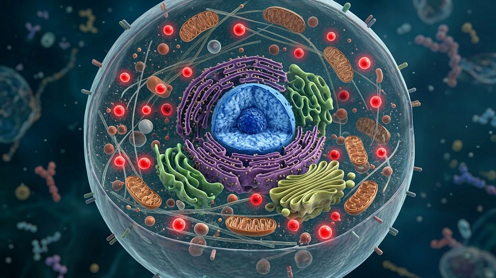
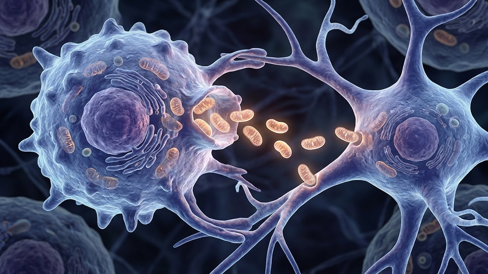
According to a new study using a mouse model of a rare disease, transplanted immune cells donate their mitochondria to neighboring donor cells, improving energy production and rescuing function [1].
Here, have some mitochondria
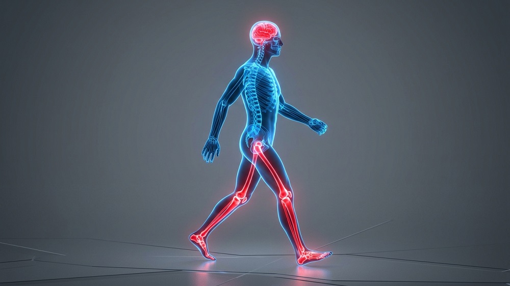
Friedreich’s ataxia is a rare genetic disease in which deficiency in the protein frataxin impairs mitochondrial energy production, especially in energy-hungry cells such as heart muscle cells (cardiomyocytes) and neurons [2]. This causes heart disease, movement problems, and other complications typical of this disease. Mitochondrial dysfunction is also an important hallmark of aging, underlying multiple age-related conditions [3].
Microglia, the brain’s resident immune cells, and macrophages, related immune cells in other tissues, also become dysfunctional in Friedreich’s ataxia. Restoring these immune cells is possible via a bone marrow transplant.
However, there might be an additional benefit: immune cells are known to transfer their mitochondria to neighboring cells [4]. What if donor immune cells could replace diseased cells and also donate mitochondria to neighboring non-immune cells, rescuing their metabolism? This was the premise of a new study from Stanford University that was published in Nature Communications.
Mitochondrial transfer confirmed
Previous bone marrow transplantation studies had shown benefits in mouse models of Friedreich’s ataxia, but replacement of brain microglia was inefficient [5]. To solve this, the authors developed a conditioning regimen that greatly improves the replacement rate.
The researchers used YG8-800 mice, which lack the mouse frataxin gene but carry a human FXN gene with a pathogenic mutation, recapitulating several features of the disease. Before transplantation, the mice were given the drug busulfan, which depletes blood-forming cells and makes room for the graft; after transplantation, they were given pexidartinib, which depletes existing microglia, helping open the brain’s microglial niche to replacement cells.
To distinguish donor cells from donated mitochondrial material, the healthy donor marrow carried two fluorescent labels: green GFP throughout the cell and red mKate2 targeted to mitochondria.
The treatment achieved extensive donor-cell engraftment in blood and brain. Around five months after transplantation, approximately 82% of the measured brain microglial/myeloid population in affected recipients was donor-derived.
The researchers then looked for recipient cells that contained the red mitochondrial label but lacked the green donor-cell label. Recipient brain cells acquired donor mitochondrial signals, and acquisition or retention was greater in affected mice than in healthy mice.
The signal was detected across several brain cell populations, including neurons and supporting cells. This showed that donor cells were distributed widely through the brain and that this distribution was accompanied by mitochondrial transfer.
Interestingly, cells outside the brain also showed signs of mitochondrial transfer from donor cells. In bone marrow, about 22% of the remaining recipient cells were positive for the mitochondrial label.
Functional improvement
Healthy marrow improved growth and reduced hair loss in affected mice. In terms of body weight, treated mice were roughly in the middle between the untreated mice (affected mice who had received affected bone marrow) and healthy controls: the treatment recovered about half the weight deficit.
Female survival improved from about 53% in affected controls to 80% with healthy marrow. Males also showed improved growth and hair condition, but their survival improvement was nonsignificant.
The researchers then tested females at approximately 29-30 weeks, when this model shows motor abnormalities. Treated mice performed better in coordination, spontaneous movement, and strength tests, although they did not quite reach healthy levels of performance.
Transfer on contact
Single-cell RNA sequencing then revealed the mitochondrial signal across a broad range of brain cell types. Signal-positive cells showed coordinated changes in energy-metabolism genes. In these cells, many nuclear genes involved in energy production were more active. Other changes involved antioxidant defenses and cellular maintenance.
However, expression of genes encoded by mitochondrial DNA often decreased rather than increased. The authors suggest possible explanations, including normalization of compensatory responses or changes in mitochondrial quality control, but these are speculative at this point.
Proteomic analysis found that healthy marrow partially restored proteins associated with synapses. Severely depleted mitochondrial respiratory-chain proteins also recovered toward healthy levels.
As cardiac disease is a major component of Friedreich’s ataxia, the researchers then examined the heart. Donor-derived macrophages populated the heart, and donor mitochondrial signals also appeared outside donor cells. Echocardiography then showed that healthy marrow partially improved cardiac pumping performance.
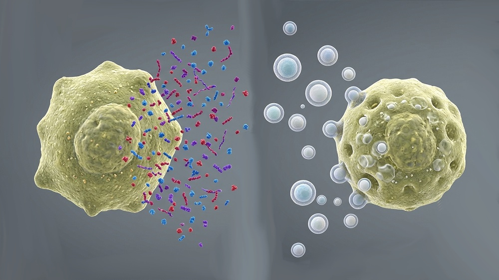
To better understand the mechanism behind mitochondrial transfer, the authors cultured healthy donor macrophages together with fibroblasts from healthy or affected mice. Affected fibroblasts acquired much more mitochondrial signal than healthy fibroblasts. No transfer was detected when the cells were physically separated under the tested conditions, suggesting a contact-dependent mechanism.
The affected cells carrying donor mitochondrial signals showed a clear improvement in respiratory capacity. However, recovery remained incomplete: recipient cells performed better than affected controls but worse than healthy cells.
“It’s a way to use the blood system to treat non-blood organs,” said senior study author Natalia Gomez-Ospina, MD, Ph.D., assistant professor of pediatrics. “It’s profound. Cells are talking to each other in ways that are more consequential than we’ve realized, and this has many implications for disease treatment. For instance, the brain can say, ‘Uh oh, my microglia have been depleted. Let’s repopulate them from the bone marrow. We can leverage that process to get healthy cells into the brain.”
Literature
[1] Cho, H., Sayana, R., Koladiya, A., Colella, P., Cho, S., Jahng, J. W., … & Gomez-Ospina, N. (2026). Myeloid cell replacement induces intercellular mitochondrial transfer and restores metabolism in a mouse model of mitochondrial disease. Nature Communications.
[2] Lynch, D. R., & Farmer, G. (2021). Mitochondrial and metabolic dysfunction in Friedreich ataxia: update on pathophysiological relevance and clinical interventions. Neuronal signaling, 5(2), NS20200093.
[3] López-Otín, C., Blasco, M. A., Partridge, L., Serrano, M., & Kroemer, G. (2023). Hallmarks of aging: An expanding universe. Cell, 186(2), 243-278.
[4] Scheiblich, H., Eikens, F., Wischhof, L., Opitz, S., Jüngling, K., Cserép, C., … & Heneka, M. T. (2024). Microglia rescue neurons from aggregate-induced neuronal dysfunction and death through tunneling nanotubes. Neuron, 112(18), 3106-3125.
[5] Rocca, C. J., Goodman, S. M., Dulin, J. N., Haquang, J. H., Gertsman, I., Blondelle, J., … & Cherqui, S. (2017). Transplantation of wild-type mouse hematopoietic stem and progenitor cells ameliorates deficits in a mouse model of Friedreich’s ataxia. Science translational medicine, 9(413), eaaj2347.
About the author

Arkadi Mazin