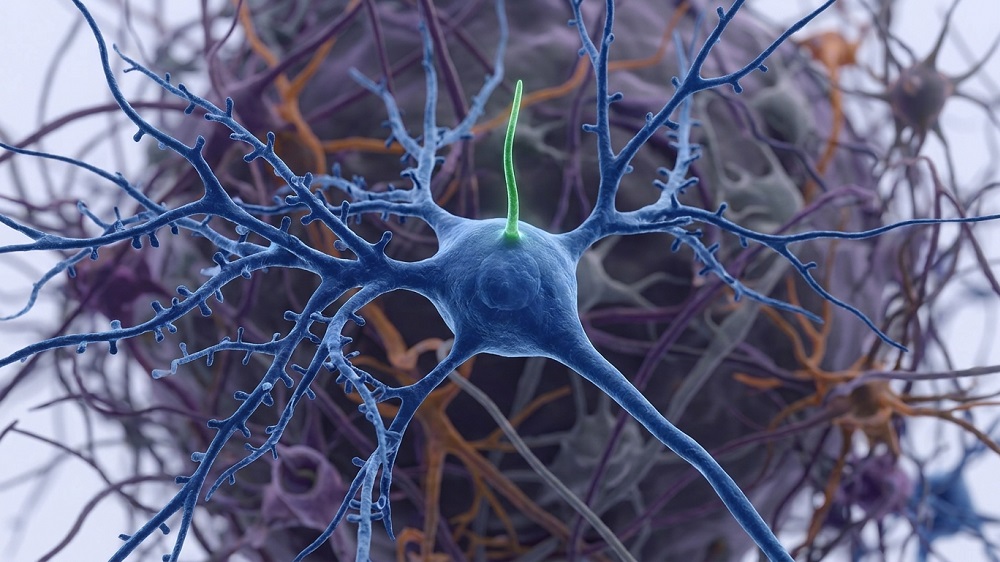

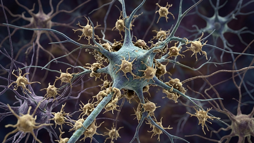
A new study suggests that in amyotrophic lateral sclerosis – and possibly other neurodegenerative diseases – the brain’s immune cells devour stressed but still living neurons due to altered signaling [1].
The living dead
Amyotrophic lateral sclerosis (ALS), an age-related neurodegenerative disease, is characterized by the progressive loss of motor neurons. Historically, much of ALS research focused on what goes wrong inside the neurons themselves. However, it is becoming clear that neighboring cells, particularly the brain and spinal cord’s resident immune cells (microglia), can strongly influence neurons’ survival.
Microglia are essentially phagocytes: among other jobs, they recognize damaged or dead cells and engulf them. Microglia activation has been linked to several neurodegenerative conditions [2] and is known to be triggered by elevated expression of the tyrosine kinases AXL and MER from the TAM receptor family. TAM machinery recognizes its targets via phosphatidylserine (PtdSer), a lipid normally confined to the inner surface of the cell membrane. During cellular death by apoptosis, it flips to the outer surface and essentially tells phagocytes to attack the cell.
The authors of a new study published in Nature Communications suspected that something in this signaling system goes wrong in ALS, making microglia attack neurons that are not actually dying. “Cells that are dying throw an ‘eat me’ sign out on their surface, and the TAM system recognizes that sign,” explains Greg Lemke, distinguished professor emeritus at Salk Institute and a lead author. “It’s an essential system that clears billions upon billions of dead and dying cells from the body daily. We wondered whether microglia were corrupting this TAM system to kill living neurons in ALS.”
Inside out
The researchers started by investigating whether this machinery is actually engaged in human disease by examining postmortem lumbar spinal cord from six people with sporadic ALS and three age-matched controls. In microglia, MER was about threefold higher in ALS than in healthy controls, while AXL was increased about 16-fold.
They then reproduced this in a mouse model of ALS (SOD1G93A mice). As the disease progressed, Axl and Mer expression rose in the spinal cord. Both receptors were concentrated mostly in microglia rather than in motor neurons or astrocytes.
To see if the ligand part of the pathway was also activated, the authors examined Gas6, one of the proteins that bridges PtdSer on a target cell to TAM receptors on a phagocyte. In human spinal cord tissue and in SOD1 mice, motor-neuron GAS6 expression was significantly higher in ALS than in controls.
Next, the team looked for externalized PtdSer, which usually appears on dying cells and signals to TAM. They detected essentially no externalized PtdSer in normal spinal cord, whereas late-stage SOD1 mice contained many PtdSer-positive cells, including motor neurons.
That could simply mean these neurons were already dying by apoptosis. So, the researchers stained for cleaved caspase-3, a marker of ongoing apoptosis, but most PtdSer-positive neurons tested negative for it. Therefore, the authors concluded that the great majority of neurons displaying the “eat-me” signal were still alive rather than truly apoptotic.
Get ill sooner, die later
When the researchers crossed SOD1 mice with mice lacking both Axl and Mer, this produced an unexpected result. These mice developed their earliest signs of illness about 30 days sooner but reached the terminal clinical endpoint roughly three weeks later than ordinary SOD1 mice.

TAM signaling is important for immune homeostasis, so, TAM-deficient mice develop autoimmunity, inflammation, and several other pathologies sooner. However, as ALS progressed, the knockout produced a large survival advantage. That led to the next question: were motor neurons actually being preserved? Apparently, they were, as SOD1 mice lacking Axl and Mer retained roughly three times as many motor neurons as ordinary SOD1 mice.
“It somehow was not devastating,” says first author Youtong Huang, Ph.D., a former graduate student researcher in Lemke’s lab. “When we looked at how many motor neurons mice without Axl and Mer had, compared to mice with Axl and Mer, we found losing the TAM proteins meant preserving muscle control.”
The team then discovered that it was more than just neuron bodies surviving: in Axl and Mer-deficient SOD1 mice, the neurons’ axons remained connected to muscles, and muscle sizes were preserved compared to regular SOD1 mice.
One design problem needed to be addressed: Axl and Mer having been deleted throughout the body from development onward. The team staged a cleaner experiment by creating a mouse model where deletion of TAM receptors was triggered by tamoxifen – mostly in microglia in adult animals rather than from every cell throughout life.
They turned microglia-confined silencing of Axl and Mer on day 100, when disease was already developing, which substantially reproduced the survival benefit of the germline knockout. On day 160, none of the tamoxifen-treated animals had reached the clinical endpoint, compared with about 60% of vehicle-treated controls. Conditional TAM deletion also preserved significantly more spinal motor neurons and partially rescued neuromuscular connections.
Finally, the authors demonstrated that knocking out the TAM receptors does reduce the “devouring” of motor neurons by microglia. In ordinary SOD1 mice, microglial lysosomes contained abundant neuronal material, but Axl/Mer deletion reduced this material by roughly tenfold.
“The bottom line is, microglia are using the TAM system to eat cells that aren’t dead,” said Lemke. “There is enormous potential for this in clinical translation. Rather than engineering entire cells as immunotherapies – a process that is far more complicated, time-consuming and invasive – we could simply design TAM-based proteins that target any cell you’d like. I’m really excited to see where this discovery goes and how it changes immunotherapy opportunities.”
Very similar TAM activation has been observed in Alzheimer’s and Parkinson’s disease models, including by the same team [3]. The authors therefore speculate that PtdSer-dependent killing of stressed-but-living neurons by microglia may be a general mechanism of neurodegeneration that is not restricted to ALS.
Literature
[1] Huang, Y., Mavinkurve, A., Sabikunnahar, B., Stevens, B., & Lemke, G. (2026). Microglia deploy TAM receptors to kill motor neurons in a mouse model of amyotrophic lateral sclerosis. Nature Communications.
[2] Hickman, S., Izzy, S., Sen, P., Morsett, L., & El Khoury, J. (2018). Microglia in neurodegeneration. Nature neuroscience, 21(10), 1359-1369.
[3] Fourgeaud, L., Través, P. G., Tufail, Y., Leal-Bailey, H., Lew, E. D., Burrola, P. G., … & Lemke, G. (2016). TAM receptors regulate multiple features of microglial physiology. Nature, 532(7598), 240-244.