

Using cohort studies and a mouse model, researchers have found that the circulating factor ANGPTL8, which is produced by senescent fat cells, is related to age-related diseases and mortality in mice and people.
Too much of a regulator leads to dysregulation

This paper introduces itself with a standard explanation of the relationship between senescence and inflammaging, including the contributions of the senescence-associated secretory phenotype (SASP) that senescent cells emit. The authors state the need for a clear understanding of the “molecular mediators” involved, focusing specifically on the contributions of fat (adipose) tissue, which has been identified in previous work as a crucial link [1].
Adipose tissue is not just energy storage; it is an endocrine organ all its own that regulates metabolism [2], and the aging of this tissue pushes it towards dysregulation and inflammation [3]. This includes SASP emissions, which senescent adipose tissue creates in sufficient quantities to stimulate systemic inflammation [4]. This paper makes it clear that aging fatty tissues are a cause, rather than just a consequence, of other aspects of aging [5].
The related molecule on which this paper focuses is ANGPTL8, a metabolic regulator that itself is upregulated when insulin is administered [6]. However, this team has previously pinpointed it as being related to multiple serious metabolic issues, such as diabetes-related kidney disease (nepropathy) [7]. Other teams have corroborated such findings, finding it to be related to diabetic atherosclerosis [8] and a higher risk of future heart attacks in existing cardiovascular disease patients [9]. Additionally, higher circulating ANGPTL8 levels have been found to be associated with all-cause mortality in diabetic patients [10].
A significant predictor
As the first part of this paper, the researchers built their own biomarker-based clock, MBA8-Clock using data from the China Cardiometabolic Disease and Cancer Cohort, which recruited nearly ten thousand people. Comparing five separate algorithms, the team found that a multilayer perceptron-based framework was the best way to utilize this clock, as it gave estimated ages with an error rate of approximately five years.
This clock contained many basic measurements of metabolic health, including ANGPTL8. The researchers found that circulating ANGPTL8 values of approximately 250 nanograms per liter of serum were associated with the lowest biological age estimations. At values higher than that, particularly in people with four times as much or more, biological age was likely to be estimated markedly higher. Removing ANGPTL8 from the clock notably diminished its predictive abilities. The researchers hold that “circulating ANGPTL8 represents a consistent, biologically meaningful contributor to age prediction across individuals.”
The next step was to create another clock, AIMR-Model, this one based on all-cause mortality over a ten-year period. The researchers used five algorithmic analyses, and this time they discovered that an extreme gradient-based framework yielded the most accurate results. Once more, ANGPTL8 was found to be a significant contributor; people who had been found to have higher ANGPTL8 levels were significantly more likely to have died within 10 years of that initial assessment. Further analysis revealed that “ANGPTL8 may amplify age-related clinical vulnerability rather than acting solely as an isolated risk marker,” as it was consistently found to occur alongside high blood pressure and other well-known risk factors for age-related disease.
Mice without ANGPTL8 live longer
The researchers then turned to mice. As in humans, ANGPTL8 naturally increases in mice with age: 4-month-old (young) wild-type mice have significantly less of it than their 22-month-old (old) counterparts. As expected, the source was found to be the adipose tissue rather than the liver or any other organs; while hepatocytes did somewhat increase their production of the protein, that was localized rather than systemic.
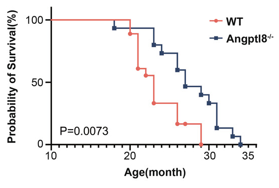
In addition, the researchers created a group of mice that do not express the murine gene Angptl8. Compared to their wild-type counterparts, the old Angptl8-less mice performed considerably better on various physical tests at older ages. They had less fat mass, less senescence in their fat, and reductions in circulating SASP factors. They were also able to hang onto a rotarod longer, performed better on a treadmill, engaged in more exploratory behavior, and had more gastrocnemius muscle mass along with more fast-twitch fibers. Finally, and perhaps most importantly, these mice also lived longer.

The specific pathway involved in the relationship between ANGPTL8 and senescence was identified. This protein was found to bind to AKT2 on the molecular level, affecting the fundamental AKT/mTOR/S6K pathway, and this finding was confirmed in mice; mice with Angptl8 turned off have significantly decreased activity of AKT, mTOR, and S6K. Deriving fat precursor cells (preadipocytes) from older mice confirmed these findings, and experiments using cells with Angptl8 overexpression found that AKT is indeed necessary for ANGPTL8 to contribute to cellular senescence.
These researchers are of the clear opinion that they have hit upon a significant target, stating that “from a translational standpoint, the dual identity of ANGPTL8 as both a circulating biomarker and a functional effector of aging is particularly compelling.” However, this paper did not test any potential method of removing circulating ANGPTL8 from the bloodstreams of wild-type animals. If such a drug can be developed and proven to work in a human clinical trial, it may be possible to grant additional healthspan and lifespan to older people suffering from metabolic issues.
Literature
[1] Hotamisligil, G. S. (2017). Inflammation, metaflammation and immunometabolic disorders. Nature, 542(7640), 177-185.
[2] Ou, M. Y., Zhang, H., Tan, P. C., Zhou, S. B., & Li, Q. F. (2022). Adipose tissue aging: mechanisms and therapeutic implications. Cell death & disease, 13(4), 300.
[3] Tchkonia, T., Morbeck, D. E., Von Zglinicki, T., Van Deursen, J., Lustgarten, J., Scrable, H., … & Kirkland, J. L. (2010). Fat tissue, aging, and cellular senescence. Aging cell, 9(5), 667-684.
[4] Dahlquist, K. J., & Camell, C. D. (2022). Aging leukocytes and the inflammatory microenvironment of the adipose tissue. Diabetes, 71(1), 23-30.
[5] Franceschi, C., Garagnani, P., Vitale, G., Capri, M., & Salvioli, S. (2017). Inflammaging and ‘Garb-aging’. Trends in Endocrinology & Metabolism, 28(3), 199-212.
[6] Abu-Farha, M., Ghosh, A., Al-Khairi, I., Madiraju, S. M., Abubaker, J., & Prentki, M. (2020). The multi-faces of Angptl8 in health and disease: Novel functions beyond lipoprotein lipase modulation. Progress in lipid research, 80, 101067.
[7] Pan, L., He, Y., Xiang, Y., Mao, B., Meng, X., Guo, Y., … & Yu, X. (2025). Angiopoietin-like protein 8 mediates inflammation and fibrosis of tubular cells in diabetic kidney disease progression by interacting with Akt2. Metabolism, 156418.
[8] Ye, H., Zhu, Q., Zong, Q., Luo, S., Ji, Z., Zhang, R., & Zou, H. (2026). Elevated ANGPTL8 (Angiopoietin‐Like Protein 8) Levels as a Novel Predictor of Atherosclerosis in Type 2 Diabetes: Beyond Lipid Metabolism. Journal of the American Heart Association, 15(3), e044806.
[9] Morinaga, J., Kashiwabara, K., Torigoe, D., Okadome, Y., Aizawa, K., Uemura, K., … & Oike, Y. (2023). Plasma ANGPTL8 levels and risk for secondary cardiovascular events in Japanese patients with stable coronary artery disease receiving statin therapy. Arteriosclerosis, thrombosis, and vascular biology, 43(8), 1549-1559.
[10] Zou, H., Xu, Y., Chen, X., Yin, P., Li, D., Li, W., … & Yu, X. (2020). Predictive values of ANGPTL8 on risk of all-cause mortality in diabetic patients: results from the REACTION Study. Cardiovascular Diabetology, 19(1), 121.