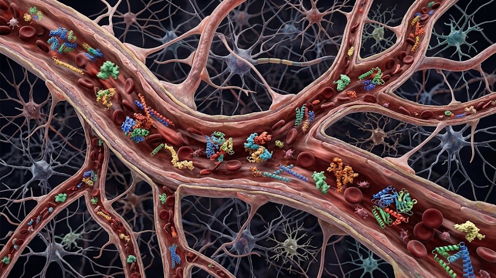

Researchers have gone into deep detail regarding how each of five senescent brain cell types expresses and receives factors that encourage other cells to become senescent.
The contagion of age

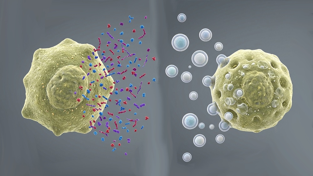
This paper revolves around paracrine senescence: SASP factors being emitted by primary senescent cells to drive secondary senescence in other cells [1]. Senescent cells differ by both tissue type and senescence source, and primary senescent cells, which were driven senescent through other stressors, do not necessarily exhibit the same features or function in the same ways as secondary senescent cells [2]. Recent work has explored many of those differences in detail [3, 4].
The molecular causes of this spreading senescence are fairly well-established. The researchers specifically name the inflammatory factors TGFβ, IGF1, IL-6, IL-8, and CCL2 as being part of the SASP [5], and they mention the role of reactive oxygen species (ROS) and the inflammatory NFκ-B pathway as mediating the process within cells [6].
However, only limited amounts of this research have been done on brain cells. In this study, the researchers sought to fill that gap by taking a closer look at how SASP factors affect various types of these cells and what might be done about them.
Five brain cell types express the SASP very differently
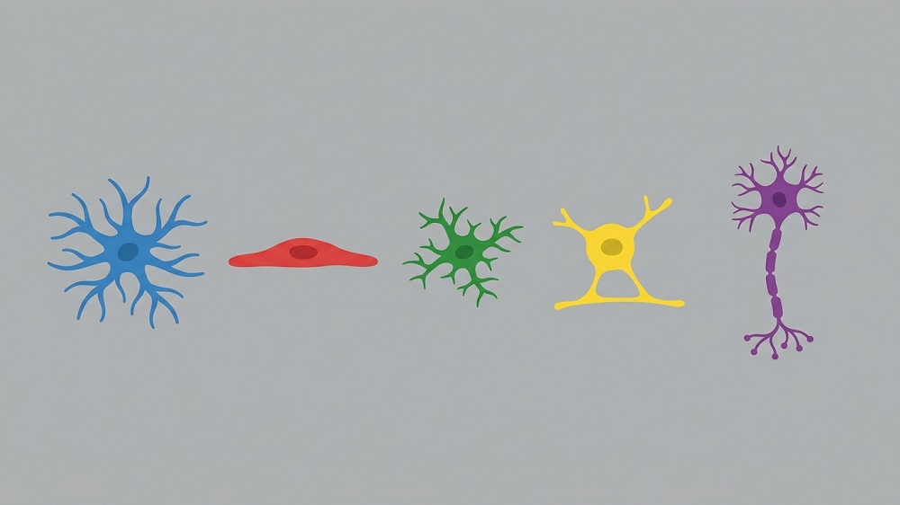
This study used the expression of the well-known biomarker SA-β-gal to determine if a cell had become senescent. It used five cell types commonly found in the brain: astrocytes, endothelial cells, microglia, oligodendrocytes, and neurons. To create primary senescent cells, the researchers treated each of these types with 5-bromodeoxyuridine (BrdU) for a week, a technique that they had used in a previous study [3].
Each of these cell types developed a specific SASP profile after senescence was induced. A panel of 286 cytokines revealed that, while a small handful of cytokines was common between cell types, each type was expressing its own combination of factors, particularly microglia, oligodendrocytes, and neurons, which expressed more factors compared to astrocytes; endothelial cells were found to express very few of these factors at all. Oligodendrocytes and microglia expressed a considerable number of factors that were only common between these two groups.
The cells also responded much differently when treated with conditioned media derived from these groups (BrdU CM), a technique that would presumably induce secondary senescence. Interestingly, this did not cause neurons and oligodendrocytes to become secondarily senescent, regardless of the source. Despite their few SASP factors under direct BrdU exposure, astrocytes and endothelial cells would still become senescent when treated with BrdU CM derived from microglia or astrocytes. Microglia were found to be particularly susceptible to secondary senescence; any BrdU CM source other than endothelial cells would drive them senescent in this way.
There was a very interesting result: When normal astrocytes were exposed to BrdU CM from other astrocytes, their expression of CDNK1A, which encodes for the senescence-associated factor p21, would significantly increase. However, when microglia were exposed to the same astrocytic SASP, their expression of CDNK1A significantly decreased instead. Unsurprisingly, however, both astrocyte and microglia SASP increased the expression of the DNA damage marker γH2AX in both types of cells.
Neurons and oligodendrocytes were also found to not be completely immune to the SASP. Despite not becoming senescent, neurons were affected by BrdU CM from astrocytes and microglia, expressing extra CDNK1A along with CDNK2A, which increases p16. Oligodendrocytes’ related biomarkers were affected by the SASP of astrocytes, microglia, and, interestingly, endothelial cells.

Potential targets
This paper goes into the relationship of various SASP factors in exacting detail. The researchers frequently noted that the CM of these senescent cells did not always induce senescence; on some occasions, it was found to downregulate inflammatory factors instead, and there were also effects on MIF, a double-edged factor whose age-related effects are dependent on context [7].
The researchers also performed an extensive series of experiments using various drugs that inhibit the spreading of the SASP. Most of these drugs found partial successes; most notable was the drug Bindarit, which had significant, if incomplete, effects on senescence transmission between astrocytes. The factors CCL2, MIF, CXCR7, and DPP4 were identified as potential targets for future therapeutic work.
This study provides an illuminating look into the complexity inherent in dealing with the wide panoply of factors that make up the overall circulating SASP. However, even this level of detail only dealt with SASP factors taken from five cell types driven primarily senescent by chemical exposure; CM derived from the secondary senescent cells themselves was not studied here.
Additionally, this was only a cellular study; these findings were not confirmed in an animal model, and the researchers noted that conditioned media do not reflect the complete variety of factors found in the human brain. Animal experiments and human trials will have to be done in order to determine which, if any, of these factors might be a valuable target for reducing unwanted senescence in the brain.
Literature
[1] Martin, L., Schumacher, L., & Chandra, T. (2023). Modelling the dynamics of senescence spread. Aging Cell, 22(8), e13892.
[2] Teo, Y. V., Rattanavirotkul, N., Olova, N., Salzano, A., Quintanilla, A., Tarrats, N., … & Chandra, T. (2019). Notch signaling mediates secondary senescence. Cell reports, 27(4), 997-1007.
[3] Russo, T., Plessis-Belair, J., Sher, R., & Riessland, M. (2025). Systematic profiling reveals distinct senescence signatures and regulators across human brain cell types. Nature Communications, 16(1), 11059.
[4] Sweeney, E. M., Abate, G., Bakker, B. R., Mastinu, A., Lai, Y., Uberti, D., … & Tambaro, S. (2026). Cell Type‐Specific Expression of p16, p21, and p53 Reveals Age‐Dependent Glial Senescence in the AppNL‐G‐F Mouse Model of Alzheimer’s Disease. Aging Cell, 25(4), e70478.
[5] Admasu, T. D., Rae, M. J., & Stolzing, A. (2021). Dissecting primary and secondary senescence to enable new senotherapeutic strategies. Ageing research reviews, 70, 101412.
[6] da Silva, P. F., Ogrodnik, M., Kucheryavenko, O., Glibert, J., Miwa, S., Cameron, K., … & von Zglinicki, T. (2019). The bystander effect contributes to the accumulation of senescent cells in vivo. Aging cell, 18(1), e12848.
[7] Altulea, A., Nehme, J., & Demaria, M. (2026). Dual role of MIF in aging and cellular senescence. Cytokine & Growth Factor Reviews.