A Tougher Extracellular Matrix Strengthens Tendons in Rats

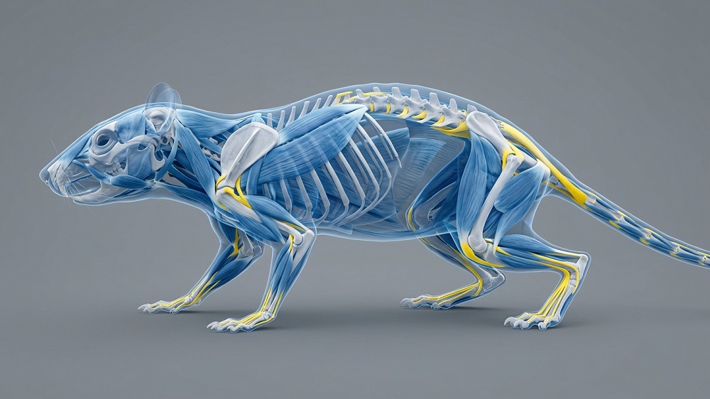
Researchers have found that an upstream promoter of two extracellular matrix proteins increases healing and strength capabilities in the tendons of rats.
Tendon injuries in older people
Previous research has found that, like with many other injuries, tendon injuries have an age-related component. The tendons of the biceps and rotator cuffs are more commonly injured in older people than in younger people [1], and older people heal slower after forearm injuries and have less range of motion in the fingers for a longer time [2].
Previous work has investigated this issue using explanted murine models, taking mouse tendons from the animals and investigating their functional abilities. Tendons from aged mice don’t handle stress deprivation as well [3], nor do they respond properly to added strain [4]. An aged tendon can often support the same amount of force as a young tendon; it simply heals slower [5].
In humans, tendon stem progrenitor cells (TSPCs) change with aging in their gene expression, including genes related to basic functions such as motility and the cellular skeleton [6], and these cells often lack self-renewal abilities [7]. Animal studies have confirmed that cell numbers along with the organization of elastin, a foundational protein in tendon function, decline as well [8].
This study began with a statistical, population-based analysis of trends in tendon injury. This analysis found that, unsurprisingly, tendon injuries, which are often caused by heavy lifting at work, decreased between 1990 and 2021. A country’s development index corroborated this idea; countries undergoing industrial development may have more than less-developed countries, but after a certain level of development, this decreases. However, despite not being under such stressful conditions, older people even today remain at a high risk of such injuries.
Male and female rat tendons age differently
The researchers then turned to rats, taking tendons from eight-week-old (young) and 18-month-old (old) groups of males and females, then stretching them a hundred times to simulate normal mechanical load. They found that in male but not female rats, tendon weight and, surprisingly, tensile strength significantly increased between the two groups; in female but not male rats, the tensile strength, force required to cause a 2-millimeter gap, and stiffness were all weakened with aging.
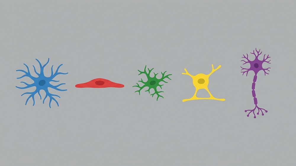
These changes occurred alongside cell type changes. In female rats, there were proportionaly more epithelial and immune cells with aging, and the numbers of fibroblasts and stromal cells decreased. In male rats, immune cells rose as well, but the proportion of fibroblasts increased rather than decreased; stromal cells also declined, but so did epithelial cells.
More extracellular matrix expression helps rat tendons
In both sexes, however, the researchers noted significant declines in the expression of two genes related to the extracellular matrix: Col1a1 and Sparc. This was also accompanied by a decrease in the transcription factor Creb3l1, which these researchers found to be a regulator of these genes, binding directly to their promoters. Transfecting rat tendon cells with a lentivirus that increases Creb3l1 was also found to increase both Col1a1 and Sparc; silencing Creb3l1 led to a substantial increase in cellular senescence.
The researchers then injected this lentivirus into living animals in order to determine its effects on tendon tissue. In males, tendon elasticity and strength was significantly increased after three weeks, and in both sexes, tendons appeared to heal better as well, with the treatment groups having tendinous tissue at the sites of injury while the control groups had granulous tissue instead.
Rats have different biomechanical stresses than people, and rat tendons taken outside the body are no substitute for human results. However, this study shines significant light on sex differences that should be examined for their relevance to humans, and it suggests a potential path forward for a treatment that encourages proper tendon repair by affecting proteins related to the extracellular matrix. Future work is required to determine how such a treatment can be developed and whether it could reduce strains and sprains in older people.
Literature
[1] Clayton, R. A., & Court-Brown, C. M. (2008). The epidemiology of musculoskeletal tendinous and ligamentous injuries. Injury, 39(12), 1338-1344.
[2] Edsfeldt, S., Eklund, M., & Wiig, M. (2019). Prognostic factors for digital range of motion after intrasynovial flexor tendon injury and repair: long-term follow-up on 273 patients treated with active extension-passive flexion with rubber bands. Journal of Hand Therapy, 32(3), 328-333.
[3] Connizzo, B. K., Piet, J. M., Shefelbine, S. J., & Grodzinsky, A. J. (2020). Age-associated changes in the response of tendon explants to stress deprivation is sex-dependent. Connective tissue research, 61(1), 48-62.
[4] Aggouras, A. N., Stowe, E. J., Mlawer, S. J., & Connizzo, B. K. (2024). Aged tendons exhibit altered mechanisms of strain-dependent extracellular matrix remodeling. Journal of Biomechanical Engineering, 146(7), 071009.
[5] Ackerman, J. E., Bah, I., Jonason, J. H., Buckley, M. R., & Loiselle, A. E. (2017). Aging does not alter tendon mechanical properties during homeostasis, but does impair flexor tendon healing. Journal of Orthopaedic Research, 35(12), 2716-2724.
[6] Kohler, J., Popov, C., Klotz, B., Alberton, P., Prall, W. C., Haasters, F., … & Docheva, D. (2013). Uncovering the cellular and molecular changes in tendon stem/progenitor cells attributed to tendon aging and degeneration. Aging cell, 12(6), 988-999.
[7] Zhou, Z., Akinbiyi, T., Xu, L., Ramcharan, M., Leong, D. J., Ros, S. J., … & Sun, H. B. (2010). Tendon‐derived stem/progenitor cell aging: defective self‐renewal and altered fate. Aging cell, 9(5), 911-915.
[8] Godinho, M. S., Thorpe, C. T., Greenwald, S. E., & Screen, H. R. (2017). Elastin is localised to the interfascicular matrix of energy storing tendons and becomes increasingly disorganised with ageing. Scientific reports, 7(1), 9713.
About the author

Josh Conway