The Blog

Building a Future Free of Age-Related Disease

Supplements

How Supplement Formulas, Including NOVOS, Affect Skin Aging

Researchers have tested several combinations of ingredients with anti-aging properties. Those combinations, including a 12-ingredient formulation created by NOVOS, helped to reduce DNA damage and oxidative stress in human skin cells in cultures [1].

Fighting multiple Hallmarks of Aging at once

Aging impacts multiple cellular processes, and these impacts can be assessed through biomarkers. The researchers of this paper used human skin cells (keratinocytes) as their targets for assessing the impact of the NOVOS supplement and six other formulations on DNA damage and oxidative stress markers. They believe that since skin is frequently exposed to DNA-damaging agents, such as UV light, it is the right choice for assessing these markers.

Supplements 1

The tested supplement formulation created by NOVOS, a New York-based company, is a mix of 12 ingredients: pterostilbene, a resveratrol-related polyphenol derived from blueberries; glucosamine sulfate, a supplement used for osteoarthritis treatment; fisetin, a senotherapeutic; glycine, an amino acid; lithium aspartate, a mineral; calcium alpha-ketoglutarate, a small molecule present in the human body; magnesium malate, a natural substance found in apples; vitamin C, also known as ascorbic acid; L-theanine, an amino acid found primarily in green and black tea; hyaluronic acid; Rhodiola rosea root extract; ginger root extract; and a boost consisting of β-nicotinamide mononucleotide.

These nature-based ingredients, all of which have good safety profiles, were selected based on their anti-aging activity, documented role in counteracting multiple aging processes, or association with aging-related mechanisms. We have previously written about them and their selection process in more detail.

The authors explained that they tested combinations of different compounds because this approach could address multiple aging processes at once. A combination of compounds could also effectively reduce unwanted side effects that can appear when a single compound is used in high concentrations.

Reduction in DNA damage

Even beneficial ingredients can be toxic in excessive concentrations. Therefore, the researchers started their study by assessing the compounds’ highest safe concentrations to use. They followed up by testing the impact of supplement formulation on protecting cells from DNA damage, using tumor p53 binding protein 1 (53BP1) as a DNA damage marker.

The researchers treated 2D cell cultures with the six formulations and NOVOS. Following this treatment, they induced DNA damage with the genotoxic agent neocarzinostatin (NCS).

Data analysis revealed that treating cells with the six different formulations caused mild adverse effects on cells, as evidenced by higher numbers of DNA damage-related foci in some cells. NOVOS treatment did not cause that.

Cells pre-treated with NOVOS were less likely to have many of these foci after NCS administration, which suggests that NOVOS helps alleviate the DNA damage caused by NCS. However, it didn’t bring the damage down to the level of control cells that were not exposed to NCS.

The six different formulations also positively affected cells exposed to NCS. Those formulas “significantly prevented DNA damage, observed as reduced foci numbers, even with a stronger effect compared to the NOVOS’ original formulation itself.”

Supplements 2

Based on this statistical analysis and the number of ingredients in each formulation, the researchers chose NOVOS, F1, F2, and F4 for the following experiments.

The first was a test of one more DNA damage marker, γ-H2AX, a marker that reflects double-strand breaks in the DNA. However, they performed it in the 3D skin cell cultures since 3D cultures better reflect the skin microenvironment and ”allow the formation of a cellular structure, like a tissue.” The results of this experiment suggested that all four of these formulations significantly reduced DNA damage compared to cells that were only exposed to NCS.

Notably, the concentration of NOVOS used in this experiment is lower than that of other formulations. Still, it can achieve similar effects in counteracting DNA damage as F1, F2, and F4. The authors suggest that this may be because of some synergistic effects between the NOVOS compounds.

Supplements 3

Decreased oxidative stress

In the following test, the researchers aimed to assess the formulations’ ability to prevent oxidative damage to the cells. Intracellular reactive oxygen species (ROS) are difficult to measure. However, since many of them are converted to hydrogen peroxide, hydrogen peroxide can be used as a proxy for intracellular ROS level changes and oxidative stress.

The researchers performed this experiment using 2D and 3D cultures. Those cells were treated with different F1, F2, F4, and NOVOS formulation concentrations. Then, the researchers used the pro-oxidant medication to induce oxidative damage.

F1 and F4 formulations were able to significantly reduce intracellular hydrogen peroxide production following menadione treatment in 2D and 3D cell cultures. NOVOS was able to achieve this in 2D but not 3D culture.

Supplements 4

Limitations

This study suggests that NOVOS, along with the F1, F2, and F4 combinations, are able to counteract some aging-related processes in skin cells.

While the researchers discussed the benefits of using a combination of compounds over a single compound, this research lacks an experiment that compares the combinations to single compounds. Such experiments would be very informative in testing whether the observed effects result from the combined effect of the multiple compounds or whether the observed effects come mainly from the action of a single compound and there is a negligible contribution from the remaining compounds, which would allow for simpler supplements. This is notable as the F1, F2, and F4 combinations with half the NOVOS compounds show similar effects as NOVOS in the experiments.

The authors also note that further investigation into specific molecular pathways involved in the observed processes is necessary.

One of the study authors is an employee of NOVOS Labs. However, in the Conflict of Interest, the authors note: ”The funder NOVOS Labs was not involved in the study design, collection, analysis, interpretation of data, of this article or the decision to submit it for publication.”

Disclosure: A portion of the profits and equity from NOVOS are being donated to nonprofits working in the longevity science space, which includes us here at lifespan.io.

Our Vice President, Dr. Oliver Medvedik, is also a scientific consultant, putting him in good company with Dr. Joao Pedro Magalhaes, Dr. Pamela Maher, Dr. Avi Rosenbaum, and Dr. Matt Kaeberlein, names with whom regular readers may be familiar.

We would like to ask you a small favor. We are a non-profit foundation, and unlike some other organizations, we have no shareholders and no products to sell you. All our news and educational content is free for everyone to read, but it does mean that we rely on the help of people like you. Every contribution, no matter if it’s big or small, supports independent journalism and sustains our future.

Literature

[1] Punzo, A., Perillo, M., Silla, A., Malaguti, M., Hrelia, S., Diogo Barardo, Cristiana Caliceti, & Lorenzini, A. (2024). Promising Effects of Novel Supplement Formulas in Preventing Skin Aging in 3D Human Keratinocytes. Nutrients, 16(16), 2770–2770.

Cholesterol

Medium LDL Levels Associated With Lowest Mortality

A new observational study suggests that lower LDL cholesterol levels are not necessarily better. Instead, the ratio of triglycerides to HDL may be more important [1].

Putting common wisdom to the test

LDL cholesterol is largely considered harmful, but lower levels might not be beneficial according to a new study by researchers from the University of Pittsburgh and the University of South Florida.

The researchers based their analysis on electronic medical records that were derived from the University of Pittsburgh Medical Center’s healthcare system and dated 2000 to 2022. This allowed them to assemble a vast cohort of 178,000 patients aged 50 to 89.

The participants had to meet several criteria, including a lack of diabetes or statin therapy at baseline and during the first year of follow-up. Patients who died during the first year of follow-up or had outlier levels of total or LDL cholesterol were also excluded to mitigate reverse causation. The researchers divided the sample into six LDL-C categories: 30-79, 80-99, 100-129, 130-159, 160-189, and over 190 mg/dL.

The golden mean

Over a 6-year average follow-up time, intriguing results emerged. A U-shaped relationship was observed between LDL-C levels and 10-year overall mortality. The highest all-cause mortality (19.8%) was found in the 30-79 group. In the other groups, it was 14.7%, 11.7%, 10.7%, 10.1%, and 14.0%, respectively.

Compared to the referent group of 80-99 mg/dL, this translated into a 23% increase in mortality risk for the lowest LDL-C group. In the following three groups, within the 100-189 mg/dL range, the mortality risk was lower than in the referent group by 13%, 12%, and 9%, respectively. In the highest LDL-C group of over 190 mg/dL, the mortality risk was again higher than in the referent group – by 19%.

The results were broadly similar for atherosclerotic cardiovascular disease (ASCVD) risk. However, in this case, the highest risk was recorded for the group with the highest LDL-C levels (over 190 mg/dL). Another interesting finding was that in men, even in this subgroup, mortality risk was not significantly higher than in the referent subgroup.

The parameter that could

However, the researchers did find a parameter with a linear relationship with mortality risk: the ratio of triglycerides and HDL cholesterol (HDL-C), which is often considered beneficial. Total cholesterol to HDL cholesterol ratio (T-C/HDL-C) also seemed to be a better predictor of mortality than LDL-C.

“Compared with patients in the highest quintile of triglycerides/HDL-C ratio (value of ≥3.44), those in the lowest quintile (value of ≤1.06) had an estimated 24% lower risk of mortality,” the paper says. “Thus, in aggregate and irrespective of age, the secondary lipid measures of T-C/HDL-C ratio and triglycerides/HDL-C ratio appeared to be more predictive of mortality than LDL-C, and a triglycerides/HDL-C ratio of about 1 or lower appears to be optimal.”

Cholesterol ratios

The researchers note that the insurance industry seems to have already discovered this relationship [2]: both T-C and HDL-C are more widely used in life insurance policy underwriting than LDL-C. This is not the first time that insurance companies, financially incentivized to discover factors related to health and mortality risks, have provided clues for scientists.

As with any observational study, this one cannot definitely prove causality. However, it aligns with some, though not all, of the previous research on this subject [3]. The authors call for more studies, including those on statin use, that would take into account both statins’ effectiveness in preventing cardiovascular disease and their side effects, such as muscle pain and increased risk of type 2 diabetes [4].

In this analysis among primary prevention-type patients without diabetes aged 50–89 years not on statin therapy at baseline or within 1 year, we found no evidence of a gradient relationship between LDL-C and long-term mortality risk. Instead, we observed that within the entire LDL-C range of 100–189 mg/dL (about two-thirds of the total patient population), mortality risk was similar and slightly lower than the referent LDL-C category of 80–99 mg/dL. These data conflict with the prevailing belief that ‘lower LDL-C is better’ yet align with results from multiple studies.

We would like to ask you a small favor. We are a non-profit foundation, and unlike some other organizations, we have no shareholders and no products to sell you. All our news and educational content is free for everyone to read, but it does mean that we rely on the help of people like you. Every contribution, no matter if it’s big or small, supports independent journalism and sustains our future.

Literature

[1] Kip, K. E., Diamond, D., Mulukutla, S., & Marroquin, O. C. (2024). Is LDL cholesterol associated with long-term mortality among primary prevention adults? A retrospective cohort study from a large healthcare system. BMJ open, 14(3), e077949.

[2] Fulks, M., Stout, R. L., & Dolan, V. F. (2009). Association of cholesterol, LDL, HDL, cholesterol/HDL and triglyceride with all-cause mortality in life insurance applicants. J Insur Med, 41(4), 244-253.

[3] Johannesen, C. D. L., Langsted, A., Mortensen, M. B., & Nordestgaard, B. G. (2020). Association between low density lipoprotein and all cause and cause specific mortality in Denmark: prospective cohort study. Bmj, 371.

[4] Mansi, I. A., Chansard, M., Lingvay, I., Zhang, S., Halm, E. A., & Alvarez, C. A. (2021). Association of statin therapy initiation with diabetes progression: a retrospective matched-cohort study. JAMA internal medicine, 181(12), 1562-1574.

Synapses

Blocking a Dopamine Receptor May Improve Memory

Neurological researchers, in Aging Cell, have deepened our understanding of the brain, explaining how turning off a dopamine receptor may lead to better memory in older people.

Sometimes it’s better for the dopamine not to hit

With aging comes changes in how the brain processes dopamine [1]. This work focuses on specific dopamine receptors called D3Rs, which are less common than related D2Rs although much stronger in binding dopamine [2]. This potency has made them attractive targets in drug discovery, and prior work has focused on D3Rs for the treatment of such disorders as schizophrenia and depression [3].

One of the brain regions that contains D3Rs is the hippocampus [4], which is responsible for the formation of memories. Rodent studies have found that, unlike D2Rs, blocking D3Rs improves memory [5] and cognition [6], and the same appears to be true for people [7]. However, the reasons why blocking a receptor would lead to cognitive benefits are not clear, which is why these researchers took a closer look.

Cells and mice agree

This work began with an examination of CA3-CA1 synapses, which are very common targets in brain research, derived from the hippocampi of mice. Without a D3R blocker, giving light stimulation to these synapses creates a weak long-term connection, which takes time and reinforcement to become a more permanent one.

However, administering a D3R blocker makes even this light stimulation form a permanent connection much more quickly. Strong stimulation does normally cause these synapses to form a permanent connection, but even this was enhanced with a D3R blocker. These findings were confirmed with a strain of mice that has the genes for D3 reception knocked out. Further work found that these behavioral changes were post-synaptic: they did not affect how the neurons worked before the stimulation.

Turning to living animals, the researchers found that D3R blocking turned short-term memory into long-term memory much more readily. Normally, a light training regimen allows mice to distinguish between familiar and unfamiliar objects for six to eight hours [8], after which they forget about it. However, administering D3R blockers to the mice allowed them to distinguish between the objects 24 hours later, as if they had gone through a heavier training regimen. The same was true of D3R knockout mice: they gained long-term memories much more quickly.

Although memories are harder to form with age, D3R numbers were found to be decreased with age, but mostly in the pre-synaptic sites. Post-synaptic D3R sites were largely similar between adult and aged mice. Comparing the hippocampal neurons of D3R knockout mice and wild-type mice, the researchers found that the age-related loss of memory formation did not occur: these neurons’ ability did not seem to have diminished. These findings were confirmed in mice that had their D3R receptors blocked through genetics or chemical intervention. No sex differences were found.

These findings are impressive, and it may be that D3 receptor blockers might become an effective “memory pill” for people seeking to learn information quickly or people suffering from age-related memory problems. However, there may be significant neurological side effects to engraving more things in long-term memory that would normally be lost. Further work needs to be done to judge the effectiveness and safety of blocking D3 in animals and, ultimately, in people.

We would like to ask you a small favor. We are a non-profit foundation, and unlike some other organizations, we have no shareholders and no products to sell you. All our news and educational content is free for everyone to read, but it does mean that we rely on the help of people like you. Every contribution, no matter if it’s big or small, supports independent journalism and sustains our future.

Literature

[1] Shohamy, D., & Wimmer, G. E. (2013). Dopamine and the cost of aging. Nature Neuroscience, 16(5), 519-521.

[2] Maramai, S., Gemma, S., Brogi, S., Campiani, G., Butini, S., Stark, H., & Brindisi, M. (2016). Dopamine D3 receptor antagonists as potential therapeutics for the treatment of neurological diseases. Frontiers in neuroscience, 10, 451.

[3] Kiss, B., Laszlovszky, I., Krámos, B., Visegrády, A., Bobok, A., Lévay, G., … & Román, V. (2021). Neuronal dopamine D3 receptors: translational implications for preclinical research and CNS disorders. Biomolecules, 11(1), 104.

[4] Li, Y., & Kuzhikandathil, E. V. (2012). Molecular characterization of individual D 3 dopamine receptor-expressing cells isolated from multiple brain regions of a novel mouse model. Brain Structure and Function, 217, 809-833.

[5] Watson, D. J., Loiseau, F., Ingallinesi, M., Millan, M. J., Marsden, C. A., & Fone, K. C. (2012). Selective blockade of dopamine D3 receptors enhances while D2 receptor antagonism impairs social novelty discrimination and novel object recognition in rats: a key role for the prefrontal cortex. Neuropsychopharmacology, 37(3), 770-786.

[6] Xing, B., Meng, X., Wei, S., & Li, S. (2010). Influence of dopamine D3 receptor knockout on age-related decline of spatial memory. Neuroscience letters, 481(3), 149-153.

[7] Gross, G., Wicke, K., & Drescher, K. U. (2013). Dopamine D 3 receptor antagonism—still a therapeutic option for the treatment of schizophrenia. Naunyn-Schmiedeberg’s archives of pharmacology, 386, 155-166.

[8] Blokland, A., & Sesia, T. (2023). Delay-dependent forgetting in object recognition and object location test is dependent on strain and test. Behavioural Brain Research, 437, 114161.

Swimmer

Associations Between Professional Sports and Longevity

A new study analyzed how being an elite athlete in various sports affects human lifespan. Some of the results were surprising [1].

Elite sports and aging

Longevity enthusiasts have long wondered if professional athletes live longer than the general population and what sports show stronger associations with lifespan. In a new study published in the journal GeroScience, a group of scientists from Holland attempted to research this using data on tens of thousands of elite athletes born between 1862 and 2002. The sample encompassed 183 countries and 44 sports disciplines.

The data was collected by the researchers themselves using public sources. The effect on lifespan was determined by comparing the ages of athletes with those of the corresponding reference populations, and the results were adjusted for sex, year of death, and country. The vast majority of the athletes were males, which limited the study’s applicability to the female population.

Volleyball, what’s wrong with you?

The study yielded both expected and intriguing results. In males, the highest life extension was observed for pole vaulting and gymnastics (8.4 and 8.2 years, respectively). Racquet sports and mixed track and field were not far behind (5.7 years both). In general, most sports were associated with statistically significant life extension, with soccer and wrestling showing some of the smallest positive effect sizes.

However, there were several outliers. Sumo was associated with a massive 9.8-year negative effect on lifespan. Surprisingly, volleyball came in second (-5.4 years), followed by mountaineering (-3.8 years), martial arts (-2.5 years), and handball (-2.2 years). Other notable sports with negative, albeit smaller, effect sizes, were ice hockey (-0.8 years) and boxing (-0.6 years).

Sports longevity 1

Since the sample contained much fewer female athletes, associations for them were not as clear-cut. From the limited list of sports in the female subset, only golf and racquet sports showed significant positive effects on lifespan (3.2 and 2.8 years, respectively).

Interestingly, contrary to what was observed in males, most sports seemed to have negative effects on female lifespan. Those with significant effect sizes included basketball (-6.7 years), table tennis (-4.8 years), cricket (-4.2 years), mixed track and field (-2.4 years), sprinting (-2.1 years), and skiing (-2 years).

Sports longevity 2

Interpreting the results

Populational studies like this one can only show correlations and not causative effects, and interpreting their results can be notoriously difficult due to the abundance of confounding factors. The researchers discuss, at length, possible explanations for their findings but admit that those are mere speculations.

One hypothesis that seems plausible is that sports that combine aerobic and anaerobic activity, both of which are associated with health and longevity [2], produce the largest positive effect. However, this was not the case for martial arts and boxing, most probably because of the high risk of injuries.

Sumo is a great illustration of the deleterious effects of a high-calorie diet and obesity that cannot be offset even by high levels of exercise. That said, the length of a professional career in sports usually does not exceed two or three decades, and in later life, the amount of physical activity is expected to decline.

The origins of the sex gap observed in this study are unclear. The researchers note that the male population, especially in earlier cohorts, is more prone to unhealthy habits, and “participation in sports may prompt males to minimize such habits, leading to a more profound impact of sports on their lifespan.”

Females, on the other hand, lead healthier lives on average, which decreases the populational effect of giving up unhealthy habits. Differences in aging processes in men and women, such as menopause, might account for some of the effect, too.

Various studies have tried to understand the health impact of extremely strenuous exercise, which elite athletes routinely engage in. The results are largely inconclusive, but some evidence suggests that too much exercise can be harmful.

The researchers were surprised by the large negative effect on lifespan associated with volleyball in both sexes and with handball in men. “We hypothesize,” they write, “that the physical trauma that volleyball players are exposed to [3] may instigate severe skeletomuscular stress, which impacts healthspan, and, in the long term, lifespan.”

In summary, this study established an association between various types of sports and the lifespan of a diverse cohort of international athletes. The impact on lifespan varies across sports, with notable differences between male and female athletes. While male athletes experienced a mostly favorable lifespan extension, with an increment of up to 8 years across different sports, female athletes had limited and scarce data, thus limiting our ability to draw definitive conclusions regarding this association.

We would like to ask you a small favor. We are a non-profit foundation, and unlike some other organizations, we have no shareholders and no products to sell you. All our news and educational content is free for everyone to read, but it does mean that we rely on the help of people like you. Every contribution, no matter if it’s big or small, supports independent journalism and sustains our future.

Literature

[1] Altulea, A., Rutten, M. G., Verdijk, L. B., & Demaria, M. (2024). Sport and longevity: an observational study of international athletes. GeroScience, 1-13.

[2] Patel, H., Alkhawam, H., Madanieh, R., Shah, N., Kosmas, C. E., & Vittorio, T. J. (2017). Aerobic vs anaerobic exercise training effects on the cardiovascular system. World journal of cardiology, 9(2), 134.

[3] Reitmayer, H. E. (2017). A review on volleyball injuries. Timisoara Physical Education and Rehabilitation Journal, 10(19), 189-194.

A4LI Logo

NILAR: A4LI’s Response to NIH Reform Proposal

The longevity advocacy and lobbying group has submitted detailed feedback to the congressional proposal to replace NIA with the National Institute on Dementia – and now you can sign the petition.

No, dementia doesn’t equal aging

Back in June, the longevity community was rattled by the news that representative Cathy McMorris-Rogers (R-WA), Chair of the Energy and Commerce Committee, proposed a transformation of the National Institute on Aging (NIA) into the National Institute on Dementia. While the proposal, part of a sprawling reform of the National Institutes of Health (NIH), simultaneously called for increasing the institute’s budget by 47 million dollars, it was slammed as misguided and dangerous by many aging experts.

Dementia, of course, is just one age-related disease. Others include cancer, cardiovascular diseases, type 2 diabetes, osteoporosis and osteoarthritis, and so on. The geroscience hypothesis postulates that by influencing common upstream causes of those diseases, such as age-related inflammation (“inflammaging”), immunosenescence, and epigenetic dysregulation, we can exert a much larger effect on healthspan and lifespan than by fighting diseases one by one as they appear.

Cramming the vast realm of aging into the confines of a single age-related disease reveals a fundamental lack of understanding of what aging is and how it relates to human health. Were this proposal to go through, it would cripple aging research.

One of its critics, longevity advocate and author Andrew Steele, quipped on X: “This would be a health research disaster—aging causes 85% of American deaths, but we wouldn’t have an NIH institute?!”

NILAR: a vision for the future

The committee asked the public to submit feedback on the proposal. One organization that answered the call was the Alliance for Longevity Initiatives (A4LI), a DC-based non-profit focused on lobbying and educating politicians on the topic of longevity. A4LI was instrumental in the creation of the Congressional Caucus for Longevity Science and has organized numerous high-profile events.

Going beyond critique of the E&C committee’s proposal, A4LI put together its own counterproposal calling for the creation of NILAR: a new National Institute for Longevity and Aging Research. In stark contrast to the committee’s proposal, the one by A4LI is crafted with deep knowledge of the longevity field. It identifies NIH’s lack of focus on longevity research and calls it “a profound misallocation of federal resources.”

“Today, less than 0.5% of NIH funding is allocated toward investigating and treating the biology of aging, despite the fact that biological aging is the primary risk factor for 9 out of 10 leading causes of death in the U.S.,” the proposal says.

The proposed list of NILAR divisions is impressive: Aging Biology; Biomarkers of Health, Function and Aging; Translational Geroscience; and Economic and Societal Impact of Aging. All of these correspond to central problems and subfields in aging research.

“As the urgency of our aging population crisis grows day by day,” the proposal concludes, “the time has come for Congress to establish an agency focused directly on researching and developing therapeutics that target the biology of aging to ameliorate multiple age-related diseases at once.”

Sign to move forward

A4LI also started a petition that you can sign here. It was already signed by about 700 influential academics, biotech executives, and policy leaders. “We aim to reach 1,000 signatures by the end of the year,” A4LI founder Dylan Livingston told lifespan.io.

“When I started A4LI in early 2021,” he added, “my first priority was to speak with as many industry experts as possible to identify the most critical policy initiatives. Refocusing and increasing funding for aging biology research and development emerged as the top priority. NILAR is the first step toward making that a reality.”

lifespan.io President Keith Comito assisted with the creation of the proposal in his role as an A4LI board member and was one of the initial group of signatories. While Keith unequivocally supports the NILAR proposal, he has thoughts on how to upgrade it.

“Rather than the agency only passively receiving grant submissions to general grant calls, it would benefit from being more targeted and prescriptive, upregulating certain areas of funding based on tactical analysis of the field,” he said. “This could be assisted by leveraging a network or council of top-class experts with diverse opinions in a model akin to bounties in IT. Instead of just a grant call looking to fund dementia-related therapies, for example, the grant call could already have a very specifically identified research target.” The well-defined NILAR proposal has a context-independent value and can inform any future administration, regardless of the results of the November elections.

“Next, we’ll monitor the upcoming E&C proposal,” Dylan said. “Given the political developments since June 14th, when the E&C Committee first proposed this, I wouldn’t be surprised if we don’t hear anything until after the election. My hope is that NILAR is included in the next proposal, but if not, we have a strong foundation to build on.”

We would like to ask you a small favor. We are a non-profit foundation, and unlike some other organizations, we have no shareholders and no products to sell you. All our news and educational content is free for everyone to read, but it does mean that we rely on the help of people like you. Every contribution, no matter if it’s big or small, supports independent journalism and sustains our future.
Pinpointing a cell

Targeting Senescent Cells by Their Surface Markers

New research in Aging Cell has suggested that targeting senescent cells based on their surface proteins (surfaceome) may be effective in dealing with them.

Focusing on what’s easiest to target

Senescence and the SASP have well-known markers, such as p16, p21, and the well-known SA-β-gal [1]. However, these markers are not particularly specific. Examination of the proteins (proteome) and RNA transcriptions (transcriptome) of these cells provides more information, but that information is not readily available from the surfaces of living cells, which provide the targets of potential drugs.

While previous research has found some molecules that become more common on cells’ surfaces as they become senescent [2, 3, 4], including PD-L2, which helps cells evade the immune system [5], it has proven difficult to translate this basic science into applied biotechnologies. These researchers, therefore, sought to create a more detailed map of the surfaceome, specifically looking for compounds that appear in considerably greater numbers on senescent cells and are feasible to target with drugs.

Different cells give different results

This study was conducted on four different cell types: human lung fibroblasts (NHLFs) and umbilical cord cells (HUVECs) along with astrocytes (ASTs) and embryonic fibroblasts (MEFs) taken from mice. Multiple methods were used to induce senescence, including genotoxins, oxidative stress, and proteosome-related stress, although not all methods were used for all cells. These inducers had mostly expected effects, upregulating well-known inflammatory SASP chemokines.

The differences in method and in cell type drastically affected the surfaceomes of the induced senescent cells. Cells that were exposed to oxidative stress had transporter-related changes, while other cells had changes relating to cell adhesion. Among the three combinations of human cell types and senescence inducements that were used, only 27 surface molecules out of hundreds were changed in common between them, and among the five combinations used for the mouse cells, there was only a single one: N(4)-(beta-N-acetylglucosaminyl)-L-asparaginase (AGA).

Cell type mattered more than the choice of senesence inducer, and the effects on human cells were much greater than on murine cells. Cell type even sometimes reversed the direction of the change in proteins (from 20% to 40% in the three head-to-head comparisons used), even when the inducer was the same. However, this also allowed these researchers to develop signatures: by examining populations of cells, researchers are able to predict what sort of stress brought them to senescence, whether through DNA damage, oxidative stress, or proteasome interference.

Towards new senotherapeutics

This information was put to use in looking for potential new drugs. Four of these proteins, PLXNA1, PLXNA3, PTK7, and CYB5R1, were found to be almost nonexistent outside of senescence and common among various populations of senescent cells, making them prime candidates for senotherapeutic targeting. Examination of 3-month-old and 20-month-old mice found that these proteins do indeed form on the surfaces of naturally senescent cells. Similarly, examining an established dataset found that expression of these proteins coincided with the well-known senescence marker p16ink4a.

These surfaceome markers are already associated with cancer. In fact, targeting PTK7 is already being explored as a cancer therapy [6]. Therapeutics that affect these surface markers, therefore, might be effective in handling some forms of both senescence and cancer: two age-related problems that are sometimes in opposition and sometimes coincide.

We would like to ask you a small favor. We are a non-profit foundation, and unlike some other organizations, we have no shareholders and no products to sell you. All our news and educational content is free for everyone to read, but it does mean that we rely on the help of people like you. Every contribution, no matter if it’s big or small, supports independent journalism and sustains our future.

Literature

[1] Demaria, H. S. A. N. J. M.(2018). Hallmarks of Cellular Senescence. Trends Cel. Biol, 28, 436-453.

[2] Kim, K. M., Noh, J. H., Bodogai, M., Martindale, J. L., Pandey, P. R., Yang, X., … & Gorospe, M. (2018). SCAMP4 enhances the senescent cell secretome. Genes & development, 32(13-14), 909-914.

[3] Kim, K. M., Noh, J. H., Bodogai, M., Martindale, J. L., Yang, X., Indig, F. E., … & Gorospe, M. (2017). Identification of senescent cell surface targetable protein DPP4. Genes & development, 31(15), 1529-1534.

[4] Mrazkova, B., Dzijak, R., Imrichova, T., Kyjacova, L., Barath, P., Dzubak, P., … & Hodny, Z. (2018). Induction, regulation and roles of neural adhesion molecule L1CAM in cellular senescence. Aging (Albany NY), 10(3), 434.

[5] Chaib, S., López-Domínguez, J. A., Lalinde-Gutiérrez, M., Prats, N., Marin, I., Boix, O., … & Serrano, M. (2024). The efficacy of chemotherapy is limited by intratumoral senescent cells expressing PD-L2. Nature cancer, 5(3), 448-462.

[6] Jie, Y., Liu, G., Feng, L., Li, Y., E, M., Wu, L., … & Gu, A. (2021). PTK7-targeting CAR T-cells for the treatment of lung cancer and other malignancies. Frontiers in Immunology, 12, 665970.

Bryan Johnson Don't Die

I Dined with Bryan Johnson and Didn’t Die

Anxious about being late and distrustful of LA’s afternoon traffic, I got to my destination quite a few minutes early and found myself in a quiet middle-class neighborhood. The house I was looking for did stand out from the rest but rather modestly so.

A week earlier, at a longevity-related event in the Bay Area, I had met Bryan Johnson, the celebrity founder of Blueprint, “the most measured person in the world,” and a self-described rejuvenation athlete. I interviewed him last year, and it was a perfect moment to ask for a follow-up interview.

After we chatted for a while, Bryan said: “Hey, you should come to one of my Don’t Die Dinners.” This is something Bryan has been doing for a couple of years now, with the guest list sometimes featuring first-tier celebrities such as the Kardashians.

Don’t Die is a concept or philosophy that Bryan has been developing. It shares some premises with Ray Kurzweil’s theory of singularity, which states that due to the ever-increasing pace of technological progress, a future is near that is completely unimaginable – and hence, unpredictable from our current standpoint. How do we prepare ourselves for such a future? How do we ensure our survival as a species? How do we find purpose?

Bryan thinks he has at least a partial answer. When we sat down for that follow-up interview in a secluded poolside patio, some half an hour before guests were supposed to start arriving for dinner, he tried to explain it all to me.

You think big. Your philosophy deals with monumental things like preparing humanity for the age of AI, but your answer is, basically, “exercise, eat healthy, sleep well.” Isn’t it a bit anticlimactic? How does all this work together?

If you take a few moments in history, like the day the atomic bomb was demonstrated, or the day before the Wright brothers’ flight, or the day before ChatGPT launched, and ask people what tomorrow would look like, they will describe certain models of the world. And then tomorrow happens, and everything’s different. Everything you thought to be the case has now changed, and that’s my fundamental premise.

Basically, it means that we’re on the eve of everything changing beyond recognition, even if today looks normal and tomorrow feels like it’s going to be normal. We think this is going to be the case, but it’s not. Sometimes, things just change dramatically.

So, in this event of a big change, what’s in our power is not to know what’s coming because we can’t predict that, it’s to get the philosophy of existence correct. Don’t Die is the philosophy, and the way it manifests itself is in our individual actions.

When I started this project, I thought, okay, we’re currently facing several existential crises. We have climate risk, AI risk, we’re at each other’s throats with nukes and biowarfare. We’re in a dangerous spot as a species. Many things could either collapse society or eliminate us.

How can we address any of these problems if a vast majority of people do not feel empowered to do anything? What can they do about AI? What can they do about climate change – recycle Amazon boxes?

Don’t Die is a way to say you’re actually not powerless because the entirety of our existence is the collection of all of our behaviors. And as irrelevant as exercise, sleep, or diet can feel, they’re not. By your decision not to go to bed on time, not to exercise, you’re basically stamping with approval everything that’s going on today. You’re participating in this “die culture.”

So, Don’t Die is meant to say that we have power, we can do something. These small actions we take cause this cascade of follow-on effects.

Do you want people to act differently or to think differently?

Both.

So, what you’re saying is that acting differently makes you think differently?

Exactly. If I step into the Don’t Die world, this means that I’m going to start eating well, exercise, become aware of whether my foods are clean or not, of what happens to my body.

And then, this might make me become aware of what my child is eating at school. Why are you feeding my child pizza, canned vegetables, and chocolate milk? How does that make any sense at all?

It’s this cascade: once you internalize it for your own life, you start seeing the world through this new frame. I believe that doing the basics of health and wellness can lead to remapping people’s understanding of the world. They start seeing everything through this new lens.

If you have some sort of respect and sympathy for your own body and health, you might project this mindset onto the outside world, including nature, right?

Absolutely. We treat planet Earth the same way we treat our body. We go YOLO with our bodies – and then we do the same to Earth, we pollute it with abandon, and so on. So, yes, when you begin caring for your body, you then, by proxy, start treating everything differently – your child, his school, politics, all of it.

Do you speak from experience here, or is this purely theoretical?

Definitely, it’s my personal experience too. I now see the world entirely through this lens. I can’t look at anything and not see this construct.

“Don’t Die” sounds great, but we don’t know if longevity escape velocity is even achievable, not to mention how far away it is. So, you talk about not dying, but then you don’t really give people a blueprint – pun intended – for not dying. Because Blueprint doesn’t seem like a recipe for not dying at this point, just a recipe for being biologically younger.

I hope you’ll find answers in our tonight’s Don’t Die conversation. A person only acquires an understanding of Don’t Die after a two-hour-long conversation. In fact, I have never succeeded in getting someone to understand Don’t Die without this talk.

How, then, do you plan to scale your philosophy? How will it take over the world?

I wrote Don’t Die host guidelines. If you want to host a Don’t Die Dinner, there’s a 35-page document and it plays out the entire event. I want eventually a million such dinners held by people all over the world. You’ll see it tonight, I don’t want to spoil it for you.

Sure, in this case, let’s move on to some practical things you did with Blueprint recently. For instance, I think it’s interesting that you cut your caloric restriction by half, from 20% to 10%.

Yes, the hypothesis was that caloric restriction is positive, but how much CR is optimal? We started with 20%. We had 1,950 calories, and my RDA (recommended daily amount) is 2,500. As a result, I got really skinny. And we wondered, do I really need to be this skinny? Is 10% CR or no CR at all just as efficacious? So, we went from 20% to 10%, and we lost nothing on the biomarkers.

And what about fat content? I think it was about 6%, and you thought at that time this was optimal.

I did put on about 18 pounds, gained a lot of muscle, but also a lot of fat. And my biomarkers are still the same. It’s an interesting experiment, and the question is, what if I went all the way up to 2,500 calories, zero CR?

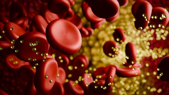
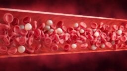
Your staying just as healthy despite gaining some fat actually aligns well with a lot of previous research. And then, there were those blood transfusions everyone’s talking about.

Yes, I did six of them. I had a donor who was eating a Blueprint diet. I did five total infusions from that donor and then one from my son. And over that time course, we couldn’t detect any changes in my biomarkers. That’s not to say there weren’t any positive or negative effects, but we just couldn’t find them.

Or isolate them?

Correct. However, at that time, we weren’t doing many new things. I did this once every three weeks, so it was a pretty short window of time. But when my father did just one infusion, when I gave him one liter of my plasma, and he removed 700 milliliters of his plasma, his speed of aging measured by DunedinPACE clock went down from the age equivalent of 71 to 46 immediately. A dramatic effect. And this is about the same effect size you see in rats; it mapped pretty closely.

We know that older and less healthy animals benefit the most from many anti-aging therapies.

Exactly.

Continuing with therapies, a few months ago, you went to Vitalia to get a follistatin gene therapy. How did that go? Do you have any preliminary results?

I’d say, three things. One, my follistatin levels increased by 160% fourteen days after the treatment. That was their hypothesis about C-max (maximum concentration). I don’t know if that’s true, because we only did one measurement. So, at least a 160% increase.

Then, my muscle mass. It was already in the top one percentile for optimal, and I got an additional increase of 7%. And finally, my speed of aging dropped from 0.7 to 0.64.

And is this because of follistatin?

That was the only thing that changed.

Blueprint going commercial surprised a lot of people. How did that happen?

I had no commercial intent when I started Blueprint. It was simply an exploration – what can a person do in this field? I didn’t expect it to go viral, but it did, and the number one thing people said is, make it easy. “I see your page, you have all these supplements, drugs, treatments, it’s so complicated, I don’t want to do it, it’s too much.”

Sort of, “Simplify this and take my money”?

Yes. “Just make it easy.” And I got this request from everybody. I didn’t want to be in the food business. It sucks. Low margins, a lot of headaches, everyone’s doing it. And I sat on it for six months. After Blueprint went viral, I didn’t do anything for six to eight months. I just tried to evaluate my next steps because once you start something, you own it, right?

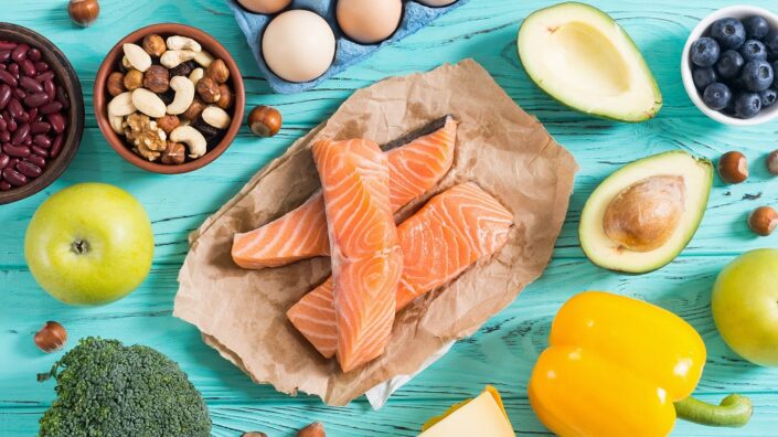
When we were doing Blueprint, we would test all the supplements, all the food I was eating, and we found that most of the food was alarmingly dirty. I’m not sure you’re aware of how dirty food is. Even in supplements, you don’t always get what’s on the label.

We tried to tackle this problem ourselves to see if we could buy ingredients ourselves, do testing, and build the whole supply chain. And my friends were like, “Can I have it too?”

This is how you source your supplements, too?

Yes, I started sourcing everything myself – the olive oil, the chocolate, all the supplements. For instance, we would buy raw batches of lentils and test them for everything. We searched the world over for clean lentils and couldn’t find any, even the ones that were labeled organic. Everything was alarmingly dirty.

Finally, I said, okay, people want it to be easy, this is legitimately a major problem. What if we could just be the source of every calorie a person wants to consume? But I’m definitely not doing it for money, otherwise I wouldn’t be doing it. There are so many things that make much more money.

Have you then thought about publishing more data or more detailed data about all your supplements and food and how they are cleaner?

Yes, I actually posted a video today on X. We’re doing it. I basically went to the grocery store, I took ten dark chocolates, and measured them for heavy metals and flavanols. So, yes, we’re starting to share this publicly.

When we talked last year, you said you didn’t want to invest in or sponsor fundamental longevity research because you have your own way of contributing, of doing things. Is this something that has changed?

No. I learned this when I was investing in deep tech. The hit rate in deep tech is very low. It’s very hard to make money in deep tech.

So, you don’t want to bet on discovery. You want to bet on companies that build picks and shovels for the entire industry. That’s why I bet on Gingko, for instance. They are building the ability to read and write DNA at a lower cost.

Basically, we think that synthetic biology is here, so, everyone will want to do all these things with biology, and the critical element we need is low-cost read and write capabilities.

If you’re trying to go for a specific therapy or indication, you only have one shot on target. You have your molecule, your FDA trials, and you either win or lose.

So, you prefer to invest in universally needed tools?

I’ve been trying to think methodically: with the limited capacity I have as a human, how do I find the biggest fulcrum? I could certainly fund non-profits and companies, but instead, I’m saying, why don’t I try to build the basics of sleep, diet, and exercise for hundreds of millions of people and a philosophy for billions of people? To me, that’s a bigger fulcrum than a single drug, or a single non-profit, or a single study, and so on.

I’m inherently an entrepreneur trying to find arbitrage in systems, I like arbitrage. And I think that the world’s systems are breaking, and new systems will have to be better. I think that it’s better to make money out of the gate than to bet on these ten-year-long things, and it’s better to empower millions of people to be part of the ecosystem than a few.

I know that people criticize me: “You’ve got money! Do this, do that!” They’re trying to be capital allocators. What they don’t understand is that they’re not me, I’m not them, I don’t think like them, and they don’t even understand my vision, what I’m trying to do.

Have you been talking to other high-net-worth individuals about longevity? I’ve heard from many people that multimillionaires and billionaires are notoriously reluctant to join our cause.

Yes, I have talked to many, and I think they get it but in a particular way. They don’t think we’re going to stop aging imminently, but they agree with the primary thesis of my argument.

I think what you’re saying is that they’re discouraged, there’s no movement. They don’t change their habits, they don’t invest in biotech or aging, and it seems like it makes no sense. Your only real asset in life is your existence. Why would you just die without a fight? You can’t take your money with you.

Basically, they’re playing the “immortality of accomplishment” game, chasing status and power. What I’m saying to them is, hey, I understand that you have your worldview, but here’s a different perspective that’s going to change everything you care about. The world is about to change fundamentally, and that affects everything you’re doing. All of it may be of no consequence.

Now, that has the potential of stopping them in their tracks, giving them this “I can’t just carry on as is now” feeling. That doesn’t mean they’re going to just drop a billion dollars on funding anti-aging research, but they get the idea that everything they care about – all their status, their power, their reputation, is potentially now at risk because of this major change that’s coming.

Is this working?

I’ve had several billionaires who, after we engaged, just said: “I’m in, what do you need?” I haven’t cashed those checks yet. I’m really trying to build a solid community, not to act hastily. I’m trying to build the infrastructure. That’s what I’ve spent the past six months on – how to transform Don’t Die into an actionable plan politically, economically, morally, ethically, socially.

The pudding is served

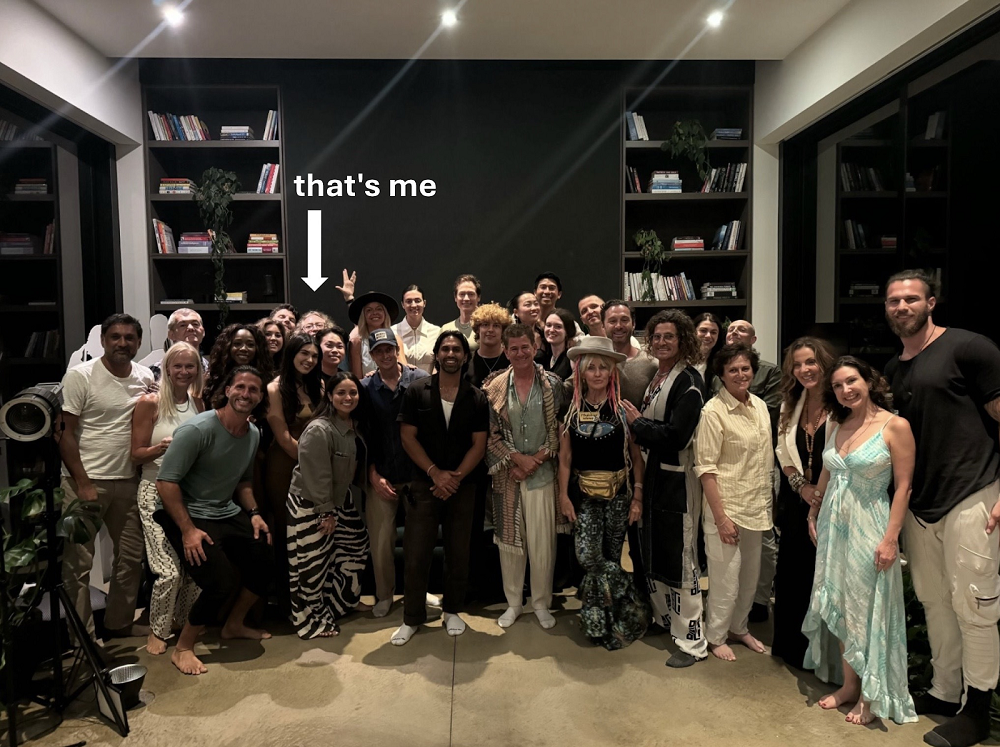
As we were talking, guests began to arrive, and the growing murmur of voices signaled that we should wrap things up and head back inside. I didn’t mind – after all I’d heard about Don’t Die, I couldn’t wait to see Bryan in action.

He told me in advance that this time, the crowd was going to be unusually large, around 30 people (no Kardashians, though). And what a motley LA crowd it was! Entrepreneurs and physicians, artists and scientists, investors and inventors, and, well, one science journalist. Most were dressed casually, while two or three looked like they came straight from Burning Man (in fact, they ended up inviting everyone to Burning Man).

Bryan Johnson Group

As guests mingled in a crowded foyer, a modest dinner was served. First, we toasted with shots of Bryan’s olive oil, which he proudly markets (with tongue in cheek) as “Snake Oil”. By then, I had already bought a couple of bottles and formed my opinion: great quality, a bit tangy for my taste.

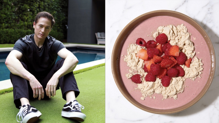
Then, each guest picked up an earthenware bowl of Nutty Pudding, a staple of Bryan’s kitchen made from macadamia milk and nuts, walnuts, chia seeds, flaxseed, cocoa, berries, cherries, cinnamon, and pomegranate juice. This super-duper-healthy slush has a distinct dusty pink color and an equally distinct taste: yummy but takes a bit of getting used to.

Clutching our bowls, we all stepped into the living room and sat down. Bryan started by asking people to introduce someone else in the audience. This proved to be a great bonding experience, albeit time-consuming. Since people were probably invited by word of mouth, many of them knew someone, so that wasn’t a problem for them. This left a handful of loners, such as myself, but I quickly struck a deal with the person sitting next to me, who turned out to be a nuclear scientist, and we introduced each other.

Do we know anything at all?

Then the action began. Bryan told us that he was going to ask us a series of questions. The first one was whether we’d agree to replace our own decisions with an algorithm if we knew for sure that it guarantees us the best physical, mental, and spiritual health we can imagine. This was a clear nod to Blueprint, which is touted by Bryan as the algorithm that should supplant your faulty decision-making mechanisms when it comes to eating, sleeping, exercising, and other types of body maintenance.

This got the room divided, with people either objecting to the idea that an algorithm can know more about our well-being than we do or pointing out that it was a trick question: if the algorithm is going to make us happy by definition, there was no reason not to succumb to it. After all, it cannot make us simultaneously happy and unhappy.

Next, Bryan invited us to think about how people from the 25th century would view our morals, ethics, and social norms. The audience generally agreed that our descendants would find objectionable our penchant for killing our bodies, our brethren, and other animals while being hopelessly divided by politics and religion. However, it was also quite clear that we had zero idea about who those descendants may be and whether they will exist at all.

Then, Bryan told us about his son, a college student. For the entirety of human history, he said, things kept changing slowly enough for people to be able to imagine their life course. Just a couple of decades ago, the path, the possibilities were understandable – you could, at least in theory, choose an occupation, learn the trade, earn your living, and retire in due course.

Today, with the advance of AI, things have changed fundamentally. What should his son study, Bryan asked? How should he chart his path?

Predictably, no one in the audience could conjure a solid answer. Bryan used this and previous questions as a segue to talk about our greatly increased inability to predict the future. Many, if not all, of our dearly held concepts might not survive in a post-AI world.

For the entire evening, Bryan subtly led us to this conclusion, showing that people tend to “consult their knowns,” but this method isn’t effective enough in an ever less-predictable world. Even thinking from the first principles won’t cut it. Instead, we have to think from the “zeroth principles,” that is, assume that we know next to nothing, and imagine something entirely new.

Don’t Die, and find out

What should we do, then? According to Bryan, Don’t Die is the solution. To be honest, I still can’t entirely wrap my head around it. Does it mean it’s wrong? Not necessarily. A hard-to-comprehend idea can be a genius one.

However, I feel that Don’t Die philosophy is still a work in progress (something Bryan himself confirmed to me), a star-forming cloud of ideas, some of which seem intriguing and profound, while others are sort of headscratchers for me at this point.

One idea that aligns well with the longevity field is that of the sanctity of life and the immense value in extending it. Bryan’s twist on it is that cherishing life, consciousness, and the body that contains it provides a comprehensible goal most people can unite behind.

“This is our sole known priority,” Brian said. “The only thing we all agree upon. It’s not a solution to everything, but it’s a framework, like democracy. Ideologies cannot provide the same level of unity. It’s our only common identity when we can’t plan forward.” His book Don’t Die: Dialogues adds, “the ‘fountain of youth’ thing is a clear and good goal because, at the very least, we get that goal. We understand it. We can define it.”

Bryan sees the world’s reaction to the COVID pandemic as a manifestation of this common goal that we all share deep inside, even if, ostensibly, it might not always align with our ideologies or religious beliefs. Again, this weird dualism, where people value life and good health but do not translate this into an anti-aging sentiment, is well known, and Bryan is trying to bridge this gap.

Bryan thinks that learning to trust algorithms, like he does with Blueprint, can potentially free up so much time and energy “to explore new frontiers” that the impact will be comparable to the invention of fire, which uniquely sped up human civilization. We would both be more mentally fit and have more time on our hands (indefinitely more if we can figure out how to stop and reverse aging).

As to abandoning our beliefs and embracing the unknown, I actually agree that not only does this nurture open-mindedness and curiosity, but also, somewhat surprisingly, quells that nagging dread of the future. We’ll just have to wait and see, right? We’re an inquisitive, fearless, and inventive species that has extricated itself from all kinds of tight places before. But to be able to stick around, we’ll have to stay alive.

On the way, we’ll also have to learn to trust algorithms that will be taking away more and more of our tasks to the point of threatening our very relevance. Building machine minds is a dangerous game, but humanity is bound to play it. There’s just no other way – again, something that most aging scientists would agree with (aging is probably just too complex for the unassisted human mind to unravel).

As I approached Bryan to thank him for the evening, it was way past his bedtime. “See,” he said, smiling, “I do sacrifice things sometimes when I feel it’s worth it.”

He asked me what I thought of the dinner. “It was very thought-provoking,” I said frankly as I shook his hand. “For me, this is one of the best feelings in the world.”

Did the dinner turn me into a Don’t Die warrior? Probably not, but on the other hand, I’m already an ally. Is Don’t Die the best way to promote “the longevity cause”? I don’t know. But it’s not a bad way, either. It’s just Bryan’s way.

We would like to ask you a small favor. We are a non-profit foundation, and unlike some other organizations, we have no shareholders and no products to sell you. All our news and educational content is free for everyone to read, but it does mean that we rely on the help of people like you. Every contribution, no matter if it’s big or small, supports independent journalism and sustains our future.
Keto diet

Keto Diets May Help Cognitive Function in Alzheimer’s

The results of 10 independently conducted clinical trials suggest that ketogenic diets have a positive impact on the cognitive function of Alzheimer’s disease patients [1].

Keto instead of a pill?

Despite extensive research, there is stilll no effective treatment for Alzheimer’s disease. Therefore, approaches that aim to delay disease through lifestyle changes, such as diet, are being investigated.

One of the possible therapeutic targets is brain energy metabolism. The authors of this article focused specifically on this metabolism and the impact of ketogenic diets on Alzheimer’s disease.

High energy demands

The brain has high energy needs. Under normal conditions, it consumes glucose, but when glucose levels are low, the brain can use ketones as an energy source. Previous studies have found that in patients with mild cognitive impairment and Alzheimer’s disease, the brain can absorb ketones more easily than glucose [2, 3].

Proper energy balance is important for brain health. Previous research suggests that prolonged brain energy shortage leads to neuronal death, impaired amyloid-β and tau protein clearance, and the development of Alzheimer’s disease [4]. This suggests that restoring the brain’s energy balance can be a potential therapeutic strategy.

Better cognitive scores

The authors of this study decided to “collect and systematically evaluate the clinical randomized controlled trials” addressing the impact of a ketogenic diet on Alzheimer’s disease. The researchers identified ten trials from the United States, Japan, Canada, China, and New Zealand. Those studies enrolled 691 participants diagnosed with Alzheimer’s disease, but due to 40 participants dropping out, 651 participants were included in the final analysis. These trials lasted between 12 to 60 weeks.

The studies differed in their exact diets. One study used a standard ketogenic diet (macronutrient ratio of 58% fat, 29% protein, 7% fiber, and 6% net carbohydrates by weight). Eight studies used the medium chain triglycerides diet (MCT), a diet that uses lab-made types of fats that produce ketones more easily than fats normally found in food. One study selected the modified Atkins diet (MAD).

The studies included in this analysis used different cognitive function assessment tests, including the Mini-Mental State Examination (MMSE), a commonly used “screening tool for cognitive disorders,” the cognitive subscale of the Alzheimer’s Disease Assessment Scale (ADAS-Cog), the Nishimura Mental State Scale (NM scale), and the Activities of Daily Living (ADL) scale. ADAS-Cog is a scale used to assess the memory of Alzheimer’s disease patients. The NM scale measures abilities in daily life. Using different assessment scales and obtaining similar results adds to the credibility of these studies.

In total, these studies concluded that ketogenic diets have an overall positive impact on Alzheimer’s disease patients’ cognitive function. A meta-analysis showed significant changes caused by the interventions in studies that used the MMSE, ADAS-Cog, and NM scales but not ADL.

Changes in blood lipids

Some of the studies also looked at the biochemical profiles of the study participants. Four out of ten studies also analyzed blood lipid profiles. A ketogenic diet resulted in significantly elevated concentrations of triglycerides and low-density lipoprotein-cholesterol (LDL-C), the “bad” cholesterol, but not total cholesterol nor high-density lipoprotein-cholesterol (HDL-C), the “good” cholesterol.

Keto’s dark side

The authors recognize that despite the positive cognitive test results and possible positive implications of ketogenic diets for Alzheimer’s disease patients, this diet and those studies have their shortcomings.

First, the researchers noted that the studies they analyzed have a relatively short duration (typically around three months). While this is convenient from a research perspective and allows for initial evaluation of the diets’ impact, there is a lack of information and monitoring of long-term health effects.

Additionally, long-term adherence to such a strict diet might be difficult; even during the short period of the study, some participants withdrew due to incompatibility.

Some other studies observed some negative health effects of keto diets. It was reported that a strict ketogenic diet could possibly lead to symptoms such as constipation [5], fatigue, headache, nausea, and low blood sugar (hypoglycemia) [6] or more serious reactions such as dehydration, hepatitis, inflammation of the pancreas (pancreatitis), disturbances in sodium, magnesium and uric acid blood levels [7], decreased bone mineral density, kidney stones, heart issues, and anemia [8]. Also, the increase in triglycerides and LDL-C might increase the risk of cardiovascular diseases.

Limitations

This is a review that is built from data gathered by other researchers. The included studies were not all conducted under the same conditions, and the differences between diets and study durations limit how the data can be interpreted. Some studies were also not conducted with large sample sizes.

Overall, this study provides compelling evidence for the potential of KD in the treatment of Alzheimer’s disease, while also highlighting the need to carefully consider potential health risks associated with its use. Future research could explore optimising KD formulations and intervention strategies to maximise cognitive improvements while minimising associated health risks.

We would like to ask you a small favor. We are a non-profit foundation, and unlike some other organizations, we have no shareholders and no products to sell you. All our news and educational content is free for everyone to read, but it does mean that we rely on the help of people like you. Every contribution, no matter if it’s big or small, supports independent journalism and sustains our future.

Literature

[1] Rong, L., Peng, Y., Shen, Q., Chen, K., Fang, B., & Li, W. (2024). Effects of ketogenic diet on cognitive function of patients with Alzheimer’s disease: a systematic review and meta-analysis. The journal of nutrition, health & aging, 28(8), 100306.

[2] Nugent, S., Croteau, E., Pifferi, F., Fortier, M., Tremblay, S., Turcotte, E., & Cunnane, S. C. (2011). Brain and systemic glucose metabolism in the healthy elderly following fish oil supplementation. Prostaglandins, leukotrienes, and essential fatty acids, 85(5), 287–291.

[3] Croteau, E., Castellano, C. A., Fortier, M., Bocti, C., Fulop, T., Paquet, N., & Cunnane, S. C. (2018). A cross-sectional comparison of brain glucose and ketone metabolism in cognitively healthy older adults, mild cognitive impairment and early Alzheimer’s disease. Experimental gerontology, 107, 18–26.

[4] Zhang, S., Lachance, B. B., Mattson, M. P., & Jia, X. (2021). Glucose metabolic crosstalk and regulation in brain function and diseases. Progress in neurobiology, 204, 102089.

[5] Dashti, H. M., Mathew, T. C., & Al-Zaid, N. S. (2021). Efficacy of Low-Carbohydrate Ketogenic Diet in the Treatment of Type 2 Diabetes. Medical principles and practice : international journal of the Kuwait University, Health Science Centre, 30(3), 223–235.

[6] Roehl, K., & Sewak, S. L. (2017). Practice Paper of the Academy of Nutrition and Dietetics: Classic and Modified Ketogenic Diets for Treatment of Epilepsy. Journal of the Academy of Nutrition and Dietetics, 117(8), 1279–1292.

[7] Włodarek D. (2019). Role of Ketogenic Diets in Neurodegenerative Diseases (Alzheimer’s Disease and Parkinson’s Disease). Nutrients, 11(1), 169.

[8] Crosby, L., Davis, B., Joshi, S., Jardine, M., Paul, J., Neola, M., & Barnard, N. D. (2021). Ketogenic Diets and Chronic Disease: Weighing the Benefits Against the Risks. Frontiers in nutrition, 8, 702802.

Ora Biomedical

Ora Biomedical Awarded Technology Development Grant

Ora Biomedical, Inc., a pioneering longevity biotechnology company, is pleased to announce that it has been awarded a Small Business Innovation Research (SBIR) Phase I grant from the National Institutes of Health (NIH). The $324,240 grant will support the development of EleGantry, an innovative software and hardware infrastructure designed to enhance rigor and reproducibility in invertebrate research, with a particular focus on longevity studies using Caenorhabditis elegans (C. elegans).

EleGantry: Revolutionizing Preclinical Drug Discovery

Ora Biomedical specializes in discovering and developing interventions that extend healthy lifespan and combat age-associated diseases by targeting the fundamental mechanisms of biological aging. The EleGantry system represents a significant advancement in the company’s high-throughput drug discovery pipeline. By automating key steps in the C. elegans research process and enhancing data collection across the entire lifespan, EleGantry will address the growing need for increased rigor and reproducibility in preclinical studies.

“With the NIH’s support, we are poised to identify and overcome major sources of variability and challenges associated with C. elegans research, contributing to our goal of creating the world’s largest and highest quality longevity interventions database,” said Dr. Mitchell Lee, CEO of Ora Biomedical. “EleGantry will not only elevate the standard of preclinical aging research but also expedite development of robust therapeutics that can significantly extend healthy human lifespan.”

Why EleGantry Matters

C. elegans has long been a mainstay in biomedical aging research because highly evolutionarily conserved features drive aging from invertebrates to mammals. However, variability in research methodologies has often led to inconsistent results, particularly in studies focused on lifespan. EleGantry will mitigate these issues by integrating advanced imaging, data collection, and analysis tools powered by state-of-the-art machine learning algorithms. The platform will also automate data reporting, making it easier for researchers to replicate studies and validate findings. Ora Biomedical’s pioneering work in this field advances non-mammal animal models for drug development and aligns closely with the FDA Modernization Act 2.0 which aims to reduce the use of live mammals in research.

“The development of EleGantry is crucial as we move towards more reliable and scalable invertebrate models for drug discovery,” added Dr. Lee. “This tool will enable us to identify and prioritize therapeutic leads with unprecedented efficiency and accuracy.”

Looking Ahead

The successful completion of this Phase I project will pave the way for the subsequent development and commercialization of EleGantry, further establishing Ora Biomedical as a leader in longevity biotechnology. The company plans to use EleGantry to expand its preclinical screening capabilities, providing a robust platform for discovering the next generation of gerotherapeutics. This project is supported by the Office Of The Director, National Institutes Of Health and the National Institute of General Medical Sciences (NIGMS), under Award Number R43OD037622. The content is solely the responsibility of the authors and does not necessarily represent the official views of the National Institutes of Health.

About Ora Biomedical

Ora Biomedical, Inc. is a longevity biotechnology company headquartered in Tukwila, WA. Launched out of the University of Washington School of Medicine in 2022, Ora Biomedical develops interventions that maximize healthy lifespan by targeting the molecular mechanisms that drive aging itself. Ora uses best-in-class robotics and AI to perform high-throughput, high-precision phenotypic testing in live animals with lifespan, healthspan, and stress resistance as primary endpoints.

www.orabiomedical.com

Company Press Contact:

Dr. Mitchell Lee

CEO & Co-Founder, Ora Biomedical, Inc.

Email: mitchell@orabiomedical.com

We would like to ask you a small favor. We are a non-profit foundation, and unlike some other organizations, we have no shareholders and no products to sell you. All our news and educational content is free for everyone to read, but it does mean that we rely on the help of people like you. Every contribution, no matter if it’s big or small, supports independent journalism and sustains our future.

Pancreas

Combination Therapy Works Against Pancreatic Cancer in Mice

In a new study, a multi-prong treatment combined with a clever delivery method has shown promise against one of the deadliest cancers [1].

Lethal and barely treatable

Pancreatic ductal adenocarcinoma (PDAC) is one of the deadliest types of cancer. It is rarely diagnosed early and very aggressive. It also responds poorly to treatment, primarily because its fibrotic tumor microenvironment (TME) hinders infiltration by immune cells.

Overall, the five-year survival rate for pancreatic cancer is slightly above 10%. Even for patients who were diagnosed early, it stands at just 37%, making this a high-priority target for cancer researchers.

One novel approach involves inducing senescence in cancer cells [2], causing them to produce the senescence-associated secretory phenotype (SASP). SASP factors are highly inflammatory, which helps remodel the TME and increase its susceptibility to infiltration by immune cells.

This approach works reasonably well in some cancer types. However, in PDAC, TME suppresses proinflammatory SASP components and the immune system activation that they are supposed to trigger.

The immune response can be bolstered by activating the stimulator of interferon genes (STING) pathway. This increases production of interferon and some other pro-inflammatory cytokines [3]. STING agonists showed some promise against PDAC, but their use is limited by their poor uptake by cells and short half-life (they are excreted quickly). The pancreas is also very hard to reach for direct intratumoral delivery.

Multiple approaches

In this study published in Science Translational Medicine, scientists from the University of Massachusetts tested a novel combination therapy in a mouse model of PDAC. One element of the combination consisted of two drugs: a STING agonist and a toll-like receptor 4 (TLR4) agonist. TLR4 agonists can additionally stimulate interferon responses. The other half was a popular duo of senescence-inducing compounds, trametinib and palbociclib (T/P).

Importantly, the STING agonist cdGMP and the TLR4 agonist MPLA were encapsulated together in nanoparticles (NPs) to overcome the usual delivery limitations. Achieving this was not an easy task since cdGMP is hydrophilic and TLR4 is hydrophobic. The researchers solved this problem by loading the two molecules into different compartments: cdGMP into the aqueous NP core and MPLA into the lipid bilayer shell.

Although NPs were administered intravenously, they found their way to the tumor area and infiltrated numerous cell types, including macrophages and antigen-presenting dendritic cells. NPs hardly accumulated in organs and did not cause toxicity.

Just as the researchers hoped, combining NPs with the pro-SASP treatment led to a synergistic effect. Pretreatment with the senescence-inducing duo T/P for 12 days increased the uptake of NPs by both tumor cells and immune cells. Moreover, it worked both ways: SASP induction was stronger with the combination therapy than with T/P alone.

“Together,” the paper says, “these findings demonstrate that combinatorial T/P and immuno-NP treatment, through both tumor cell autonomous and non–cell autonomous molecular mechanisms, enhances IFN signaling, proinflammatory cytokine production, and antigen presentation in the PDAC TME.”

An encouraging proof of concept

However, the important test is the therapy’s anti-tumor effectiveness. Here, too, the combination was more effective than NP or T/P treatments alone, leading to massive tumor necrosis in just 48 hours and significantly improved survival times. Moreover, some mice had complete response (full eradication of tumors), although the cancer returned after the treatment stopped.

“This is pretty striking,” said Marcus Ruscetti, assistant professor of molecular, cell and cancer biology at UMass Chan Medical School, and a corresponding author on the study. “We’ve never seen that in this model before.”

Ruscetti said that although all animals eventually succumbed to cancer, the results still were “a very encouraging step toward a cure.”

“If you go beyond pancreas cancer to other cancer types, you need a combination therapy to target the tumor and the immune system,” he added. “This is a strategy to be able to do that.”

Collectively, our results suggest that engineering approaches to target multiple cell types and immune suppressive barriers through induction of type I IFN signaling in the PDAC TME could pave the way for coordinated innate and adaptive immune responses to achieve immunotherapy successes that have thus far been elusive for patients with PDAC.

We would like to ask you a small favor. We are a non-profit foundation, and unlike some other organizations, we have no shareholders and no products to sell you. All our news and educational content is free for everyone to read, but it does mean that we rely on the help of people like you. Every contribution, no matter if it’s big or small, supports independent journalism and sustains our future.

Literature

[1] Chibaya, L., DeMarco, K. D., Lusi, C. F., Kane, G. I., Brassil, M. L., Parikh, C. N., … & Ruscetti, M. (2024). Nanoparticle delivery of innate immune agonists combined with senescence-inducing agents promotes T cell control of pancreatic cancer. Science Translational Medicine, 16(762), eadj9366.

[2] Ruscetti, M., Morris, J. P., Mezzadra, R., Russell, J., Leibold, J., Romesser, P. B., … & Lowe, S. W. (2020). Senescence-induced vascular remodeling creates therapeutic vulnerabilities in pancreas cancer. Cell, 181(2), 424-441.

[3] Vonderhaar, E. P., Barnekow, N. S., McAllister, D., McOlash, L., Eid, M. A., Riese, M. J., … & Dwinell, M. B. (2021). STING activated tumor-intrinsic type I interferon signaling promotes CXCR3 dependent antitumor immunity in pancreatic cancer. Cellular and molecular gastroenterology and hepatology, 12(1), 41-58.

Ground coffee

Caffeic Acid Variant May Suppress Bone Deterioration

In Aging, researchers have described how a derivative of caffeic acid inhibits osteoclasts, the cells that break down bone.

A modification of a tested compound

We have recently published new information on what triggers the imbalance between bone-destroying osteoclasts and bone-building osteoblasts. This is a well-known problem with aging, and researchers around the world have looked into ways of rectifying this imbalance. Caffeic acid, which is found in coffee, tea, fruits, vegetables, and some extracts, has, among other positive effects, been previously found to suppress osteoclasts [1], including in a rat study [2].

These researchers have previously modified caffeic acid into N-(4-methoxyphen) methyl caffeamide (MPMCA), a compound that they have found to be more effective than regular caffeic acid in protecting the liver against oxidative stress [3]. Therefore, they conducted this study in order to determine if it could also be more effective in suppressing osteoclast activity.

Broad effects against precursors and mature cells

Interestingly, MPMCA does not inhibit osteoclast precursors under normal circumstances at any of the tested doses, from 0.1 to 3 μM. However, when these precursors are stimulated to become osteoclasts through the RANKL pathway for seven days, MPMCA reduces the number of osteoclasts, markedly reduces their average size, and suppresses the production of F-actin, which is required for differentiated osteoclasts to attach to bone. These effects are dose-dependent.

Gene expression analysis confirmed these findings. The genes TRAP, CTSK, and NFATC1, all of which are related to osteoclast differentiation, were suppressed by MPMCA. Testing at higher doses, the researchers found that MAP kinase pathways were similarly suppressed, as was the well-known NF-κB pathway.

MPMCA also encouraged differentiated osteoclasts to die by apoptosis. Cleaved caspase-3 and DAP, both of which are apoptosis markers, were increased with MPMCA. Just like with precursors, both the number and size of these mature osteoclasts were reduced.

MPMCA osteoclasts

However, MPMCA, unlike caffeic acid [2], did nothing to osteoblasts. Osteoblast differentiation markers, including gene expression, were unaffected.

These findings are very positive, but they were only in cells. If the results of this study can be replicated, and this compound can be confirmed to be safe and effective in animal models, MPMCA may be a candidate for clinical trials against osteoporosis and age-related bone loss.

We would like to ask you a small favor. We are a non-profit foundation, and unlike some other organizations, we have no shareholders and no products to sell you. All our news and educational content is free for everyone to read, but it does mean that we rely on the help of people like you. Every contribution, no matter if it’s big or small, supports independent journalism and sustains our future.

Literature

[1] Ekeuku, S. O., Pang, K. L., & Chin, K. Y. (2021). Effects of caffeic acid and its derivatives on bone: A systematic review. Drug design, development and therapy, 259-275.

[2] Tang, Q. Y., Kukita, T., Ushijima, Y., Kukita, A., Nagata, K., Sandra, F., … & Iijima, T. (2006). Regulation of osteoclastogenesis by Simon extracts composed of caffeic acid and related compounds: successful suppression of bone destruction accompanied with adjuvant-induced arthritis in rats. Histochemistry and cell biology, 125, 215-225.

[3] Tsai, T. H., Yu, C. H., Chang, Y. P., Lin, Y. T., Huang, C. J., Kuo, Y. H., & Tsai, P. J. (2017). Protective effect of caffeic acid derivatives on tert-butyl hydroperoxide-induced oxidative hepato-toxicity and mitochondrial dysfunction in HepG2 cells. Molecules, 22(5), 702.

Rejuvenation Roundup August 2024

Rejuvenation Roundup August 2024

Summer’s at its end, but rejuvenation biotechnology is still heating up, with researchers discovering ways of vastly improving mouse lifespan and conducting a recruitment drive for young scientists. Here’s how the field was advanced in August.

Interviews

Michael Antonov InterviewMichael Antonov: from Oculus to Longevity Biotech: After Oculus was sold to Facebook for two billion dollars a decade ago, Michael Antonov, one of the founders, could have become a major tech investor, a popular podcaster with his own mega-theory of everything, a hedonist, or all of the above. Instead, he gravitated towards the emerging field of longevity biotech.

Peter Diamandis: “Stay Healthy, Anti-Aging Tech is Coming”: Peter is a serial entrepreneur and investor in several fields, from commercial space flight to longevity biotech; a visionary speaker and author of several best-selling books, including Abundance, The Future is Faster Than You Think, and Life Force.

Research Roundup

Black lab miceRapamycin + Trametinib Increase Mouse Lifespan by up to 35%: Combining rapamycin and trametinib seemed to have a cumulative effect in both sexes but mostly in females. These results are probably due to a reduction in both cancer and inflammation.

Vegan Diet Lowers Biological Age in a New Study: In a study that involved pairs of identical twins, Stanford scientists have shown that a healthy vegan diet leads to a decrease in biological age and possibly to other health benefits.

Hidden minefieldNew Epigenetic Clock Built on Lurking DNA Fragments: In Aging Cell, a team of researchers has announced Retroelement-Age, a novel clock that focuses on the expression of buried pieces of DNA that are normally suppressed. The natural human genome has a large number of artifacts left over from ancient viral infections along with pieces of DNA that transpose themselves into the genome.

Hyperbaric Oxygen Therapy Increases Fitness in Elderly: In a new study, a 12-week protocol of hyperbaric oxygen therapy (HBOT) resulted in marked increases in aerobic capacity and cardiac blood flow. HBOT involves placing patients in a tightly sealed chamber where they breathe pure oxygen under higher-than-normal atmospheric pressure.

Intestines and blood vesselsFecal Microbiota Transplants Reduce Vascular Aging in Mice: Fecal microbiota transplantation from young mice to older mice improved multiple metabolic parameters and some hallmarks of aging, such as inflammation and telomere shortening. Dysbiosis, an imbalance in the gut microbiome, is a newly recognized hallmark of aging.

Clinical Trial Reveals a Foe of Fatty Liver Disease: A highly anticipated clinical trial was just published by the prestigious Journal of Nutrition with a surprising finding: that C15:0, a trace dietary saturated fat present in butter, can lower liver enzymes in young adults susceptible to fatty liver disease.

Flowing blood with white blood cellsImmune Peptide Might Keep White Blood Cells Contained: In npj Aging, researchers have described how immune cell infiltration in inflammaging can be reduced with an immune-related peptide in a mouse model. These researchers employed a mouse model of peritonitis, the inflammation of the abdominal lining, in order to determine how PEPITEM influences white blood cells in differently aged animals.

A Ketosis-Related Compound Alleviates Arthritis in Rats: In Aging Cell, researchers have published details on the pathway by which a compound commonly found in ketone bodies ameliorates osteoarthritis in a rat model. The cartilage holding joints together is built and maintained by chondrocytes, which synthesize such necessary proteins as collagen and aggrecan but deteriorate with aging.

Black 6 looking at viewerNMN Increases Lifespan in Female Mice: A new preprint from David Sinclair’s lab has discovered that NMN supplementation started in midlife increases median and maximal lifespan in female mice and boosts healthspan in males. NMN’s effect on mammalian lifespan has not been tested until now.

A Botanical Extract Extends Lifespan and Healthspan in Mice: According to a new study, a polyphenol-rich natural extract positively impacts lifespan, healthspan, and cellular senescence. These results were observed in both cell culture and a mouse model. Traditional and folk medicines offer many botanical extracts that can be tested by modern science for their medicinal properties.

Elderly man with large waistlineInflammation May Drive Fat Distribution in Older People: A new study published in Aging Cell has detailed what happens to individual fat cells in white adipose tissue (WAT) as they age. Previous research has described WAT as an organ in its own right, functioning not only as an energy storage source but as a regulator of metabolism.

Anti-Inflammatory Diets Mitigate Risk of Dementia: A new study suggests that an anti-inflammatory diet can significantly reduce the risk of dementia and delay its onset even in people with existing cardiometabolic diseases.

Tricky balanceFighting Inflammation May Cause More Senescence: A researcher publishing in Biogerontology has reviewed the literature on the relationship between cellular senescence and the immune system, finding that dwindling immune surveillance allows senescent cells to accumulate.

High-Intensity Interval Training Improves Cognition: Cognitive decline is one of the risks associated with getting older, but new research suggests there may be a way to fight back. A new study suggests that high-intensity interval training improves spatial learning and maintains brain volume and connectivity.

Manuka TreeManuka Honey Effective Against Breast Cancer in Mice: UCLA scientists have shown that Manuka, an exclusive honey variety, can quench estrogen receptor-positive breast cancer in vitro and in vivo. Breast cancer remains the deadliest type of cancer in women, claiming more than 40 thousand victims a year in the US alone.

Why Bone Destruction Is Accelerated with Aging: Researchers have described a relationship between the NAD+ regulator CD38, mitochondrial function, and the gradual destruction of bone in Aging Cell. Not only do older mice have more osteoclasts, their osteoclasts are bigger and more aggressive in wearing down bone.

Weekend backpackersWeekend-Oriented Physical Activity Superior for Brain Health: According to a new study, “weekend warriors”, people who have most of their physical activity over one or two days a week, enjoy the most robust risk reduction for several brain health-related conditions.

NNMT Inhibitor Improves Muscle Function in Aged Mice: A recent paper has reported on improving muscle function in aged mice through combined treatment of exercise and the inhibition of NNMT, an enzyme involved in NAD+ metabolism.

Aged skinSkin Aging and Cellular Mobility at the Protein Level: In Aging, researchers have described how the changing production of skin cells’ proteins is a core part of their age-related decline. In healthy skin, dermal fibroblasts produce the proteins needed to maintain the extracellular matrix (ECM), the network of collagen and elastin that holds soft tissues together, but this ability declines with age.

Some Cells Age Much Faster Than Others: In Stem Cell Reports, researchers have described how cell subpopulations and division affect the results of epigenetic clocks. Some cells and tissues accumulate epigenetic aging much more rapidly than others.

News Nuggets

A4LI LogoA4LI Responds to NIH Reform Proposal: The Alliance for Longevity Initiatives (A4LI) is proposing a new National Institute for Longevity and Aging Research (NILAR) in response to the recent NIH reform proposed by House Energy and Commerce (E&C) Chair Cathy McMorris-Rodgers (R-WA).

AlopeciaA Phase 2 Clinical Trial for Hair Loss: Los Angeles-based Pelage Pharmaceuticals has just announced a Phase 2a clinical trial for the treatment of androgenetic alopecia, also known as pattern baldness. It is thought that up to 50% of men experience hair loss as a result of pattern baldness by age 50.

Coming Up

40+ Experts Gather in Shanghai for TimePie Longevity Forum: From November 1st to 3rd, 2024, the 5th TimePie Longevity Forum will take place in Shanghai, China. Bringing together 40+ leading speakers from around the globe and attracting over 1,000 attendees, this event is set to be one of the most significant platforms for international anti-aging research, academic exchange, and industry collaboration in China.

Xplore ProgramYoung Longevity Talent from the 2024 Xplore Program: LongX, an initiative dedicated to providing global early-career avenues into longevity, has concluded Cohort 1 of its first fully remote educational venture, the Xplore Program. The longevity community is welcome to attend the Xplore Program showcase event on Sep 7, 2024 to support the next generation of longevity talent.

We would like to ask you a small favor. We are a non-profit foundation, and unlike some other organizations, we have no shareholders and no products to sell you. All our news and educational content is free for everyone to read, but it does mean that we rely on the help of people like you. Every contribution, no matter if it’s big or small, supports independent journalism and sustains our future.

DNA hourglass

Some Cells Age Much Faster Than Others

In Stem Cell Reports, researchers have described how cell subpopulations and division affect the results of epigenetic clocks.

Good, but never perfect

As these researchers note, epigenetic clocks have had successes in analyzing the impact of things that impact biological age, such as the effects of stress and stress reduction [1] and of progeria [2]. However, such things as differentiation of stem cells are known to change the epigenetic markers used by these clocks [3], raising questions about their reliability.

One way of avoiding this is to use single-cell clocks, but those lack the overall precision of other clocks [4] and have not yet been developed for every cell type. Additionally, some tissues have been found to age more slowly than others [5]. Therefore, these researchers dived more deeply into this subject, attempting to find out how clocks are affected by differentiation, tissue type, and cellular proliferation.

Even similar cells age at different rates

The researchers used three types of clocks to analyze tissues in this study: tissue-specific, pan-tissue for a single species, and two universal clocks that measure age for any mammalian species.

Their first experiment was on muscle tissue derived from mice. The stem cells of muscle tissue were calculated as being less than half of the epigenetic age of differentiated muscle cells, even using a sample taken from a single mouse. The universal clocks also found that stem cells were epigenetically younger, although this effect was far less striking.

Even the type of stem cell has an effect. In bone marrow, hematopoietic stem and progenitor cells (HSPCs), which can differentiate towards multiple fates, were examined alongside progenitors committed to lymph or blood cell (myeloid) fates along with differentiated cells. Curiously, HSPCs trended towards being the most aged of this group, while the lymphoid group trended towards being the least. This difference reached statistical significance in the universal clocks, with lymphoid-fated stem cells being reported as the epigenetically youngest with the other three clustered together.

While previous work has found differences in human intestinal tissue between stem cell-rich crypts and differentiated cell-rich villi [6], these researchers found no difference in their sample. They suggest that this difference may be species-specific or due to sampling issues.

Then, the researchers turned to epithelial tissues: specifically, the esophagus, tongue, skin, and lungs. Skin and tongue cells had largely similar measurements between stem cells and differentiated cells, while esophageal stem cells trended towards being slightly older. However, in lung tissue, the pan-tissue clock reported that differentiated cells were twice as old as the stem cells, and the universal clocks reported ages in the decades, far longer than how long any mouse has ever actually lived.

Dividing cells age much faster

With these results in hand, the researchers then looked at cellular division. Epithelial issues that had more cellular divisions were epigenetically older than cells with fewer divisions. Repeatedly dividing stem cells from the antral gland in the stomach, the researchers found that cells that had divided only once were younger than cells that had divided 10 times and far younger than cells that had divided 19 times, according to both pan-tissue and universal clocks. Similar results were found with stem cells that had been allowed to repeatedly divide.

The researchers then went back to whole tissue and found that these results also applied there. In muscle tissue, cell types that do not divide often (quiescent cells) remained relatively young in older animals, while precursor cells that divide frequently were much older. This was compounded by the fact that the number of such rapidly dividing cells declines with age. In muscle tissue, these cells are rare enough that they are unlikely to affect a whole-tissue clock; however, their effects on blood tissue, despite efforts to account for this fact [7], remain “experimentally unverified” according to these researchers.

These results have led the researchers to hypothesize that accumulated DNA damage, and not just epigenetic alterations, leads to an increase in epigenetic age according to clocks. As previous research has found that at least some of this damage can be repaired [8], this offers an important insight for people developing epigenetic interventions.

We would like to ask you a small favor. We are a non-profit foundation, and unlike some other organizations, we have no shareholders and no products to sell you. All our news and educational content is free for everyone to read, but it does mean that we rely on the help of people like you. Every contribution, no matter if it’s big or small, supports independent journalism and sustains our future.

Literature

[1] Poganik, J. R., Zhang, B., Baht, G. S., Tyshkovskiy, A., Deik, A., Kerepesi, C., … & Gladyshev, V. N. (2023). Biological age is increased by stress and restored upon recovery. Cell Metabolism, 35(5), 807-820.

[2] Horvath, S., Oshima, J., Martin, G. M., Lu, A. T., Quach, A., Cohen, H., … & Raj, K. (2018). Epigenetic clock for skin and blood cells applied to Hutchinson Gilford Progeria Syndrome and ex vivo studies. Aging (Albany NY), 10(7), 1758.

[3] Bock, C., Beerman, I., Lien, W. H., Smith, Z. D., Gu, H., Boyle, P., … & Meissner, A. (2012). DNA methylation dynamics during in vivo differentiation of blood and skin stem cells. Molecular cell, 47(4), 633-647.

[4] Hernando-Herraez, I., Evano, B., Stubbs, T., Commere, P. H., Jan Bonder, M., Clark, S., … & Reik, W. (2019). Ageing affects DNA methylation drift and transcriptional cell-to-cell variability in mouse muscle stem cells. Nature communications, 10(1), 4361.

[5] Horvath, S., Mah, V., Lu, A. T., Woo, J. S., Choi, O. W., Jasinska, A. J., … & Coles, L. S. (2015). The cerebellum ages slowly according to the epigenetic clock. Aging (Albany NY), 7(5), 294.

[6] Lewis, S. K., Nachun, D., Martin, M. G., Horvath, S., Coppola, G., & Jones, D. L. (2020). DNA methylation analysis validates organoids as a viable model for studying human intestinal aging. Cellular and molecular gastroenterology and hepatology, 9(3), 527-541.

[7] Zhang, Z., Reynolds, S. R., Stolrow, H. G., Chen, J. Q., Christensen, B. C., & Salas, L. A. (2024). Deciphering the role of immune cell composition in epigenetic age acceleration: Insights from cell‐type deconvolution applied to human blood epigenetic clocks. Aging Cell, 23(3), e14071.

[8] Beerman, I., Seita, J., Inlay, M. A., Weissman, I. L., & Rossi, D. J. (2014). Quiescent hematopoietic stem cells accumulate DNA damage during aging that is repaired upon entry into cell cycle. Cell stem cell, 15(1), 37-50.

Aged skin

Skin Aging and Cellular Mobility at the Protein Level

In Aging, researchers have described how the changing production of skin cells’ proteins is a core part of their age-related decline.

Understanding why skin cells lose their function

In healthy skin, dermal fibroblasts produce the proteins needed to maintain the extracellular matrix (ECM), the network of collagen and elastin that holds soft tissues together [1]. When skin is damaged, they secrete matrix metalloproteinases (MMPs), which destroy the damaged tissue. They summon macrophages to damaged areas to clear away debris and bring in cells to repair the area [2].

Unsurprisingly, with aging, this process is significantly impeded [3]. Aged skin has deteriorated ECM, which is the direct cause of wrinkles [4]. The destructive factors that young skin cells secrete as part of wound healing are constantly secreted in the aged [5]. Older people also have fewer dermal fibroblasts, and they are less able to migrate [6].

This paper takes a molecular look at the fibroblasts, focusing on what proteins they secrete and determining the role that cellular senescence plays in their deterioration.

Proteins and the cytoskeleton

This study compared skin cells derived from younger (under 35) and older (over 55) donors. While the researchers found nearly a thousand differences in secreted proteins (the secretome), under normal circumstances, only 16 of these secreted proteins were more than doubled or halved between the young and the old. When stimulated with the cytokine TGF-β, only 11 of these proteins met this threshold.

Many of the involved proteins were related to the ECM, senesence, and inflammation, and a particularly large fraction was related to enzymatic function. Some were related to actin, the protein responsible for the cells’ structure (cytoskeleton). The protein ACTC1, one of the six isoforms of actin, was very substantially decreased with age.

The total number of proteins (the proteome) that was different between the young and the old was more than four times as large as the secretome: over four thousand proteins were different, with 63 of them being doubled or halved under normal circumstances and 73 with the application of TGF-β. Like the secretome, many of these proteins were related to actin and to the ECM. Curiously, nearly a full quarter of the proteins found in older fibroblasts were ones for which the researchers did not know the functions, if any.

The researchers focused on four specific actin-related proteins. CFL1 was substantially decreased in older people, and this protein is also related to wound healing: removing CFL1 from younger fibroblasts sharply increases the amount of time it takes for these cells to engage in healing in vitro. CORO1C was multiplied substantially in older cells, and the researchers were unable to conclude a reason why this was the case: the related mRNA was not very different between younger and older cells. The same was found to be true for another actin-related protein, FLNB.

ARPC3 may be a key part of this puzzle. This protein was found to be necessary for fibroblast migration, and the researchers believe that it regulates CFL1 and CORO1C. They further hold that the combination of the three acts “together to regulate the actin cytoskeleton and the cytoskeleton regulation itself.” This protein is secreted less by aging cells.

In total, the researchers believe that the proteins involved in the cytoskeleton of skin cells, and their related ability to move, are a core part of skin aging and the related loss of wound healing ability. They hold that further study of these proteins might be productive in learning to better control skin aging.

We would like to ask you a small favor. We are a non-profit foundation, and unlike some other organizations, we have no shareholders and no products to sell you. All our news and educational content is free for everyone to read, but it does mean that we rely on the help of people like you. Every contribution, no matter if it’s big or small, supports independent journalism and sustains our future.

Literature

[1] Braverman, I. M., & Fonferko, E. (1982). Studies in cutaneous aging: I. The elastic fiber network. Journal of Investigative Dermatology, 78(5), 434-443.

[2] Brun, C., Demeaux, A., Guaddachi, F., Jean-Louis, F., Oddos, T., Bagot, M., … & Michel, L. (2014). T-plastin expression downstream to the calcineurin/NFAT pathway is involved in keratinocyte migration. PloS one, 9(9), e104700.

[3] Brun, C., Jean‐Louis, F., Oddos, T., Bagot, M., Bensussan, A., & Michel, L. (2016). Phenotypic and functional changes in dermal primary fibroblasts isolated from intrinsically aged human skin. Experimental dermatology, 25(2), 113-119.

[4] Makrantonaki, E., & Zouboulis, C. C. (2008). Skin alterations and diseases in advanced age. Drug Discovery Today: Disease Mechanisms, 5(2), e153-e162.

[5] Lupa, D. M. W., Kalfalah, F., Safferling, K., Boukamp, P., Poschmann, G., Volpi, E., … & Krutmann, J. (2015). Characterization of skin aging–associated secreted proteins (SAASP) produced by dermal fibroblasts isolated from intrinsically aged human skin. Journal of Investigative Dermatology, 135(8), 1954-1968.

[6] Reed, M. J., Ferara, N. S., & Vernon, R. B. (2001). Impaired migration, integrin function, and actin cytoskeletal organization in dermal fibroblasts from a subset of aged human donors. Mechanisms of ageing and development, 122(11), 1203-1220.

Lab mouse

NNMT Inhibitor Improves Muscle Function in Aged Mice

A recent paper has reported on improving muscle function in aged mice through combined treatment of exercise and the inhibition of NNMT, an enzyme involved in NAD+ metabolism [1].

Age-related muscle loss

Muscle loss is a common problem in the aging population. Studies estimate that “after age 50, adults lose approximately 1% of their muscle mass and strength each year.” After turning 60, the annual muscle strength loss is even higher [2, 3].

Great effort has been invested in identifying possible treatments for age-related muscle decline and the resulting loss of skeletal muscle and strength (sarcopenia), with over 100 clinical trials in the past decade [4]. The authors of this study investigated nicotinamide N-methyltransferase (NNMT), which is involved in NAD+ metabolism and plays a role in the methylation of nicotinamide and DNA [5].

The group that conducted this study recently reported that in aged mice, the inhibition of NNMT increased contractile muscle function following injury [6]. This made them hypothesize that inhibiting NNMT in sedentary mice should improve muscle function similarly to exercise. On the molecular level, if the mechanisms behind exercise and NNMT inhibition are different, combining the treatments should have an additive effect on muscle function.

Increasing strength and running capacity

The researchers used aged (22-month-old) mice and divided them into sedentary and exercise groups. The exercise group was engaged in progressive weighted wheel running (PoWeR). Each group was further divided into two subgroups. One subgroup received a daily NNMT inhibitor for eight weeks, and the second was a control.

NNMT Exercise Mice

For their indicator of muscle function, the researchers chose forelimb grip strength. According to this measurement, both exercise and NNMT inhibition positively affected strength. Compared to untreated sedentary mice, the mice treated with an NNMT inhibitor had ~40% higher grip strength and the PoWeR group had a 20% increase in grip strength. Combining the NNMT inhibitor with exercise gave an even bigger effect than single treatments, and the researchers observed approximately 60% greater grip strength than the sedentary, untreated control group. A similar effect was observed when grip strength was normalized to body weight.

The results of this research agree with the results from a different group, which observed that NNMT inhibition ameliorated the loss of grip strength in aged mice.

The researchers also measured average weekly running distances in week one of the experiment and analyzed how the treatments influenced it. The NNMT inhibitor-treated PoWeR group’s running capacity increased, and this was maintained throughout the eight weeks of the experiment. In contrast, the inhibitor-untreated PoWeR group’s running capacity first increased; however, it was not increased as much as in the NNMT inhibitor-treated PoWeR group, and at week seven, it went back to the week one level.

According to the authors, this suggests “that NNMTi treatment improved tolerance to, or recovery from, intensive exercise.” The authors also note that mice treated with an NNMT inhibitor had reduced susceptibility to fatigue compared to controls.

Additionally, the researchers observed changes in muscle mass and composition. They observed that the mass of a few muscles, including the heart, increased. NNMT inhibitor treatment didn’t have such an effect on muscle mass.

On the molecular level

To understand the molecular mechanisms behind the observed effects of NNMT inhibitor treatment and exercise, the authors analyzed the proteins and metabolites produced in one of the large leg muscles of the mice.

The bulk analysis of the protein profiles of different treatment groups showed that the sedentary and exercise groups’ protein profiles were distinct. However, the protein profile of the NNMT inhibitor-treated sedentary group overlapped with that of the exercise group. Over three-quarters of proteins differentially regulated in the NNMT inhibitor-treated and exercise groups were regulated in the same direction (either increased or decreased). This suggests the involvement of many similar cellular processes.

The protein profile of the NNMT inhibitor-treated exercising group overlapped partly with that of the untreated exercise group. The researchers noted that only 15% of those proteins were regulated in the same direction (either increased or decreased) in both groups, suggesting that treating the exercising mice with an NNMT inhibitor might have modulated additional molecular processes and pathways.

Metabolite analysis identified a similar pattern to the protein analysis of the sedentary and exercise groups. Metabolites from those two cohorts created separate clusters, indicating great differences between these cohorts and exercise’s great impact on muscle metabolism.

Further analysis suggested that treating the sedentary mice with an NNMT inhibitor might have shifted the metabolic profile of these mice closer to that of the exercising mice.

The researchers also noted differences between the metabolic and protein profiles of the NNMT inhibitor-treated exercising cohorts. Contrary to its protein profile, the metabolic profile of that group was separate from that of other groups, suggesting that the combination of an NNMT inhibitor and exercise has a profound impact.

On a more granular level, when comparing the sedentary group treated with an NNMT inhibitor and the exercise group to sedentary controls, the researchers noted an increase in proteins involved in the protein production process (protein translation), which can explain the increased muscle weight. AMP, a low-energy signal that activates AMPK, was also increased in the mice in the exercise and NNMT inhibitor-treated groups. AMPK is a protein known for its energy-sensing properties, muscle hypertrophy, and protein translation [7, 8], linking those two observations.

Both interventions also reduced fat content in the muscle cells, which suggests improvements in muscle quality since high fat content in muscles “correlates with poorer muscle quality, functional performance, increased skeletal muscle insulin resistance.” [9, 10]

Possible treatment improvements

These researchers point out that using NNMT inhibitors could be an avenue for improving the current standard of care for age-associated loss of muscle mass and strength, which includes resistance or a combination of resistance and endurance exercise. However, bringing this to the clinic would require further testing in humans.

We would like to ask you a small favor. We are a non-profit foundation, and unlike some other organizations, we have no shareholders and no products to sell you. All our news and educational content is free for everyone to read, but it does mean that we rely on the help of people like you. Every contribution, no matter if it’s big or small, supports independent journalism and sustains our future.

Literature

[1] Dimet-Wiley, A. L., Latham, C. M., Brightwell, C. R., Neelakantan, H., Keeble, A. R., Thomas, N. T., Noehren, H., Fry, C. S., & Watowich, S. J. (2024). Nicotinamide N-methyltransferase inhibition mimics and boosts exercise-mediated improvements in muscle function in aged mice. Scientific reports, 14(1), 15554.

[2] Beaudart, C., Locquet, M., Reginster, J. Y., Delandsheere, L., Petermans, J., & Bruyère, O. (2018). Quality of life in sarcopenia measured with the SarQoL®: impact of the use of different diagnosis definitions. Aging clinical and experimental research, 30(4), 307–313.

[3] von Haehling, S., Morley, J. E., & Anker, S. D. (2010). An overview of sarcopenia: facts and numbers on prevalence and clinical impact. Journal of cachexia, sarcopenia and muscle, 1(2), 129–133.

[4] Lo, J. H., U, K. P., Yiu, T., Ong, M. T., & Lee, W. Y. (2020). Sarcopenia: Current treatments and new regenerative therapeutic approaches. Journal of orthopaedic translation, 23, 38–52.

[5] Roberti, A., Fernández, A. F., & Fraga, M. F. (2021). Nicotinamide N-methyltransferase: At the crossroads between cellular metabolism and epigenetic regulation. Molecular metabolism, 45, 101165.

[6] Neelakantan, H., Brightwell, C. R., Graber, T. G., Maroto, R., Wang, H. L., McHardy, S. F., Papaconstantinou, J., Fry, C. S., & Watowich, S. J. (2019). Small molecule nicotinamide N-methyltransferase inhibitor activates senescent muscle stem cells and improves regenerative capacity of aged skeletal muscle. Biochemical pharmacology, 163, 481–492.

[7] Kong, L., Zhang, H., Lu, C., Shi, K., Huang, H., Zheng, Y., Wang, Y., Wang, D., Wang, H., & Huang, W. (2021). AICAR, an AMP-Activated Protein Kinase Activator, Ameliorates Acute Pancreatitis-Associated Liver Injury Partially Through Nrf2-Mediated Antioxidant Effects and Inhibition of NLRP3 Inflammasome Activation. Frontiers in pharmacology, 12, 724514.

[8] Attwaters, M., & Hughes, S. M. (2022). Cellular and molecular pathways controlling muscle size in response to exercise. The FEBS journal, 289(6), 1428–1456.

[9] Distefano, G., Standley, R. A., Zhang, X., Carnero, E. A., Yi, F., Cornnell, H. H., & Coen, P. M. (2018). Physical activity unveils the relationship between mitochondrial energetics, muscle quality, and physical function in older adults. Journal of cachexia, sarcopenia and muscle, 9(2), 279–294.

[10] Stefan, N., Kantartzis, K., Machann, J., Schick, F., Thamer, C., Rittig, K., Balletshofer, B., Machicao, F., Fritsche, A., & Häring, H. U. (2008). Identification and characterization of metabolically benign obesity in humans. Archives of internal medicine, 168(15), 1609–1616.