The Blog

Building a Future Free of Age-Related Disease

How Spaceflight Alters the Rate of Aging

A review published in Aging Cell has explored the impact of spaceflight on human and other organisms’ health and aging [1].

Hazards of spaceflight

Will sending humans to space make them age slower or faster? Will they be healthier, or will their health suffer? As space exploration progresses, researchers are working to find answers to those questions.

It is no surprise that NASA is investigating the impact of spaceflight on human health. Its Human Research Program has reported the five hazards of human spaceflight. Those hazards are space radiation, isolation, distance from Earth, altered gravity fields, and hostile and closed environments, all of which pose risks to human health. The most dangerous risks were categorized based on the systems they affect: visual, cardiovascular, central nervous, and musculoskeletal.

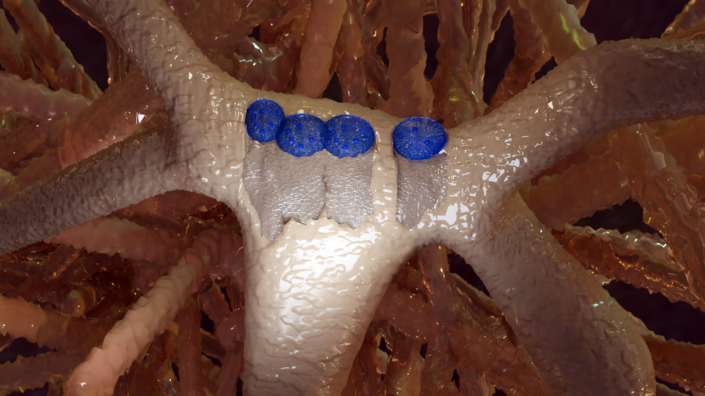
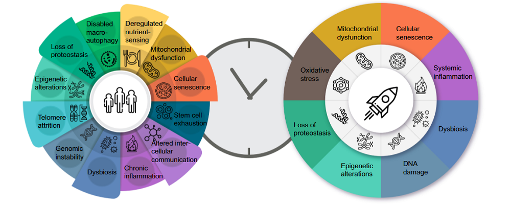
As shown below, previous research has summarized common features of aging on Earth and in space [2]. However, there is still a need to determine how long-term space missions would affect human health and, consequently, what the effect would be on aging.

Aging Spaceflight

Circadian rhythms in humans

Humans evolved to live on Earth. Our circadian rhythms reflect the Earth’s 24-hour light and dark cycles. Spaceflight disrupts those natural cycles; the sunrise-sunset cycle in Earth’s orbit lasts only 45 minutes.

Those circadian rhythms have an impact on health, including retinal function, cardiac metabolism, neural activity, and the formation of skeletal muscular tissue.

The circadian clock regulates some of the essential mechanisms of the cardiovascular system, such as blood pressure and heart rate [3, 4]. Research also suggests that an altered clock-associated mechanism is a risk factor for cardiovascular disease.

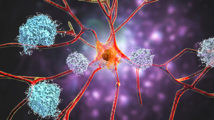
Additionally, circadian dysfunction occurs frequently in some aging-related neurodegenerative diseases, such as Alzheimer’s and Parkinson’s diseases [5]. Disruptions in circadian rhythms also result in such problems as sleep disruptions, sleep fragmentations, and reduced daytime alertness [6].

Scientists have been aware of space’s effect on circadian rhythms for over three decades. The molecules that control those cycles are called core-clock genes. Research has shown that in mammals on Earth, nearly 50% of expressed genes show rhythmic ~24-hour changes in expression in at least one organ [7].

There are also studies in fruit flies and rodents that were taken on space missions showing that clock genes play a significant role in pathologies associated with spaceflight. However, there is still a lack of thorough understanding of the molecular mechanisms of these conditions in humans.

Effects on aging and health

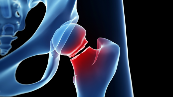
Some dysfunctions accelerate when people are in space. Changes in cardiovascular and musculoskeletal systems can be accelerated even 10-fold in space compared to on Earth [8].

Some of these changes are due to the lack of Earth gravity, such as muscle atrophy caused by muscle disuse. Astronauts can experience cephalad fluid shifts, which occur when fluids shift from the lower body toward the head and the heart. This results in smaller blood production, abnormal blood pressure, lower heart rate, and other cardiac issues. To prevent these changes from happening, astronauts need to follow strict exercise routines in space.

Astronauts also experience a condition known as Spaceflight-Associated Neuro-ocular Syndrome (SANS). SANS includes swelling of a portion of the optic nerve, retinal nerve fiber layer thickening, and other related conditions.

To compare organisms on Earth and during spaceflight, NASA performed a unique experiment. One of the astronauts sent for spaceflight had an identical twin. This way, researchers could monitor both siblings: one on a one-year space mission, the second on Earth.

Researchers observed multiple changes in the space-faring astronaut compared to the twin, including increased retinal thickness and changes in arterial dimensions. These changes reversed after six months upon return to Earth.

Researchers also observed a change in one of the Hallmarks of Aging: telomere length. While the astronaut was in space, researchers observed the astronaut’s telomeres lengthen, which suggests an anti-aging effect. However, 6 months after returning to Earth, the changes reversed. Moreover, following six months after the astronaut’s return to Earth, researchers observed shorter telomeres and more DNA damage, which suggests accelerated aging [9].

On the other hand, there are several reported anti-aging effects of spaceflight on humans and animals. A 520-day Space mission had an anti-aging effect on DNA methylation patterns in astronauts [10]. C. elegans worms and fruit flies in space showed slower aging [11, 12]. On the other hand, yeast fungi show signs of accelerated aging when exposed to simulated microgravity [13].

Longer and further space missions are planned for the future. As a part of the preparation for those missions, researchers need to determine in more detail how spaceflight influences the health and lifespan of astronauts.

We would like to ask you a small favor. We are a non-profit foundation, and unlike some other organizations, we have no shareholders and no products to sell you. All our news and educational content is free for everyone to read, but it does mean that we rely on the help of people like you. Every contribution, no matter if it’s big or small, supports independent journalism and sustains our future.

Literature

[1] Malhan, D., Schoenrock, B., Yalçin, M., Blottner, D., & Relόgio, A. (2023). Circadian regulation in aging: Implications for spaceflight and life on earth. Aging cell, e13935. Advance online publication.

[2] Biolo, G., Heer, M., Narici, M., & Strollo, F. (2003). Microgravity as a model of ageing. Current opinion in clinical nutrition and metabolic care, 6(1), 31–40.

[3] Costello, H. M., & Gumz, M. L. (2021). Circadian Rhythm, Clock Genes, and Hypertension: Recent Advances in Hypertension. Hypertension (Dallas, Tex. : 1979), 78(5), 1185–1196.

[4] El Jamal, N., Lordan, R., Teegarden, S. L., Grosser, T., & FitzGerald, G. (2023). The Circadian Biology of Heart Failure. Circulation research, 132(2), 223–237.

[5] Leng, Y., Musiek, E. S., Hu, K., Cappuccio, F. P., & Yaffe, K. (2019). Association between circadian rhythms and neurodegenerative diseases. The Lancet. Neurology, 18(3), 307–318.

[6] Czeisler, C. A., Chiasera, A. J., & Duffy, J. F. (1991). Research on sleep, circadian rhythms and aging: applications to manned spaceflight. Experimental gerontology, 26(2-3), 217–232.

[7] Zhang, R., Lahens, N. F., Ballance, H. I., Hughes, M. E., & Hogenesch, J. B. (2014). A circadian gene expression atlas in mammals: implications for biology and medicine. Proceedings of the National Academy of Sciences of the United States of America, 111(45), 16219–16224.

[8] Vernikos, J., & Schneider, V. S. (2010). Space, gravity and the physiology of aging: parallel or convergent disciplines? A mini-review. Gerontology, 56(2), 157–166.

[9] Garrett-Bakelman, F. E., Darshi, M., Green, S. J., Gur, R. C., Lin, L., Macias, B. R., McKenna, M. J., Meydan, C., Mishra, T., Nasrini, J., Piening, B. D., Rizzardi, L. F., Sharma, K., Siamwala, J. H., Taylor, L., Vitaterna, M. H., Afkarian, M., Afshinnekoo, E., Ahadi, S., Ambati, A., … Turek, F. W. (2019). The NASA Twins Study: A multidimensional analysis of a year-long human spaceflight. Science (New York, N.Y.), 364(6436), eaau8650.

[10] Nwanaji-Enwerem, J. C., Nwanaji-Enwerem, U., Van Der Laan, L., Galazka, J. M., Redeker, N. S., & Cardenas, A. (2020). A Longitudinal Epigenetic Aging and Leukocyte Analysis of Simulated Space Travel: The Mars-500 Mission. Cell reports, 33(10), 108406.

[11] Honda, Y., Higashibata, A., Matsunaga, Y., Yonezawa, Y., Kawano, T., Higashitani, A., Kuriyama, K., Shimazu, T., Tanaka, M., Szewczyk, N. J., Ishioka, N., & Honda, S. (2012). Genes down-regulated in spaceflight are involved in the control of longevity in Caenorhabditis elegans. Scientific reports, 2, 487.

[12] Ma, L., Ma, J., & Xu, K. (2015). Effect of spaceflight on the circadian rhythm, lifespan and gene expression of Drosophila melanogaster. PloS one, 10(3), e0121600.

[13] Fukuda, A. P. M., Camandona, V. L., Francisco, K. J. M., Rios-Anjos, R. M., Lucio do Lago, C., & Ferreira-Junior, J. R. (2021). Simulated microgravity accelerates aging in Saccharomyces cerevisiae. Life sciences in space research, 28, 32–40.

Effect of AMD

A Protective Mechanism Against AMD

A paper published in Aging explains the relationship of long noncoding RNAs, which change with aging, to age-related macular degeneration (AMD).

A disease of deposits and aging

This paper begins with a discussion of AMD and its prevalence. AMD is the most common cause of vision loss in people over 70 years old [1]. Its main characteristic is the formation of drusen, yellow-brown deposits within the eye. These deposits have been found to contain a wide variety of proteins associated with the complement pathway, a feature of the immune system. They even include amyloid beta, the same protein associated with Alzheimer’s disease [2].

AMD has been shown to be caused by the aging of retinal pigmental epithelial (RPE) cells, and it is possible to model AMD by inducing senescence in these cells [3] or by inducing oxidative stress through sodium iodate. Interestingly, quercetin has been reported to be effective against that stress in a model [4].

While most RNA strands are meant to execute code stored in DNA, long noncoding RNAs (lncRNAs) are different. Despite their lack of transcription ability, these molecules are associated with multiple aspects of aging. Their relationship to AMD has been previously, but lightly, explored [5]. One group of these lncRNAs, called NORAD, is triggered by DNA damage. NORAD sequesters two key proteins in order to maintain genomic stability [6], and mice without NORAD age more rapidly [7].

The value of NORAD

For the first part of this study, the researchers induced senescence in RPE cells by subjecting them to irradiation. Then, they created a different group of NORAD-deficient RPE cells and subjected them to the same irradiation. As expected, the lack of DNA protection from NORAD drove these cells senescent more quickly, increasing known aging biomarkers along with many of the secreted proteins associated with the drusen of AMD.

The researchers then turned to a mouse model. Along with a control group, NORAD-deficient mice were injected with sodium iodate in the tail vein. As expected, the NORAD-deficient mice suffered more damage, lost more function, and had more age-related proteins concentrated in the eyes, even though there was no significant difference before the sodium iodate injection.

This loss of function was matched by mitochondrial tests. After sodium iodate, reactive oxygen species were considerably more prevalent in the NORAD-deficient mice than their unaltered counterparts. The researchers found that this was true for the NORAD-deficient cells in their culture as well.

Finally, the researchers examined the relationship of NORAD to the well-known sirtuin SIRT1 and the metabolic regulator PGC-1α, whose acetylation is increased in AMD. They found that knocking out NORAD had similar effects to knocking out SIRT1 and that NORAD facilitates the binding of SIRT1 to PGC-1α, which may explain some of its AMD-related effects.

A potential new target

The researchers hold that their mouse and cellular models are very similar to human AMD in the clinic. While the researchers would have liked to overexpress NORAD in order to determine if that would have protective effects against AMD, they were unable to, as overexpressing NORAD caused cellular disruption. The researchers also note that NORAD has different effects on different organs.

However, if a future treatment can increase the amount of NORAD in aging eyes, it may be possible to directly affect a key cause of AMD at its root. Further studies will need to be conducted to determine if such a treatment can be viable.

AMD Progression
We would like to ask you a small favor. We are a non-profit foundation, and unlike some other organizations, we have no shareholders and no products to sell you. All our news and educational content is free for everyone to read, but it does mean that we rely on the help of people like you. Every contribution, no matter if it’s big or small, supports independent journalism and sustains our future.

Literature

[1] Wong, W. L., Su, X., Li, X., Cheung, C. M. G., Klein, R., Cheng, C. Y., & Wong, T. Y. (2014). Global prevalence of age-related macular degeneration and disease burden projection for 2020 and 2040: a systematic review and meta-analysis. The Lancet Global Health, 2(2), e106-e116.

[2] Ohno-Matsui, K. (2011). Parallel findings in age-related macular degeneration and Alzheimer’s disease. Progress in retinal and eye research, 30(4), 217-238.

[3] Chae, J. B., Jang, H., Son, C., Park, C. W., Choi, H., Jin, S., … & Chung, H. (2021). Targeting senescent retinal pigment epithelial cells facilitates retinal regeneration in mouse models of age-related macular degeneration. Geroscience, 43, 2809-2833.

[4] Chang, Y. Y., Lee, Y. J., Hsu, M. Y., Wang, M., Tsou, S. C., Chen, C. C., … & Lin, H. W. (2021). Protective effect of quercetin on sodium iodate-induced retinal apoptosis through the reactive oxygen species-mediated mitochondrion-dependent pathway. International Journal of Molecular Sciences, 22(8), 4056. [5] Blasiak, J., Hyttinen, J. M., Szczepanska, J., Pawlowska, E., & Kaarniranta, K. (2021). Potential of long non-Coding RNAs in age-related macular degeneration. International Journal of Molecular Sciences, 22(17), 9178.

[6] Munschauer, M., Nguyen, C. T., Sirokman, K., Hartigan, C. R., Hogstrom, L., Engreitz, J. M., … & Lander, E. S. (2018). The NORAD lncRNA assembles a topoisomerase complex critical for genome stability. Nature, 561(7721), 132-136.

[7] Kopp, F., Elguindy, M. M., Yalvac, M. E., Zhang, H., Chen, B., Gillett, F. A., … & Mendell, J. T. (2019). PUMILIO hyperactivity drives premature aging of Norad-deficient mice. Elife, 8, e42650.

Osteoblasts

Figuring Out Why Stem Cells Won’t Build Bone

Researchers publishing in Aging have uncovered some of the molecular mechanisms behind why mesenchymal stem cells (MSCs) do and don’t differentiate into bone-building osteoblasts, unlocking a potential new approach to osteoporosis.

A matter of cellular fate

MSCs are downstream of pluripotent stem cells: they can differentiate into multiple cell types, but not every somatic cell in the body. MSCs’ potential somatic cell fates include cartilage cells (chondrocytes), fat cells (adipocytes), and, germane to this study, osteoblasts: the cells that are responsible for building bone.

Previous research has found that MSCs differentiating into too many adipocytes and not enough osteoblasts is a key part of osteoporosis [1], and many of the gene expression and protein networks have already been extensively explored [2]. This focus on proteomics has yielded many valuable insights into MSCs’ behavior [3]. However, the technologies used in these studies did not fully describe the early stages of differentiation, and the many proteins involved in the differentiation process have not been entirely described.

Screening a very broad protein base

To further investigate proteins during differentiation, these researchers used Tandem Mass Tags (TMT), a process that used reflective spectra to identify 609,196 secondary proteins and screen 96,006 peptides. Only 6,543 of the proteins were informative, and of those, only a few hundred were found to significantly change during the differentiation process at 3 or 7 days.

For bone cells, the proteins that were upregulated were focused on cellular adhesion and the extracellular matrix, while for fat cells, the upregulated proteins were related to metabolism and PPAR, a known pathway that fat cells use. The Western blot, a very old method of protein analysis, was used to verify these findings with four random proteins from each cell type, and they were found to be accurate.

Two proteins in focus

Two proteins stood out as being of particular interest: FBLN2 and NPR3. FBLN2 has been previously described in the literature as being related to the extracellular matrix and bone formation [4] and is well-known for being strongly expressed in osteoblasts [5]. Using silencing RNA to knock out this protein in MSCs reduced their ability to build bone by nearly 60%. The downstream effects of this silencing had effects on over a thousand, generally scattered, proteins.

Knocking out NPR3 in the same way reduced osteogenesis by 65%. This protein has been discussed more in the context of the cardiovascular system than osteogenesis [6]. While an excess of this protein’s pathway has been previously linked to harmful overgrowth of bone [7], no other research had previously connected its lack to osteoporosis. A relatively large number of NPR3’s downstream effects were found to be related to metabolism.

Enlightening, but incomplete

This is a cellular and proteomic study rather than an animal study. While very valuable for future research, it does not answer, nor attempt to answer, what might cause FBLN2 or NPR3 to be less expressed. However, its novel findings open up an entirely new avenue into preclinical studies that might pave the way towards fundamental osteoporosis treatments that treat this crippling disease at its root.

We would like to ask you a small favor. We are a non-profit foundation, and unlike some other organizations, we have no shareholders and no products to sell you. All our news and educational content is free for everyone to read, but it does mean that we rely on the help of people like you. Every contribution, no matter if it’s big or small, supports independent journalism and sustains our future.

Literature

[1] Pino, A. M., Rosen, C. J., & Rodríguez, J. P. (2012). In osteoporosis, differentiation of mesenchymal stem cells (MSCs) improves bone marrow adipogenesis. Biological research, 45(3), 279-287.

[2] Kasoju, N., Wang, H., Zhang, B., George, J., Gao, S., Triffitt, J. T., … & Ye, H. (2017). Transcriptomics of human multipotent mesenchymal stromal cells: retrospective analysis and future prospects. Biotechnology Advances, 35(4), 407-418.

[3] Moravcikova, E., Meyer, E. M., Corselli, M., Donnenberg, V. S., & Donnenberg, A. D. (2018). Proteomic profiling of native unpassaged and culture‐expanded mesenchymal stromal cells (MSC). Cytometry Part A, 93(9), 894-904.

[4] Bardai, G., Lemyre, E., Moffatt, P., Palomo, T., Glorieux, F. H., Tung, J., … & Rauch, F. (2016). Osteogenesis imperfecta type I caused by COL1A1 deletions. Calcified tissue international, 98, 76-84.

[5] Hergeth, S. P., Aicher, W. K., Essl, M., Schreiber, T. D., Sasaki, T., & Klein, G. (2008). Characterization and functional analysis of osteoblast-derived fibulins in the human hematopoietic stem cell niche. Experimental hematology, 36(8), 1022-1034.

[6] Pandey, K. N. (2005). Biology of natriuretic peptides and their receptors. Peptides, 26(6), 901-932.

[7] Hisado-Oliva, A., Ruzafa-Martin, A., Sentchordi, L., Funari, M. F., Bezanilla-López, C., Alonso-Bernáldez, M., … & Heath, K. E. (2018). Mutations in C-natriuretic peptide (NPPC): a novel cause of autosomal dominant short stature. Genetics in Medicine, 20(1), 91-97.

The Journal Club is a monthly livestream hosted by Dr. Oliver Medvedik which covers the latest aging research papers.

Join us for Journal Club August 2023

The Journal Club for August returns live to the lifespan.io Facebook channel on September 5th at 12:00 Eastern hosted by Dr. Oliver Medvedik. This time we are taking a look at a paper where researchers have transfered a lifespan and healthspan boosting mechanism from the naked mole rat, a very long lived rodent, to mice. The paper includes researchers such as Vera Gorbunova, Steve Horvath, and Vadim Gladyshev and demonstrates that longevity is something that can be emulated from one species to another.

This study raises interesting questions and possibilities. Perhaps we can learn new tricks from species like the naked mole rat that do not age the same way we do.

Abstract

Abundant high-molecular-mass hyaluronic acid (HMM-HA) contributes to cancer resistance and possibly to the longevity of the longest-lived rodent-the naked mole-rat. To study whether the benefits of HMM-HA could be transferred to other animal species, we generated a transgenic mouse overexpressing naked mole-rat hyaluronic acid synthase 2 gene (nmrHas2). nmrHas2 mice showed an increase in hyaluronan levels in several tissues, and a lower incidence of spontaneous and induced cancer, extended lifespan and improved healthspan. The transcriptome signature of nmrHas2 mice shifted towards that of longer-lived species. The most notable change observed in nmrHas2 mice was attenuated inflammation across multiple tissues. HMM-HA reduced inflammation through several pathways, including a direct immunoregulatory effect on immune cells, protection from oxidative stress and improved gut barrier function during ageing. These beneficial effects were conferred by HMM-HA and were not specific to the nmrHas2 gene. These findings demonstrate that the longevity mechanism that evolved in the naked mole-rat can be exported to other species, and open new paths for using HMM-HA to improve lifespan and healthspan.

Join the Journal Club live

Lifespan Heroes can join us live on the call and join in the discussion directly using the details below:

Join Zoom Meeting Meeting ID: 834 1705 9396 Passcode: 209384

Literrature

Zhang, Z., Tian, X., Lu, J. Y., Boit, K., Ablaeva, J., Zakusilo, F. T., Emmrich, S., Firsanov, D., Rydkina, E., Biashad, S. A., Lu, Q., Tyshkovskiy, A., Gladyshev, V. N., Horvath, S., Seluanov, A., & Gorbunova, V. (2023). Increased hyaluronan by naked mole-rat Has2 improves healthspan in miceNature, 10.1038/s41586-023-06463-0. Advance online publication. https://doi.org/10.1038/s41586-023-06463-0
Curcumin

Antioxidants Rescue Cognitive Decline in Mice

Scientists have found that EGCG and curcumin, two well-known antioxidants, are effective both alone and especially in combination in a mouse model of cognitive decline induced by oxidative stress [1].

Not the usual stress

Oxidative stress results mostly from molecules called reactive oxygen species (ROS), which are byproducts of normal chemical reactions in the body. It is an aspect of aging that exerts wide-ranging adverse effects on numerous organs and tissues and is known to play a role in neurodegeneration and dementia [2].

The body produces endogenous antioxidants, the most important one being glutathione. People also consume potent antioxidants with food, including curcumin, the orange compound found in turmeric, and epigallocatechin 3 gallate (EGCG), a polyphenol that is especially abundant in green tea. Both have shown health benefits in animal and human studies [3].

What happens if we combine those two ROS-targeting compounds? This question was at the center of a new study published in Nature Scientific Reports.

The combination brings a win

The researchers used a mouse model of age-related cognitive decline induced by D-galactose (D-gal), a compound that exacerbates oxidative stress. D-gal-treated animals exhibit numerous symptoms of aging, including mitochondrial dysfunction, inflammation, and neuronal degeneration, highlighting the broad range of ROS’ effects.

Previous studies have shown that both curcumin and EGCG improve memory function in aged mice, at least partially via antioxidative pathways. However, the results of a very limited number of existing studies that considered the cumulative effects of those two molecules were inconclusive.

The researchers ran a battery of behavioral tests on several groups of mice, such as the passive avoidance task, which capitalizes on a rodent’s natural preference for darkness and its ability to learn to avoid an environment in which it has experienced an aversive stimulus. In this test, naturally aged and D-gal-treated mice showed less retention time and lower rate of freezing response, which is indicative of poorer memory.

Antioxidant Effectiveness

In all tests, young mice scored highest, while mice treated with D-gal and naturally aged mice demonstrated similar deterioration of cognitive function, showing that D-gal indeed emulates age-related cognitive decline.

Both antioxidants effectively elevated memory function, including in healthy young controls. Curcumin alone was less effective than EGCG alone. However, the two molecules showed a synergistic effect, at times elevating memory function markers higher than in young healthy controls.

The researchers then measured three markers of oxidative stress: glutathione along with the crucial antioxidant enzymes superoxide dismutase and catalase. The results mirrored those of the memory tests: in naturally aged mice and mice treated with D-gal, the levels of the three markers were much lower than in healthy young controls. Both EGCG and curcumin alone were able to significantly improve them.

The more robust effect of EGCG vs curcumin was even more evident here: while the latter was not able to fully rescue the three markers’ levels, after EGCG treatment, they actually surpassed those of healthy young controls. Yet, the cumulative effect of the combination treatment was even more robust.

The third contender

Another interesting feature of this study was that the EGCG and curcumin were tested against a positive control: astaxanthin, also a powerful antioxidant. Astaxanthin is what gives salmon its pink hue and it is found mostly in seafood. However, the richest source of astaxanthin is certain algae, the same ones that help give flamingoes their majestic pink plumage. Astaxanthin alone matched the results of the combined treatment with EGCG and curcumin.

This study pitted three antioxidants and one combination treatment against each other with some interesting results, highlighting the promise of combination therapies, even if both molecules are in the same ballpark, as EGCG and curcumin are. Of course, it would be interesting to see the results of a combination that includes astaxanthin as well as studies in humans. All three molecules can be found as supplements, and while there are bioavailability issues, all three are very safe.

In the current study, we noted that combined EGCG and curcumin-treated mice showed powerful protection against the decreased activity of GSH, SOD, CAT (Fig. 6), and increasing level of AOPP, NO, and MDA (Fig. 7) in the brain compared to the monotherapy of EGCG or curcumin. Moreover, combined EGCG and curcumin were comparable to Ast, a standard antioxidant, in modulating the antioxidant level.

We would like to ask you a small favor. We are a non-profit foundation, and unlike some other organizations, we have no shareholders and no products to sell you. All our news and educational content is free for everyone to read, but it does mean that we rely on the help of people like you. Every contribution, no matter if it’s big or small, supports independent journalism and sustains our future.

Literature

[1] Rahman, M. A., Shuvo, A. A., Apu, M. M. H., Bhakta, M. R., Islam, F., Rahman, M. A., … & Reza, H. M. (2023). Combination of epigallocatechin 3 gallate and curcumin improves d-galactose and normal-aging associated memory impairment in mice. Scientific Reports, 13(1), 12681.

[2] Kim, G. H., Kim, J. E., Rhie, S. J., & Yoon, S. (2015). The role of oxidative stress in neurodegenerative diseases. Experimental neurobiology, 24(4), 325.

[3] Nagle, D. G., Ferreira, D., & Zhou, Y. D. (2006). Epigallocatechin-3-gallate (EGCG): chemical and biomedical perspectives. Phytochemistry, 67(17), 1849-1855.

Biomarkers

Biomarker Study Returns Surprising Results

Researchers publishing in Biogerontology have tested multiple biomarkers in an effort to use them as proxies for aging, and some of their findings were surprising.

A simple study with direct measurements

Unlike other association studies that are used to evaluate biomarkers of aging, this study did not use data from a pre-existing biobank or related database. Instead, the authors recruited a total of 169 people, divided evenly between the sexes, and grouped them into under-35s, ages 35 to 50, and over-50s. There was an extensive list of exclusion criteria, including pregnancy, recent viral infection, recent surgery, and age-related morbidities such as stroke and heart attack.

Many positive results

Growth differentiation factor 15 (GDF15), which can be used to measure stress and mitochondrial dysfunction, was very significantly associated with age. Interestingly, though, the highest levels were found in the 35-50 group, with the over-50 group dropping slightly but not to the level of the under-35 group.

While the related molecule GDF11 is associated with regeneration, it increases with age, according to this study. The researchers note prior work showing that people whose GDF11 levels decrease with age are more likely to present with age-related diseases [1].

Measurements of the inflammatory biomarker NLRP3 also had positive results: people under 35 had significantly less NLRP3 than people over 50. The researchers note that using NLRP3 as a biomarker is difficult because most of the recorded values were very small.

Advanced glycation end-products (AGEs) had a significant, although nonlinear, association. The under-35 group had significantly fewer AGEs than the 35-50 group. Strangely, AGEs were reported to go back down with further aging, as the over-50 group had fewer AGEs than the 35-50s. Men also had significantly more AGEs than women.

AGE groups

Surprising negative findings

This study’s negative findings may be of greater scientific interest than its positive ones.

These researchers report that telomerase itself is not a good biomarker of aging, because while telomerase activity decreases with aging [2] and older people have shorter telomeres than younger people, telomerase itself did not have statistically significant differences between the age groups studied. While counterintuitive and perhaps controversial, this finding has similarities with previous research, which found that people in their 30s have telomerase levels that decrease with age but very old people have increasing levels with age [3]. High telomerase in young people is also associated with cancer [4].

DNA and RNA oxidative damage was also not reported to be statistically different between the groups. NAD+ was, interestingly, also not found to be statistically different, which corroborates some research [5] but contradicts many other studies [6].

Klotho, another well-known compound that is associated with aging, was also not found to have significant differences between each group, although klotho was noted to decline with age within the groups. However, the measurements for both NAD+ and klotho were wildly different within each group, with some people’s values being very small while others’ were hundreds or thousands of times greater. This individual variance, itself, implies that these may be poor biomarkers in practice.

Ascertaining validity

As much of this eyebrow-raising data seems to fly in the face of generally accepted knowledge about biomarkers of aging, further investigation seems necessary, particularly given this study’s relatively small sample size, its extensive exclusion criteria, and the wide age range of the oldest group. Epigenetics and gene expression were also not tested.

Scientific teams do not always agree with one another, and this disagreement can be the result of multiple causes. Other research groups are encouraged to corroborate, explain, contradict, or outright refute this data.

Literature

[1] Schafer, M. J., Atkinson, E. J., Vanderboom, P. M., Kotajarvi, B., White, T. A., Moore, M. M., … & LeBrasseur, N. K. (2016). Quantification of GDF11 and myostatin in human aging and cardiovascular disease. Cell metabolism, 23(6), 1207-1215.

[2] Vaiserman, A., & Krasnienkov, D. (2021). Telomere length as a marker of biological age: state-of-the-art, open issues, and future perspectives. Frontiers in Genetics, 11, 630186.

[3] Hertzog, R., Popescu, D., & Călborean, O. (2021). Telomerase level: a useful tool to predict longevity. J Aging Sci 9(3):1–4.

[4] Noureen, N., Wu, S., Lv, Y., Yang, J., Alfred Yung, W. K., Gelfond, J., … & Zheng, S. (2021). Integrated analysis of telomerase enzymatic activity unravels an association with cancer stemness and proliferation. Nature communications, 12(1), 139.

[5] Schwarzmann, L., Pliquett, R. U., Simm, A., & Bartling, B. (2021). Sex-related differences in human plasma NAD+/NADH levels depend on age. Bioscience Reports, 41(1), BSR20200340.

[6] Karas, A., Holmannova, D., Borsky, P., Fiala, Z., Andrys, C., Hamakova, K., … & Borska, L. (2022). Significantly Altered Serum Levels of NAD, AGE, RAGE, CRP, and Elastin as Potential Biomarkers of Psoriasis and Aging—A Case-Control Study. Biomedicines, 10(5), 1133.

Flavonoid Consumption Correlates With Less Biological Aging

Researchers investigated how much the consumption of flavonoids impacts the biological age of the heart, kidney, liver, and whole body [1].

Chronological age, biological age, and diet

Chronological age only refers to the number of years that have passed, but biological age refers to the lasting damage accumulated over those years. Unhealthy habits and behaviors, such as smoking and drinking alcohol, accelerate biological age. Biological age can also be accelerated due to chronic diseases and cancer. On the other hand, healthy dietary and exercise habits can decelerate biological age.

The Mediterranean diet is a known method of slowing down biological aging. Previous research has shown that adherence to the Mediterranean diet is associated with a biological age that is one year below chronological age [2, 3].

Fruits and vegetables are essential components of the Mediterranean diet, and the flavonoids found in those foods are the focus of this recent study. Flavonoids possess many beneficial properties, and previous research has found them to protect from oxidation, inflammation, mutagens, and carcinogens [4]. Due to these beneficial properties, the authors hypothesized that flavonoids may reduce the rate of biological aging.

To test their hypothesis, they used data from the National Health and Nutrition Examination Survey and the Food and Nutrient Database for Dietary Studies. They analyzed the flavonoid intake of 3,193 study participants who described their diets in a 24-dietary recall interview.

Assessing biological age

The authors categorized the study participants into three groups based on flavonoid intake. They determined participants’ whole body, cardiovascular, renal, and liver biological age using several biomarkers, including blood pressure, fasting glucose, fasting cholesterol levels, and fasting triglycerides. This study used a metric called ∆age to represent the difference between the biological and chronological ages of a single person.

The researchers observed some confounding factors, noting that participants with the highest flavonoid intake were less likely to be smokers or heavy alcohol users and were less likely to have hypertension, diabetes, or chronic kidney disease. However, they were more likely to be overweight. People with the highest flavonoid intake also ate more fiber, total fat, protein, and carbohydrates.

While people not suffering from chronic kidney disease had a ∆age of -0.62 years, the biological age of people with chronic kidney disease was higher than their chronological age, and their ∆age was 7.41 years.

Finding the correlation

The researchers observed lower ∆ages for the whole body and liver in the medium and highest intake groups. The group with the lowest flavonoid intake had a whole-body ∆age of 0.22 and a liver ∆age of 0.44. The medium group had -0.51 for the whole body and -4.15 for the liver. The group with the highest flavonoid intake had the greatest differences, as they ranged from -4.69 for the liver to -1.10 for the whole body.

Flavonoids are a diverse group of compounds that have different impacts on biological age. For example, isoflavones and flavones had a significant impact on delaying whole-body and cardiovascular aging.

Additionally, anthocyanidins showed a profound impact on the ∆age of the study participants. Participants with the highest intakes of anthocyanidins had the lowest ∆ages for the whole body (-1.45), cardiovascular system (-1.87), renal system (-1.10), and liver (-6.31). Differences between sexes were also observed, with females showing higher liver ∆age but lower whole body, cardiovascular, and renal ∆ages.

Overall, the authors reported that higher flavonoid intake was inversely correlated with whole body, cardiovascular, and liver ∆age. After analysis of analyzed multiple variables, they found that “higher intake of total flavonoids was found to be associated with a lower biological age of the cardiovascular, hepatic, and whole body among White participants who did not smoke, consume alcohol, and were free from chronic diseases.”

This analysis aligns with previous studies that link compounds like flavonoids and improvement in various markers of aging. While this study does not prove causation, the researchers hold that this correlation results from the antioxidant and anti-inflammatory properties of flavonoid-rich diets.

Literature

[1] Xing, W., Gao, W., Zhao, Z., Xu, X., Bu, H., Su, H., Mao, G., & Chen, J. (2023). Dietary flavonoids intake contributes to delay biological aging process: analysis from NHANES dataset. Journal of translational medicine, 21(1), 492.

[2] Esposito, S., Gialluisi, A., Costanzo, S., Di Castelnuovo, A., Ruggiero, E., De Curtis, A., Persichillo, M., Cerletti, C., Donati, M. B., de Gaetano, G., Iacoviello, L., Bonaccio, M., & On Behalf Of The Investigators For The Moli-Sani Study (2021). Dietary Polyphenol Intake Is Associated with Biological Aging, a Novel Predictor of Cardiovascular Disease: Cross-Sectional Findings from the Moli-Sani Study. Nutrients, 13(5), 1701.

[3] Gialluisi, A., Di Castelnuovo, A., Costanzo, S., Bonaccio, M., Persichillo, M., Magnacca, S., De Curtis, A., Cerletti, C., Donati, M. B., de Gaetano, G., Capobianco, E., Iacoviello, L., & Moli-sani Study Investigators (2022). Exploring domains, clinical implications and environmental associations of a deep learning marker of biological ageing. European journal of epidemiology, 37(1), 35–48.

[4] Panche, A. N., Diwan, A. D., & Chandra, S. R. (2016). Flavonoids: an overview. Journal of nutritional science, 5, e47.

Extracellular Vesicles from Stem Cells Reverse Senescence

Scientists have found that bubbles secreted by embryonic stem cells counter cellular senescence, in large part due to just two tiny snippets of RNA [1].

Helpful bubbles

Extracellular vesicles (EVs) are membrane-bound tiny bubbles that are loaded with various molecular cargo, such as proteins, DNA, or RNA, that cells secrete as a method of intercellular communication. According to numerous studies, EVs can recapitulate many effects of cellular therapies, such as stem cell treatments [2].

In this new study, the researchers used EVs derived from human embryonic stem cells (ESC) against cellular senescence. ESCs are considered a potent therapeutic, tool but, as the study’s authors note in the introduction, their use “is limited by immune rejection, tumorigenicity and ethical issues”. If we could culture ESCs and use EVs secreted by them to the same effect, this would solve many problems.

Smashing senescence in vitro

First, the researchers confirmed that ESC-derived EVs ameliorate cellular senescence in vitro. Cellular senescence is a state in which the cell stops proliferating due to any of several senescence-inducing stressors. Such cells secrete molecules that can induce inflammation and senescence in neighboring cells. While senescence has a role in development and wound healing, it is widely accepted that age-related excessive senescence is harmful [3].

Since cells become senescent after a certain number of divisions, the researchers used mouse embryonic fibroblasts (MEF) that accumulated seven divisions as their in vitro senescence model. EV treatment ameliorated several markers of senescence, not just chemical ones (p21, β-galactosidase) but also senescence-typical morphology, which is when cells become flat and shapeless. Moreover, EVs increased the percentage of proliferative cells to the levels comparable with young control cells.

Tiny snippets of RNA

The question is, then, what cargo were those EVs carrying, and which part of it was responsible for ameliorating senescence in MEFs? Previous research has demonstrated that EVs contain large amounts of microRNA (miRNA): short RNA snippets, typically about 22 nucleotides in length. Despite their tiny size, miRNAs can play important roles, such as regulating protein production by repressing messenger RNA (mRNA). One study found that EVs secreted by healthy cells in the hypothalamus can slow aging and that this effect is partially mediated by miRNA contained in those EVs [4].

The researchers, hence, focused their attention on miRNAs and performed miRNA sequencing of the EVs. Compared to mouse fibroblasts, in ESC-EVs, 128 miRNAs were upregulated and 110 were downregulated. Hypothesizing that the anti-aging effect is most probably produced by enriched miRNAs, the researchers scrutinized the first group.

Many of those miRNAs are known to participate in longevity-related pathways, such as mTOR and insulin signaling. Eventually, the researchers narrowed the list down to just six miRNAs that were both highly enriched in ESC-EVs and downregulated in aged cells.

When the researchers transfected aged MEFs with synthetic RNA molecules that mimic the function of a naturally occurring miRNA (miRNA mimics), two of these mimics, those of miR-15b-5p and miR-290a-5p, were especially effective in alleviating the cells’ senescent phenotype.

Down to one gene

One last remaining question was, which genes are affected by those two beneficial miRNAs? The researchers identified 11 target genes of miR-15b-5p and 10 target genes of miR-290a-5p, but only three were overlapping, and only two of them, Ccn2 and Lurap1, are known to be involved in age-related pathways. Lurap1 activates the potent inflammatory transcription factor NF-κB, and Ccn2 plays a major role in cellular senescence. However, only the latter was significantly downregulated in aged MEFs by the EV treatment.

Inhibiting either of the two miRNAs reduced the anti-senescence potential of MSC-EVs only slightly. However, inhibiting both abrogated it almost completely. Genetic knockdown of Ccn2 significantly decreased the expression of several age-related genes in MEFs, reversed their aged phenotype, and restored their proliferative ability, confirming Ccn2’s important role in senescence.

Finally, the researchers found that MSC-derived EVs induce signs of rejuvenation in aged mice. The treatments caused a major shift in the transcriptome, bringing it close to a youthful one. EVs also induced youthful histological changes in the liver and the kidneys.

Here, we investigate the antisenescence effects of ESC-EVs in vivo using aged mice. Our data show that ESC-EVs treatment rescues the transcriptome profile of aged mice and ameliorates the senescence status of several aged organs, providing evidence that ESC-EVs may be candidates for the therapy of various age-related diseases. Furthermore, we identify miR-15b-5p and miR-290a-5p, which are enriched in ESC-EVs and exert rejuvenating effects by silencing of the Ccn2-mediated AKT signaling pathway.

We would like to ask you a small favor. We are a non-profit foundation, and unlike some other organizations, we have no shareholders and no products to sell you. All our news and educational content is free for everyone to read, but it does mean that we rely on the help of people like you. Every contribution, no matter if it’s big or small, supports independent journalism and sustains our future.

Literature

[1] Yu, L., Wen, H., Liu, C., Wang, C., Yu, H., Zhang, K., … & Liu, N. (2023). Embryonic stem cell-derived extracellular vesicles rejuvenate senescent cells and antagonize aging in mice. Bioactive Materials, 29, 85-97.

[2] Weng, Z., Zhang, B., Wu, C., Yu, F., Han, B., Li, B., & Li, L. (2021). Therapeutic roles of mesenchymal stem cell-derived extracellular vesicles in cancer. Journal of hematology & oncology, 14, 1-22.

[3] Di Micco, R., Krizhanovsky, V., Baker, D., & d’Adda di Fagagna, F. (2021). Cellular senescence in ageing: from mechanisms to therapeutic opportunities. Nature reviews Molecular cell biology, 22(2), 75-95.

[4] Zhang, Y., Kim, M. S., Jia, B., Yan, J., Zuniga-Hertz, J. P., Han, C., & Cai, D. (2017). Hypothalamic stem cells control ageing speed partly through exosomal miRNAs. Nature, 548(7665), 52-57.Chicago

Amyloid beta plaques

Restoring Helper Cell Function to Fight Alzheimer’s

In a study published in Cell Stem Cell, researchers have investigated a way to repopulate the brain with functional cells in order to fight back against Alzheimer’s disease.

A gene with a microglial function

Various allelles of the apolipoprotein E (APOE) genes are well-known for their contributions to, or defenses against, Alzheimer’s, as they affect how the brain processes the amyloids associated with the disease. However, that is not the only gene being explored. Here, the researchers focus on TREM2, a gene whose R47H variant has been identified as a strong risk factor for Alzheimer’s [1].

A complete loss of TREM2 is strongly associated with dysfunctional microglia, the cells that perform maintenance and immune functions in the brain. It results in Nasu-Hakola disease, a form of dementia that can cause bone cysts to form in the brain [2]. Other mutations of TREM2 can cause dementia even without bone cysts [3].

Since properly functioning TREM2 is so necessary for brain health, the researchers considered ways of restoring it to the brain. Rather than attempting to genetically modify cells in situ, they decided that introducing functional cells into the brain would be a more effective approach.

A process with doubly afflicted mice

Since mice do not naturally develop Alzheimer’s disease, genetically modified models are always required for studies. To proceed with this study, the researchers crossbred 5xFAD, a mouse model that exhibits Alzheimer’s at 6 weeks of age, with mice that express a reporter gene. The bone marrow of these mice was used in a technique called microglia replacement by bone marrow transplantation (mrBMT), which, true to its name, introduces circulation-derived myeloid cells (CDMCs) to serve the functions of microglia.

CDMCs are not the same as microglia, being smaller and more compact. However, the researchers found that they behave similarly towards amyloid beta plaques, wrapping tightly around them to prevent them from forming tendrils. CDMC-recipient mice and regular microglial mice had similar levels of amyloid beta load, which the researchers take as evidence that CDMCs are effective replacements for microglia in this model.

Next, the researchers used a doubly afflicted mouse model: mice that suffer from Alzheimer’s disease and are negative for the TREM2 gene. As expected, the microglia of these mice failed to respond properly to the amyloid plaques. CDMCs from other mice without TREM2, as expected, also failed to respond.

Effective on multiple levels

CDMCs from Alzheimer’s mice that did produce TREM2 were found to be effective. These transplanted cells rapidly wrapped around the amyloid plaques, slowing the spread of Alzheimer’s pathology within the brain, and were showing signs of phagocytosis: the attempt to engulf and consume these plaques. However, these cells were considerably more effective on 3-week-old mice than 8-week-old mice, suggesting that they are more effective when given early to suppress the disease.

Biomarkers of axonal damage, such as Lamp1, were also found to be relatively suppressed with this CDMC treatment. One biomarker of damage, the disease-associated microglia (DAM) program [4], was significantly upregulated; this represents the helper cells attempting to protect the brain. Signaling molecules and other effects of downstream gene expression was also found to be restored by CDMC treatment, including genes that handle the lipoproteins associated with brain function.

Potential for translational research

Obviously, this line of basic research is not going to directly lead to a cure for Alzheimer’s, especially in people without TREM2 deficiencies. However, it opens multiple avenues for translational research, the most obvious being that CDMCs may be effective in replacing microglia for this and other conditions. If the helper cells of the brain can be supplemented in ways that restrict the pathology of Alzheimer’s and other diseases, it might be possible to delay neurodegeneration and give people more healthy years of life.

We would like to ask you a small favor. We are a non-profit foundation, and unlike some other organizations, we have no shareholders and no products to sell you. All our news and educational content is free for everyone to read, but it does mean that we rely on the help of people like you. Every contribution, no matter if it’s big or small, supports independent journalism and sustains our future.

Literature

[1] Jonsson, T., Stefansson, H., Steinberg, S., Jonsdottir, I., Jonsson, P. V., Snaedal, J., … & Stefansson, K. (2013). Variant of TREM2 associated with the risk of Alzheimer’s disease. New England Journal of Medicine, 368(2), 107-116.

[2] Paloneva, J., Autti, T., Raininko, R., Partanen, J., Salonen, O., Puranen, M., … & Haltia, M. (2001). CNS manifestations of Nasu–Hakola disease: a frontal dementia with bone cysts. Neurology, 56(11), 1552-1558.

[3] Chouery, E., Delague, V., Bergougnoux, A., Koussa, S., Serre, J. L., & Mégarbané, A. (2008). Mutations in TREM2 lead to pure early‐onset dementia without bone cysts. Human mutation, 29(9), E194-E204.

[4] Keren-Shaul, H., Spinrad, A., Weiner, A., Matcovitch-Natan, O., Dvir-Szternfeld, R., Ulland, T. K., … & Amit, I. (2017). A unique microglia type associated with restricting development of Alzheimer’s disease. Cell, 169(7), 1276-1290.

Hip fracture

Study: Vegetarians Have Higher Risk of Hip Fracture

Studying a large British cohort, scientists have found that vegetarians have an increased risk of hip fracture compared to regular meat eaters, but pescatarians and occasional meat eaters do not [1].

Diet and brittle bones

Hip fracture is one of the injuries most associated with aging. While hip fracture itself is treatable, it can easily set people on a slippery slope to frailty. Even after a successful surgery, patients often suffer from limited mobility and increased fear of movement, which leads to an overall decline in quality of life and an increased risk of chronic illness and death. Hip fractures are also a heavy burden on the healthcare system, costing 6 billion dollars annually in the US alone [2].

Dietary choices influence almost every aspect of health. Vegetarian and vegan diets, while having numerous health benefits, have long been suspected to increase risk of fractures [3] due to lower intakes of certain nutrients such as protein, vitamin D, and vitamin B12. Some studies have linked these diets to lower muscle mass and bone density, both of which are risk factors for fractures [4]. However, studies of hip fracture with sufficient numbers of vegetarians and vegans in the sample are scarce.

Higher risk for vegetarians

This new study, like many others, uses data from UK Biobank, a repository of health information on about half a million British citizens. Due to its enormous size, Biobank has made a lot of quality research possible. The analysis was adjusted for many confounding variables, including sex, ethnicity, socioeconomic status, tobacco and alcohol consumption, physical activity, body mass index, and history of disease.

The study used regular meat eaters as a baseline. Occasional meat eaters had virtually the same prevalence of hip fractures (a hazard ratio of 0.99: 1% lower than in regular meat eaters). Pescatarians had statistically insignificantly elevated risk (a hazard ratio of 1.08). Vegetarians fared the worst, with a massive 50% increase in hip fracture incidence.

Further analysis revealed BMI to be a significant part of the relationship between diet and hip fracture, as it statistically explained 28% of the effect. Lean mass and vitamin D levels in blood could not account for the difference between vegetarians and regular meat-eaters.

Diet hazard ratios

Initially, the vegetarian category included vegans. When vegans were considered separately, the risk estimate for vegetarians was attenuated (hazard ratio of 1.38), while vegans emerged as the riskiest category by far, with a hazard ratio of 3.26. However, due to the small number of vegans in the sample (400 participants and only 10 cases of hip fracture), these results should be taken with a grain of salt.

Consider and mitigate the risks

The results of this study are mostly in line with the scarce existing research on the subject, and they serve as a reminder that vegetarians and especially vegans that their diets must contain sufficient nutrients that are more plentiful in other diets. However, as the researchers note, “whilst the relative increase in hip fracture risk for vegetarians was high (50%), this represents an absolute difference of only 3.2 more cases per 1000 people over 10 years. This modest absolute risk difference should be weighed against the potential associated health benefits of vegetarian diets for more common conditions, including 13 fewer cancers per 1000 people over 10 years and a 9% lower risk of CVD observed previously in the UK Biobank.”

An additional strength of this study was that it considered pescatarians: people who eat fish but not other animals. Several studies have suggested that this diet, when combined with other healthy practices, reaps most of the benefits of animal-based foods while avoiding most of the risks, although this too is still being investigated.

Vegetarian men and women had a higher risk of hip fracture than regular meat-eaters and were in part explained by their lower BMI, but absolute risk differences were small and should be weighed against the potential health benefits of vegetarian diets. Further work is needed to fully understand mechanisms underpinning risk differences; diet planning and weight management could help to mitigate the risk difference, and warrant exploration in further studies so that policy recommendations can advance.

We would like to ask you a small favor. We are a non-profit foundation, and unlike some other organizations, we have no shareholders and no products to sell you. All our news and educational content is free for everyone to read, but it does mean that we rely on the help of people like you. Every contribution, no matter if it’s big or small, supports independent journalism and sustains our future.

Literature

[1] Webster, J., Greenwood, D. C., & Cade, J. E. (2023). Risk of Hip Fracture in Meat Eaters, Pescatarians, and Vegetarians: A Prospective Cohort Study of 413,914 UK Biobank Participants. Pescatarians, and Vegetarians: A Prospective Cohort Study of, 413.

[2] Manetti, S., Turchetti, G., & Fusco, F. (2020). Determining the cost-effectiveness requirements of an exoskeleton preventing second hip fractures using value of information. BMC health services research, 20, 1-11.

[3] Tong, T. Y., Appleby, P. N., Armstrong, M. E., Fensom, G. K., Knuppel, A., Papier, K., … & Key, T. J. (2020). Vegetarian and vegan diets and risks of total and site-specific fractures: results from the prospective EPIC-Oxford study. BMC medicine, 18(1), 1-15.

[4] Iguacel, I., Miguel-Berges, M. L., Gómez-Bruton, A., Moreno, L. A., & Julián, C. (2019). Veganism, vegetarianism, bone mineral density, and fracture risk: a systematic review and meta-analysis. Nutrition reviews, 77(1), 1-18.

Advanced Clock

Inflammation as a Biomarker of Mortality

In a paper published in Geroscience, researchers have developed a metric of systemic inflammation and used it to estimate mortality, finding that it performs better than epigenetic clocks.

Evaluating a danger

This paper begins with familiar discussions about inflammaging and epigenetic clocks, tools that use DNA methylation to estimate biological age. These clocks are also used to measure age acceleration: the difference between biological and chronological age [1].

These researchers note that while immune cells are used for both epigenetic and inflammatory clocks, only a small number of studies have evaluated this connection [2]. Some of this previous research has shown that the two are largely independent [3]. This work builds upon its predecessors with an attempt at building a latent variable built upon known inflammatory biomarkers.

Latent variables and their analysis

After curation, this study used data from 3,311 participants in the Health and Retirement Study, a sample of people from across the United States. All participants were at least 50 years old.

A latent variable is one that cannot be directly observed but has observable effects. In this case, that variable is systemic inflammation as a whole, which was combined from analyzing C-reactive protein (CRP), three different interleukins, tumor necrosis factor (TNF), insulin growth factor (IGF-1), and the neutrophil-to-lymphocyte ratio.

The researchers mathematically determined the relationships of these biomarkers to the latent inflammation variable. The relationships were not linear, and in the case of IGF-1, it was inverse: more IGF-1 was correlated with less inflammation.

A great many covariates were included in this study, including such standards as age, sex, and ethnicity along with cytomegalovirus (CMV) infection, alcoholism, obesity, and smoking. Multimorbidity, which was defined as having at least two of eight known risk factors, was also included.

This analysis compared systemic inflammation to several age acceleration measurements gleaned from epigenetic clocks, including GrimAge, Hannum, Levine, the Methylation Pace of Aging (MPOA, based on a Dunedin clock), and the original Horvath clock.

Significant correlations

Even after adjustment for all of the various comorbidities, inflammaging was found to be heavily correlated with 4-year mortality risk and significantly correlated with multiple epigenetic clocks. While a couple of lesser-known clocks were not correlated, and the magnitude of the associations was not particularly strong, GrimAge, MPOA, and many others were found to be significantly associated, and the power of this expansive cohort resulted in small p-values. The researchers note that their inflammation variable has stronger associations with clocks that are more geared towards mortality than chronological age.

However, the most crucial finding from this study was that systemic inflammation is more strongly correlated with mortality than any of the epigenetic clocks are. In accordance with previous research [3], the authors hold that inflammation and epigenetic aging may represent independent processes that lead to mortality. Of course, this is only an association study, and in this paper, the authors did not attempt any biological understanding of the underlying processes.

We would like to ask you a small favor. We are a non-profit foundation, and unlike some other organizations, we have no shareholders and no products to sell you. All our news and educational content is free for everyone to read, but it does mean that we rely on the help of people like you. Every contribution, no matter if it’s big or small, supports independent journalism and sustains our future.

Literature

[1] Levine, M. E., Lu, A. T., Quach, A., Chen, B. H., Assimes, T. L., Bandinelli, S., … & Horvath, S. (2018). An epigenetic biomarker of aging for lifespan and healthspan. Aging (albany NY), 10(4), 573.

[2] Irvin, M. R., Aslibekyan, S., Do, A., Zhi, D., Hidalgo, B., Claas, S. A., … & Arnett, D. K. (2018). Metabolic and inflammatory biomarkers are associated with epigenetic aging acceleration estimates in the GOLDN study. Clinical epigenetics, 10, 1-9.

[3] Cribb, L., Hodge, A. M., Yu, C., Li, S. X., English, D. R., Makalic, E., … & Dugué, P. A. (2022). Inflammation and epigenetic aging are largely independent markers of biological aging and mortality. The Journals of Gerontology: Series A, 77(12), 2378-2386.

Robot detective

New Senolytics from Artificial Intelligence

Recent research published in Nature Communications has used machine learning algorithms to find new compounds that can eliminate senescent cells [1].

Searching for new senolytics

Senolytics are molecules that destroy senescent cells. Only a small number of such molecules have been identified, and only two have shown efficacy in clinical trials: dasatinib and quercetin in combination [2]. One of the biggest challenges is that senolytics often only work against specific types of cells. Additionally, some senolytics may work well for one cell type while being toxic to other, non-senescent cell types [3].

There is also a group of senolytics that are used in cancer therapies. However, most of them target pathways that are mutated in cancer. Therefore, they cannot be used as therapeutic agents in different contexts.

These limitations highlight the need to identify new senolytics that can be safely applied in therapies. Sometimes, this search involves panel screens [4]. Other times, it involves targeting the proteins upregulated in senescence. The authors of this paper used a different approach: AI-based computational screens that detect hidden patterns in chemical data.

From big datasets to a few hits

First, the authors assembled an extensive dataset for training the algorithms. Based on published data, they identified 58 senolytics. They also added 19 senolytics reported in commercial patents.

The authors then combined their list of senolytic compounds with a wide variety of compounds that were never described in the literature as having senolytic properties, with the final list consisting of 2,523 compounds. They used this list to train machine learning models to predict whether or not a compound may be a senolytic.

First, researchers focused on two models that both showed poor performance, but the errors they displayed were different. The first model, while returning a few false positives, gave a high number of false negatives. The second model returned the opposite. Other models showed even worse performance.

These researchers then decided to choose a model that turns fewer false positives, with the reason being that false positives are worse than false negatives for early-stage drug discovery, as higher number of false positives will increase the number of predicted hits. More predicted hits result in more compounds that need to be experimentally validated, thus increasing the cost and time.

After choosing their initial model, the researchers used different tools to optimize its performance. Following optimization, they screened a 4,340-compound library and identified 21 hits, then experimentally validated them, first in cells with oncogene-induced senescence.

Three natural products found

The initial experiment identified three compounds with senolytic properties: the natural products ginkgetin, oleandrin, and periplocin, which can be found in traditional herbal medicines. Ginkgetin is a product of Ginkgo biloba, commonly known as ginkgo or maidenhair tree. Oleandrin is a product of Nerium oleander, known as oleander or nerium, and periplocin is a product of Periploca sepium, a Chinese silk vine.

These identified compounds, when added in proper concentrations, have minimal effect on normal cells but decrease the number of senescent cells.

This paper’s authors further validated their hits using a second cell line. This time, they induced senescence in human cancerous cell lines by adding etoposide, a cancer treatment medication, and treated them with the 21 candidates. Again, the same three compounds showed senolytic activity toward the senescent cells but not the normal cells.

The results of this screening suggested that oleandrin is a more potent senolytic than the known senolytic ouabain. Therefore, the researchers decided to focus on this compound and investigate it more thoroughly.

One experiment compared periplocin, oleandrin, and ouabain using low concentrations of compounds. In the cell lines used here, ouabain and periplocin was not cytotoxic. Oleandrin, on the other hand, displayed highly specific senolytic activity. Further testing of oleandrin has shown that it has greater potency and activity regarding the molecule it targets when compared to similar senolytic compounds.

Authors caution that compounds from the same chemical group as oleandrin are known for their toxicity, so oleandrin may be found to be toxic as well. Further research is necessary to determine how and when this compound can be safely used.

Utilizing artificial intelligence in the screens for new compounds

These researchers have shown how machine learning can utilize published screening data and find new therapeutic molecules. This approach reduced the number of potential hits by 200-fold. That efficiency resulted in fewer compounds that proceeded to experimental testing, thus reducing time and cost.

The authors applied some new approaches in the presented research. For example, they used only already published data to train their model. This saves the time and cost that would have been invested in conducting experiments to create training data.

There are relatively few compounds that are known senolytics, so the authors only had a limited number of compounds they could use to train the model. Despite this limitation, they were able to train the model to identify new senolytics. This shows that artificial intelligence can take full advantage of even a small data set.

While this is not the first published paper on AI and senolytics, the authors believe their work created a ”methodological groundwork for a new open science approach to drug discovery and repurposing.”

We would like to ask you a small favor. We are a non-profit foundation, and unlike some other organizations, we have no shareholders and no products to sell you. All our news and educational content is free for everyone to read, but it does mean that we rely on the help of people like you. Every contribution, no matter if it’s big or small, supports independent journalism and sustains our future.

Literature

[1] Smer-Barreto, V., Quintanilla, A., Elliott, R. J. R., Dawson, J. C., Sun, J., Campa, V. M., Lorente-Macías, Á., Unciti-Broceta, A., Carragher, N. O., Acosta, J. C., & Oyarzún, D. A. (2023). Discovery of senolytics using machine learning. Nature communications, 14(1), 3445.

[2] Hickson, L. J., Langhi Prata, L. G. P., Bobart, S. A., Evans, T. K., Giorgadze, N., Hashmi, S. K., Herrmann, S. M., Jensen, M. D., Jia, Q., Jordan, K. L., Kellogg, T. A., Khosla, S., Koerber, D. M., Lagnado, A. B., Lawson, D. K., LeBrasseur, N. K., Lerman, L. O., McDonald, K. M., McKenzie, T. J., Passos, J. F., … Kirkland, J. L. (2019). Senolytics decrease senescent cells in humans: Preliminary report from a clinical trial of Dasatinib plus Quercetin in individuals with diabetic kidney disease. EBioMedicine, 47, 446–456.

[3] Zhu, Y., Doornebal, E. J., Pirtskhalava, T., Giorgadze, N., Wentworth, M., Fuhrmann-Stroissnigg, H., Niedernhofer, L. J., Robbins, P. D., Tchkonia, T., & Kirkland, J. L. (2017). New agents that target senescent cells: the flavone, fisetin, and the BCL-XL inhibitors, A1331852 and A1155463. Aging, 9(3), 955–963.

[4] Yousefzadeh, M. J., Zhu, Y., McGowan, S. J., Angelini, L., Fuhrmann-Stroissnigg, H., Xu, M., Ling, Y. Y., Melos, K. I., Pirtskhalava, T., Inman, C. L., McGuckian, C., Wade, E. A., Kato, J. I., Grassi, D., Wentworth, M., Burd, C. E., Arriaga, E. A., Ladiges, W. L., Tchkonia, T., Kirkland, J. L., … Niedernhofer, L. J. (2018). Fisetin is a senotherapeutic that extends health and lifespan. EBioMedicine, 36, 18–28.

Gallium

Liquid Metal Nanoparticles Eradicate Colon Cancer in Mice

Scientists have combined gallium nanoparticles with immunotherapy agents to deliver a decisive blow to colon cancer tumors in mice [1].

Cancer, you are terminated

Most moviegoers are familiar with the liquid metal T-1000 from the Terminator series, and everyone knows about mercury, a liquid metal that is a very real poison. However, gallium, with a melting point of 86°F (30°C), will melt in your hand and is considerably less toxic. Many of its alloys are also well below body temperature. Combined with its high biocompatibility, this has made gallium one of the most promising nanomaterials in medicine [2].

Gallium-based nanoparticles have already shown promise against cancer due to their photothermic properties. In essence, when such particles are delivered to the tumor area and irradiated by penetrating light, such as from a laser beam, they convert this light into heat that “fries” the tumor [3].

Combined approach

The authors of this new study went even further, creating light-activated multifunctional liquid metal nanoparticles that combine photothermal therapy with immunotherapy. First, they solved the problem of poor dispersibility of gallium-based alloys in water by encasing nanoparticles made of gallium and indium in a layer of lipids, shown below in blue. Between the liquid metal and the lipid lies another layer composed of the immunomodulator IMIQ and the reporter molecule indocyanine green (ICG), which allowed the researchers to trace the nanoparticle’s movement and location.

Finally, antibodies called PD-L1 checkpoint inhibitors were attached to the outer layer. Those antibodies, already widely used in cancer therapy, block the PD-L1 receptor that cancer cells display on their membranes to trick T cells into not attacking them.

Gallum Nanoparticle

Experimenting in vitro, the researchers first confirmed that the nanoparticles did not affect the viability of healthy human cells. When irradiated by laser, the nanoparticles did become cytotoxic, albeit more to cancer cells than to healthy cells, decreasing the viability of murine colon carcinoma cells to below 20%. The researchers note that cancer cells are inherently more heat-sensitive due to the peculiarities of their metabolism.

Metal nanoparticles. injected systemically, successfully located and targeted colon tumors in mice. After a brief irradiation by laser, tumor surface temperature increased to 55 degrees Celsius. According to the authors, tumors “totally disappeared” after six rounds of treatment, with a 100% complete response rate. Nanoparticles without immunotherapy elements also demonstrated a strong anti-tumor effect due to phototherapy, but only the combination of phototherapy and immunotherapy led to complete elimination of tumors:

Gallium Nanoparticle Effectiveness

100% survival

Blood tests showed that the treatment was not just effective but safe, with no statistically significant difference in the complete blood count or biochemical parameters detected. During the 30-day period of follow-up (after which the mice were sacrificed for histological analysis), not a single mouse died in the group that had been treated with “fully loaded” liquid metal nanoparticles. Conversely, in every other group, all of the mice died. However, it would be informative to see the dynamic over a longer period as well as the effect of the treatment on lifespan.

Gallium Nanoparticles Survival

The researchers confirmed that all the elements of their combination treatment did their job. The PD-L1 checkpoint inhibitors led to an increase in T cell reaction, while IMIQ triggered activation of dendritic cells, another important element of the immune system that also activates T cells. As a result of this synergistic effect, cancer cells immediately experienced upregulation of the apoptosis-related enzyme Caspase 3 and were dead in no time.

While this design will have to be validated in other settings, such as other cancer types and metastatic cancer, the results look very promising. Eijiro Miyako of Japan Advanced Institute of Science and Technology, the lead investigator on the study, said: “We believe that the convergence of nano-immunoengineering and liquid metal technology could provide a promising modality to trigger ideal immune responses for advancing cancer immunotherapy.”

We synthesized the multifunctional LM nanocomplexes, which are highly water-dispersible with high biocompatibility, by simple pulse-type sonication. Moreover, the NIR laser-triggered LM nanocomplex can be used to effectively eliminate cancer cells and spheroids due to its powerful photothermal properties. For immunological modulations, further functionalization of the LM nanocomplex with an immune checkpoint inhibitor (Anti-PD-L1) and DC activator (IMIQ) was also developed by sonication and one-step chemical reaction. The light-activatable functional immunogenic LM nanocomplex can effectively eradicate colorectal tumors in living mice due to the synergistic anticancer performances of immunomodulators, powerful photo-exothermicity of LM, unique remote drug-releasing effect, and excellent tumor targeting effect.

Literature

[1] Qi, Y., Miyahara, M., Iwata, S., & Miyako, E. (2023). Light‐Activatable Liquid Metal Immunostimulants for Cancer Nanotheranostics. Advanced Functional Materials, 2305886.

[2] Yu, Y., & Miyako, E. (2018). Recent advances in liquid metal manipulation toward soft robotics and biotechnologies. Chemistry–A European Journal, 24(38), 9456-9462. Chicago

[3] Sun, X., Sun, M., Liu, M., Yuan, B., Gao, W., Rao, W., & Liu, J. (2019). Shape tunable gallium nanorods mediated tumor enhanced ablation through near-infrared photothermal therapy. Nanoscale, 11(6), 2655-2667.

Sweetener Packets

Artificial Sweeteners Associated with Fat, Weight Gain

A longitudinal analysis of a large cohort suggests an association between artificial sweetener intake and several markers of adiposity, such as visceral fat [1].

Safely sweet?

Artificial sweeteners were invented to provide a healthier alternative to sugar and other high-calorie sweeteners, especially in the wake of the obesity epidemic, which causes more than 300 thousand deaths per year in the US alone. Those compounds were touted as safe, but with time, concerns about their potential health risks began to emerge. Alarmingly, there is a possibility that artificial sweeteners might exacerbate the same conditions that they were designed to alleviate, such as weight gain [2].

In May, World Health Organization (WHO) issued a new guideline on artificial sweeteners, which advises against using them to control body weight or reduce the risk of diseases. Two months later, in July, the organization singled out the artificial sweetener aspartame as a possible carcinogen. However, the scientific debate is ongoing.

Longitudinal data

This new study, published in Nature, aims to elucidate the connection between artificial sweeteners and body composition and stands out due to its interesting design. The researchers used data from the Coronary Artery Risk Development in Young Adults (CARDIA) study, which began in the 80s and followed more than 5000 people for 20 years.

About 3000 people from the study also completed diet history questionnaires at baseline, year 7, and year 20, which enabled the researchers to perform longitudinal analyses and discover trends. The mean age of this cohort was 25 years, and their mean body-mass index (BMI) was a borderline normal 24.5. Calculations were adjusted for demographic and lifestyle factors, total energy intake, and diet quality.

Endpoints included BMI, incidence of obesity, and gain in three types of adipose tissue: visceral (VAT), intermuscular (IMAT), and subcutaneous (SAT). Subcutaneous fat is considered the least dangerous, while the two other types, especially VAT, have been implicated in a variety of health problems [3]. The researchers investigated the endpoints’ associations with total artificial sweetener intake along with consumption of aspartame, saccharine, sucralose, and artificially sweetened drinks.

Numerous positive associations

Total intake of artificial sweeteners, as well as intake of aspartame, saccharin, and diet beverages, showed significant positive associations with VAT, SAT, and IMAT volumes. While no associations were observed for sucralose intake, this sweetener was recently implicated in accelerating aging.

Saccharin, aspartame, diet beverages, and overall sweetener intake were also associated with greater BMI, body weight, waist circumference, and their increases over a 25-year period. All artificial sweeteners and diet soda, but not saccharine, were associated with greater risks of becoming obese over the follow-up period (median follow-up time was 17.5 years):

Artificial Sweeteners

Like any observational study, this one cannot establish a causal relationship. However, it has many strengths such as its longitudinal character and a substantially long follow-up period. Importantly, the effect sizes were not huge, with approximately 8–15% greater volumes of fat of all types in people in the top quintiles compared to people in the bottom quintiles.

Reason to be more cautious

The researchers note that their results, like the results of several previous observational studies, contradict the conclusions of most randomized controlled trials, which tend to report a negative correlation between artificial sweetener intake and adiposity [4]. However, this could be due to study design problems, such as a healthy diet or calorie control being imposed on the participants; while this study tried to control for diet quality and calorie intake, no control is perfect. It is also possible that the clinical trials were too short in duration to reflect the potential long-term effects of artificial sweetener intake.

“This is an especially timely study, given the World Health Organization’s recent warning of the potential health risks of aspartame,” said Lyn Steffen, a professor in the School of Public Health in the University of Minnesota and a principal investigator on the study. “These findings underscore the importance of finding alternatives to artificial sweeteners in foods and beverages, especially since these added sweeteners may have negative health consequences.”

While it is probably too early to give up artificial sweeteners altogether, it may be wise to follow this research closely and substitute sugar substitutes with even healthier dietary choices.

Literature

[1] Steffen, B. T., Jacobs, D. R., Yi, S. Y., Lees, S. J., Shikany, J. M., Terry, J. G., … & Steffen, L. M. (2023). Long-term aspartame and saccharin intakes are related to greater volumes of visceral, intermuscular, and subcutaneous adipose tissue: the CARDIA study. International Journal of Obesity, 1-9.

[2] Azad, M. B., Abou-Setta, A. M., Chauhan, B. F., Rabbani, R., Lys, J., Copstein, L., … & Zarychanski, R. (2017). Nonnutritive sweeteners and cardiometabolic health: a systematic review and meta-analysis of randomized controlled trials and prospective cohort studies. Cmaj, 189(28), E929-E939.

[3] Stefan, N. (2020). Causes, consequences, and treatment of metabolically unhealthy fat distribution. The lancet Diabetes & endocrinology, 8(7), 616-627.

[4] Miller, P. E., & Perez, V. (2014). Low-calorie sweeteners and body weight and composition: a meta-analysis of randomized controlled trials and prospective cohort studies. The American journal of clinical nutrition, 100(3), 765-777.

Brain Inflammation

The Inflammatory Pathway to Brain Damage

Scientists publishing in Nature have explained a key pathway that leads to immune dysfunction and neurodegeneration in aging.

Feeling the STING

Throughout this paper, the researchers cite other papers demonstrating a cause-and-effect relationship between initial damage and long-term effects. Perturbed mitochondria release their mitochondrial DNA (mtDNA) into the internal liquid (cytosol) of microglia, the helper cells that are responsible for necessary brain functions [1]. This activates the cyclic GMP–AMP synthase (cGAS) found in these cells’ cytosol [2], which leads to the activation of immune-related STING signals. This activation drives inflammation, senescence [3], and ultimately inflammaging, the age-related chronic inflammation that drives many other aspects of aging [4].

To tie these facts together and more thoroughly understand the role of STING, the researchers performed experiments on human cells and on mice.

Directly blocking STING is effective in models

The first experiment discussed in this study used human fibroblasts that had been exposed to strong radiation, which is a known cause of senescence. They then treated a group of these cells with H-151, a compound that was previously noted to block STING [5].

The compound was found to be effective, as it decreased the gene expression of many pro-inflammatory factors, such as interleukins IL-6 and IL-8 along with CCL2 and CXCL10. However, it did not decrease these factors to the level of cells that had not been irradiated in the first place.

In a further experiment, the kidneys of young mice were compared to the kidneys of aged mice that had, and had not, received H-151. The results here were similar to that of human cells: interleukins, CXCL variants, and tumor necrosis factor (Tnf) were all substantially reduced. The same was found to be true for animals that had been genetically modified not to express STING.

STING Gene Expression

Significant physical effects

The downstream effects of blocking STING in mice were clear. Whether older mice had been treated with H-151 or genetically modified, they were able to perform better on strength, endurance, and memory tests, including the well-known Morris water maze test.

A closer look at the brain cells revealed why. The treated or genetically modified mice had fewer overactivated microglia and more neurons in the hippocampus, and in this way, their brains were substantially closer to those of young mice than their aged, untreated, counterparts.

A further experiment confirmed the role of mitochondria DNA in activating STING within cells, and yet another line of experimentation used mice that had been genetically modified to express cGAS when triggered to do so. Triggering cGAS in these mice caused their neurons to die, with correspondingly worse performance on cognitive tests. This research confirms that the cGAS-STING pathway is indeed the culprit.

Is there a solution?

It is not clear in this study whether or not H-151 could be developed or modified into a drug that can proceed through the clinical trial process. There has been previous research into cGAS inhibitors that can disrupt this pathway; although such work was focused on autoimmune diseases and cancer [6]. As it appears that this pathway is a large part of brain inflammaging and accompanying cognitive decline, we look forward to a clinical trial of a drug that can target it.

We would like to ask you a small favor. We are a non-profit foundation, and unlike some other organizations, we have no shareholders and no products to sell you. All our news and educational content is free for everyone to read, but it does mean that we rely on the help of people like you. Every contribution, no matter if it’s big or small, supports independent journalism and sustains our future.

Literature

[1] West, A. P., Khoury-Hanold, W., Staron, M., Tal, M. C., Pineda, C. M., Lang, S. M., … & Shadel, G. S. (2015). Mitochondrial DNA stress primes the antiviral innate immune response. Nature 520 (Apri (7548)), 553–557.

[2] Ablasser, A., & Chen, Z. J. (2019). cGAS in action: Expanding roles in immunity and inflammation. Science, 363(6431), eaat8657.

[3] Dou, Z., Ghosh, K., Vizioli, M. G., Zhu, J., Sen, P., Wangensteen, K. J., … & Berger, S. L. (2017). Cytoplasmic chromatin triggers inflammation in senescence and cancer. Nature, 550(7676), 402-406.

[4] Franceschi, C., Garagnani, P., Parini, P., Giuliani, C., & Santoro, A. (2018). Inflammaging: a new immune–metabolic viewpoint for age-related diseases. Nature Reviews Endocrinology, 14(10), 576-590.

[5] Haag, S. M., Gulen, M. F., Reymond, L., Gibelin, A., Abrami, L., Decout, A., … & Ablasser, A. (2018). Targeting STING with covalent small-molecule inhibitors. Nature, 559(7713), 269-273.

[6] Li, Q., Tian, S., Liang, J., Fan, J., Lai, J., & Chen, Q. (2021). Therapeutic development by targeting the cGAS-STING pathway in autoimmune disease and cancer. Frontiers in Pharmacology, 12, 779425.