The Blog

Building a Future Free of Age-Related Disease

Creatine

Creatine on the Brain: A Review

A review recently published in Sports Medicine has discussed a considerable number of research papers that describe the effects of the muscle-building supplement creatine on the brain.

Broadening the studies of a well-known supplement

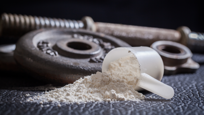
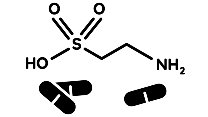
Creatine is a natural compound that is naturally formed in the human body from other amino acids and is found in meat and fish [1]. Most of the research around creatine has been on its use as a bodybuilding supplement that is predominantly consumed by, and tested on, young male athletes [2]. It appears to have effects on women [3], including older women [4], although those effects may be blunted [5].

While this team has previously reviewed the effects of creatine supplementation on the brain [6], that review did not take sex- and age-related differences into account. Therefore, these authors have revisited the topic, including aging and its associated disorders.

Uptake is vital for functioning

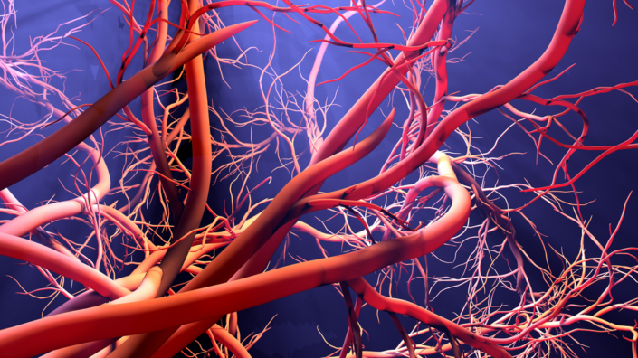
The human brain requires creatine to function properly, and children who cannot properly process creatine suffer from a wide range of crippling developmental disorders [7]. Previous research suggests that both natively produced and consumed creatine are important for function, and creatine has its own specialized transporter, CT1, to cross the blood-brain barrier [6].

The specific biochemistry of creatine in the brain is highly complex, as not all brain cells process creatine in the same way [8]. Some disorders associated with low brain creatine are unaffected by creatine supplements [9], and a lack of CT1 seems to be to blame for that [10]. Other compounds that lead to creatine have been suggested in these cases [11].

Cognitive results

Creatine supplementation does not seem to benefit the standard cognitive performance of young people [12]. However, 68- to 85-year-olds showed improvements in memory after taking a much larger dose for a week [13].

Creatine has also been shown to reduce mental fatigue in a way that suggests better handling of oxygen [14], and one clinical trial suggests that it might aid in recovery from traumatic brain injuries in children and adolescents [15]. Other research leads these authors to conclude that creatine aids the brain in responding to stressors, such as sleep and oxygen deprivation.

Neurodegenerative disorders

The researchers lament the lack of creatine studies in human beings with Alzheimer’s. One rat study suggested that creatine supplementation may be negative in this case [16], with this disease causing creatine to become toxic.

There was a randomized clinical trial conducted on people with Parkinson’s disease. While creatine was well-tolerated, that trial found no benefit [17]. A different trial used creatine along with coenzyme Q10, another well-known supplement, and found that this combination maintained cognitive performance after a year [18].

The effects on creatine on multiple sclerosis were little studied. Here, the studies focused on the muscles rather than the brain, but there was no benefit found in creatine uptake [19]. While mouse studies suggested that creatine might have benefits for amyotrophic lateral sclerosis, most human studies have found no benefit [20].

Mood disorders

Because of creatine reuptake, some researchers consider creatine to be a neurotransmitter itself [21]. Lower brain creatine is associated with mood disorders, including social anxiety disorder [22]. Research into the effects of creatine on these disorders has been mixed, with some research finding that it has a statistically significant effect in combination with an antidepressant drug [23] while a different study shows no effect [24].

The lesson learned

While the research included in this review suggests that creatine supplements appear to have limited effects against brain stress, possibly including age-related brain stress, these broadly mixed and inconclusive results showcase an important lesson. Simply because a disorder is linked to a decrease in a vital compound does not mean that supplementing that compound is going to be an effective treatment, particularly when there are significant hurdles to getting the compound to its proper place.

Additionally, the depletion of creatine, in many cases, appears to be an effect rather than a cause, and finding and ameliorating the upstream causes of these disorders is likely to be considerably more effective than creatine supplementation. However, human research is extremely limited in most cases, and it may be that creatine has a significant effect on a specific disease.

We would like to ask you a small favor. We are a non-profit foundation, and unlike some other organizations, we have no shareholders and no products to sell you. All our news and educational content is free for everyone to read, but it does mean that we rely on the help of people like you. Every contribution, no matter if it’s big or small, supports independent journalism and sustains our future.

Literature

[1] Wyss, M., & Kaddurah-Daouk, R. (2000). Creatine and creatinine metabolism. Physiological reviews, 80(3), 1107-1213.

[2] Kreider, R. B., & Stout, J. R. (2021). Creatine in health and disease. Nutrients, 13(2), 447.

[3] Smith-Ryan, A. E., Cabre, H. E., Eckerson, J. M., & Candow, D. G. (2021). Creatine supplementation in women’s health: a lifespan perspective. Nutrients, 13(3), 877.

[4] Candow, D. G., Forbes, S. C., Chilibeck, P. D., Cornish, S. M., Antonio, J., & Kreider, R. B. (2019). Effectiveness of creatine supplementation on aging muscle and bone: focus on falls prevention and inflammation. Journal of clinical medicine, 8(4), 488.

[5] Syrotuik, D. G., & Bell, G. J. (2004). Acute creatine monohydrate supplementation: a descriptive physiological profile of responders vs. nonresponders. The Journal of Strength & Conditioning Research, 18(3), 610-617.

[6] Forbes, S. C., Cordingley, D. M., Cornish, S. M., Gualano, B., Roschel, H., Ostojic, S. M., … & Candow, D. G. (2022). Effects of creatine supplementation on brain function and health. Nutrients, 14(5), 921.

[7] Fons, C., & Campistol, J. (2016, November). Creatine defects and central nervous system. In Seminars in pediatric neurology (Vol. 23, No. 4, pp. 285-289). WB Saunders.

[8] Braissant, O., & Henry, H. (2008). AGAT, GAMT and SLC6A8 distribution in the central nervous system, in relation to creatine deficiency syndromes: a review. Journal of inherited metabolic disease, 31, 230-239.

[9] Bender, A., & Klopstock, T. (2016). Creatine for neuroprotection in neurodegenerative disease: end of story?. Amino Acids, 48, 1929-1940.

[10] Christie, D. L. (2007). Functional insights into the creatine transporter. Subcellular Biochemistry, 46, 99.

[11] Ostojic, S. M., Stojanovic, M., Drid, P., Hoffman, J. R., Sekulic, D., & Zenic, N. (2016). Supplementation with guanidinoacetic acid in women with chronic fatigue syndrome. Nutrients, 8(2), 72.

[12] Rawson, E. S., Lieberman, H. R., Walsh, T. M., Zuber, S. M., Harhart, J. M., & Matthews, T. C. (2008). Creatine supplementation does not improve cognitive function in young adults. Physiology & behavior, 95(1-2), 130-134.

[13] McMorris, T., Mielcarz, G., Harris, R. C., Swain, J. P., & Howard, A. (2007). Creatine supplementation and cognitive performance in elderly individuals. Aging, Neuropsychology, and Cognition, 14(5), 517-528.

[14] Watanabe, A., Kato, N., & Kato, T. (2002). Effects of creatine on mental fatigue and cerebral hemoglobin oxygenation. Neuroscience research, 42(4), 279-285.

[15] Sakellaris, G., Kotsiou, M., Tamiolaki, M., Kalostos, G., Tsapaki, E., Spanaki, M., … & Evangeliou, A. (2006). Prevention of complications related to traumatic brain injury in children and adolescents with creatine administration: an open label randomized pilot study. Journal of Trauma and Acute Care Surgery, 61(2), 322-329.

[16] Alimohammadi-Kamalabadi, M., Eshraghian, M., Zarindast, M. R., Aliaghaei, A., & Pishva, H. (2016). Effect of creatine supplementation on cognitive performance and apoptosis in a rat model of amyloid-beta-induced Alzheimer’s disease. Iranian Journal of Basic Medical Sciences, 19(11), 1159.

[17] Kieburtz, K., Tilley, B. C., Elm, J. J., Babcock, D., Hauser, R., Ross, G. W., … & Wills, A. M. (2015). Effect of creatine monohydrate on clinical progression in patients with Parkinson disease: a randomized clinical trial. Jama, 313(6), 584-593.

[18] Li, Z., Wang, P., Yu, Z., Cong, Y., Sun, H., Zhang, J., … & Ju, X. (2015). The effect of creatine and coenzyme q10 combination therapy on mild cognitive impairment in Parkinson’s disease. European neurology, 73(3-4), 205-211.

[19] Lambert, C. P., Archer, R. L., Carrithers, J. A., Fink, W. J., Evans, W. J., & Trappe, T. A. (2003). Influence of creatine monohydrate ingestion on muscle metabolites and intense exercise capacity in individuals with multiple sclerosis. Archives of physical medicine and rehabilitation, 84(8), 1206-1210.

[20] Groeneveld, G. J., Veldink, J. H., & van der Tweel, J. (2002). A randomized placebo-controlled trial of creatine in amyotrophic lateral sclerosis. Amyot Lat Scler, 3, 23.

[21] van de Kamp, J. M., Pouwels, P. J., Aarsen, F. K., ten Hoopen, L. W., Knol, D. L., de Klerk, J. B., … & Mancini, G. M. (2012). Long-term follow-up and treatment in nine boys with X-linked creatine transporter defect. Journal of inherited metabolic disease, 35, 141-149.

[22] Yue, Q., Liu, M., Nie, X., Wu, Q., Li, J., Zhang, W., … & Gong, Q. (2012). Quantitative 3.0 T MR spectroscopy reveals decreased creatine concentration in the dorsolateral prefrontal cortex of patients with social anxiety disorder.

[23] Lyoo, I. K., Yoon, S., Kim, T. S., Hwang, J., Kim, J. E., Won, W., … & Renshaw, P. F. (2012). A randomized, double-blind placebo-controlled trial of oral creatine monohydrate augmentation for enhanced response to a selective serotonin reuptake inhibitor in women with major depressive disorder. American Journal of Psychiatry, 169(9), 937-945.

[24] Nemets, B., & Levine, J. (2013). A pilot dose-finding clinical trial of creatine monohydrate augmentation to SSRIs/SNRIs/NASA antidepressant treatment in major depression. International clinical psychopharmacology, 28(3), 127-133.

Microbes

Microbial Composition May Affect Lifespan

New research published in Scientific Reports suggests that microbes in the human gut and mouth can impact how long people live [1].

Bacteria and other microbes are often associated with diseases, but disease-causing microbes are only a minority. The majority of microbes are harmless or beneficial to humans, and we have millions of them living inside and outside us. Researchers refer to this community as the microbiota.

In previous research, scientists had noticed an association between microbiota and longevity [2]. However, the association between two things does not necessarily mean that one is causing the other. Therefore, in this new paper, researchers explored potential causal relationships between gut and mouth microbes’ composition and longevity in order to determine what compositions of microbiota result in increases or decreases in lifespan.

The good, the bad, and the ugly microbes

These researchers conducted a complex analysis that allowed them to define many causal interactions between microbes and longevity. According to their analysis, one of the microbes that positively influenced longevity was gut probiotic bacteria, which impact body fat [3].

One of the microbes in the saliva that was found to increase the odds of living longer is known to reduce the risk of gastric cancer. Different microbes identified in the analysis were also reported in previous research to have a potential negative correlation with congestive heart failure but a positive correlation with longevity. On the other hand, a gut bacterium that negatively influences longevity is also a pathogen that promotes colorectal cancer [4].

In saliva, researchers also identified microbes that decreased longevity. Some of those were known to increase the risk of diseases such as biliary tract cancer, lung cancer, asthma, obstructive pulmonary disease, and osteoporosis. The researchers concluded that decreased longevity could be a result of those diseases.

These researchers also studied people who are expected to live a long time and analyzed which bacteria tended to live in their bodies. The kinds of microbes residing in the long-lived people were associated with plant-rich diets.

On the other hand, microbes linked to a Western diet, high in fat and salt, were not common in those people [5]. Therefore, the authors speculate that long-lived people are more likely to prefer healthy diets rich in complex carbohydrates, fruit, and vegetables instead, which promote the observed microbiotal composition.

Researchers also analyzed microbiota diversity in the gut, saliva, and tongue. An in-depth analysis indicated a causal link between smaller microbes’ diversity and longevity. This result was rather unexpected as previous research had indicated the opposite. Future studies will need to address differences in the results obtained between different research groups.

Same microbes, different outcomes

The authors of this study performed the initial analysis on a Chinese cohort and replicated it on an European cohort. This investigation led to interesting observations.

The researchers identified specific microbes that were negatively correlated with longevity in people from the Netherlands but positively in the Chinese cohort. Other microbes had a negative effect on longevity in Chinese people but had no impact on longevity in Europeans. The researchers also identified groups of microbes that are more abundant in Chinese people who were predicted to live a long time but not in similar Europeans.

Such differences are likely due to different geographical populations’ diets, lifestyles, and genetic backgrounds. However, there is a need for extensive studies with people of varying ethnicities to find specific reasons why such differences exist.

Different outcomes of the same microbes are not limited to different populations. Even the same bacteria on the same person can lead to different outcomes, depending on where in the body they reside.

Researchers identified bacteria associated with decreased longevity when found in the gut. However, they were associated with increased longevity when in the mouth. They point out that it is because some of those microbes are normally part of the mouth microbiota, and that is where they are supposed to be residing. More research is needed to study how microbes spread through different body parts and affect lifespan and health.

Microbes are a piece in the bigger longevity puzzle

Authors mention that previous studies show that some of the most impactful factors influencing longevity are sex, educational level, or diseases. In this study, researchers did not have data regarding economic, behavioral, or environmental factors in the analyzed population. That limits the analysis as researchers couldn’t analyze those factors in connection with the microbial profiles of study participants. Future studies will have to analyze whether such factors influence longevity independent of microbiota.

We would like to ask you a small favor. We are a non-profit foundation, and unlike some other organizations, we have no shareholders and no products to sell you. All our news and educational content is free for everyone to read, but it does mean that we rely on the help of people like you. Every contribution, no matter if it’s big or small, supports independent journalism and sustains our future.

Literature

[1] Liu, X., Zou, L., Nie, C., Qin, Y., Tong, X., Wang, J., Yang, H., Xu, X., Jin, X., Xiao, L., Zhang, T., Min, J., Zeng, Y., Jia, H., & Hou, Y. (2023). Mendelian randomization analyses reveal causal relationships between the human microbiome and longevity. Scientific reports, 13(1), 5127.

[2] Biagi, E., Franceschi, C., Rampelli, S., Severgnini, M., Ostan, R., Turroni, S., Consolandi, C., Quercia, S., Scurti, M., Monti, D., Capri, M., Brigidi, P., & Candela, M. (2016). Gut Microbiota and Extreme Longevity. Current biology : CB, 26(11), 1480–1485.

[3] Omar, J. M., Chan, Y. M., Jones, M. L., Prakash, S., & Jones, P. J. (2013). Lactobacillus fermentum and Lactobacillus amylovorus as probiotics alter body adiposity and gut microflora in healthy persons. Journal of functional foods, 5(1), 116-123.

[4] Kostic, A. D., Chun, E., Robertson, L., Glickman, J. N., Gallini, C. A., Michaud, M., Clancy, T. E., Chung, D. C., Lochhead, P., Hold, G. L., El-Omar, E. M., Brenner, D., Fuchs, C. S., Meyerson, M., & Garrett, W. S. (2013). Fusobacterium nucleatum potentiates intestinal tumorigenesis and modulates the tumor-immune microenvironment. Cell host & microbe, 14(2), 207–215.

[5] Wu, G. D., Chen, J., Hoffmann, C., Bittinger, K., Chen, Y. Y., Keilbaugh, S. A., Bewtra, M., Knights, D., Walters, W. A., Knight, R., Sinha, R., Gilroy, E., Gupta, K., Baldassano, R., Nessel, L., Li, H., Bushman, F. D., & Lewis, J. D. (2011). Linking long-term dietary patterns with gut microbial enterotypes. Science (New York, N.Y.), 334(6052), 105–108.

Online courses about aging research.

Free Courses in Longevity Medicine

One of the challenges of making longevity and rejuvenation research a mainstream field is getting it accepted into healthcare practice. It turns out that healthcare professionals are often not aware of the amazing things being developed in labs right now.

This is no surprise, quite frankly. Rejuvenation research is a fairly niche topic within the broad size and scope of the entire medical and healthcare system. A lot of the technology being developed to directly address aging is relatively new and is not well known in the wider medical community.

To address this problem and to help disseminate information about our field, a team of doctors and researchers have created a series of free courses focused on longevity medicine. The goal is to help the medical community incorporate biomarkers of aging and longevity, along with the latest advancements in aging research, into their work.

Alex Zhavoronkov, Evelyne Bischof, Alexey Moskalev, Morten Scheibye-Knudsen, and Laura Margarint are some of the researchers and doctors behind these courses.

The courses are all free and help to build understanding and ultimately cooperation among researchers in our field and the wider medical profession. Even if you are not a medical professional, just a supporter of longevity medicine and rejuvenation, you may want to take these courses to increase your knowledge.

CME accredited

Both the 101 and 201 courses are Continuing Medical Education (CME) accredited. CME consists of educational activities that serve to maintain, develop, or increase the knowledge, skills, and professional performance and relationships that a physician uses to provide services for patients, the public, or the profession.

Aging and Longevity 101 Box
Longevity Medicine 101.
Lifestyle Hub Box
Longevity Medicine 201.
The Rejuvenation Roadmap Box
Investing in Longevity Course.

We highly recommend taking the opportunity to register and pass the 101 and 102 longevity medicine courses and the longevity investment course if you are interested in investing in rejuvenation biotechnology. Knowledge is power, and a well-informed community is all part of the drive to make our field part of mainstream medicine. The world of healthcare and how we treat aging is going to change massively in the next few decades; get ahead of the curve and start these free courses today!

We would like to ask you a small favor. We are a non-profit foundation, and unlike some other organizations, we have no shareholders and no products to sell you. All our news and educational content is free for everyone to read, but it does mean that we rely on the help of people like you. Every contribution, no matter if it’s big or small, supports independent journalism and sustains our future.
Taurine

Taurine Increases Median Lifespan in Mice by 12%

In a new study published in Science, taurine supplementation led to a significant lifespan extension in mice and health benefits in non-human primates [1].

What is taurine?

Taurine is an amino acid, but it does not participate in protein building. Instead, it has various other functions, such as helping create bile salts, which is important for digestion. Taurine has also been linked to immune system health and mitochondrial function [2]. Taurine is virtually absent from plant-based foods, but humans produce it endogenously from another amino acid, cysteine, so even if vegetarians and vegans can have lower levels of this amino acid, serious taurine deficiency is rare. Taurine is popular among athletes, although the outcomes of the relevant studies vary [3].

Lifespan extension in mice

The authors of this new study started with the fact that levels of taurine go down dramatically with age in many species. In the study, the researchers focused on mice, macaques, and humans. In those three species, a similar around 80% decline in taurine blood levels occurs with aging:

Taurine species

When middle-aged B6 mice were put on lifelong taurine supplementation, it led to a 12% increase in median lifespan. Moreover, the mortality curve shifted to the right, which suggests a “true” anti-aging effect. The sample size was relatively large (about 60 individuals in each group). The control group had normal lifespans and was fed the same diet as the study group. The effect was slightly smaller (but still significant) in male mice, which is consistent with many life-prolonging interventions in mice being sex-specific.

Taurine mice

In female mice, the most noticeable effect on healthspan was the attenuation of age-related weight gain. Taurine also increased bone mass and quality as well as the animals’ fitness. It attenuated age-related depression-like behaviors such as disinterest in exploration. Taurine-fed mice were healthier metabolically, with improved insulin sensitivity.

In male mice, taurine did not affect body mass gain (which might explain the attenuated effect on lifespan), but it improved body composition and reduced adiposity, probably via increased energy expenditure. Taurine-treated males showed greater muscle strength, neuromuscular coordination, bone density, glucose tolerance, and memory as well as reduced anxiety.

Exploring taurine’s effects

Keeping in mind that reduced adiposity alone may explain many of the observed benefits, the researchers also conducted a series of in vitro experiments to elucidate the cellular mechanisms of taurine supplementation. The genes that code for p16 and p21, common markers of cellular senescence, were downregulated by taurine in osteoblasts. Concordantly, cells cultured with taurine were four times less susceptible to irradiation-induced cellular senescence. Taurine supplementation also reduced senescence markers in mice.

Taurine positively affected additional hallmarks of aging, including telomere shortening in telomerase-deficient mice and DNA damage. Taurine-treated mice had better autophagy and decreased levels of certain pro-inflammatory cytokines, such as TNFa and IL-1a. The researchers also found signs of improved mitochondrial function.

The monkeys that the researchers put on taurine had attenuated age-related body weight gain, lower fat percentage, lower fasting glucose levels, and reduced liver damage markers.

For data in humans, researchers analyzed a dataset featuring 12,000 people. They found that higher taurine levels were associated with lower BMI, glucose levels, C-reactive protein (a marker of inflammation), and lower prevalence of diabetes. They also determined that taurine levels go up following a bout of exercise.

The researchers concluded that “taurine supplementation improved the function of every organ investigated in middle-aged female and male mice and likely increased overall health span.” and that “taurine appeared to affect all the established hallmarks of aging.” Just like the impact on the mortality curve, those too are preliminary signs of a “true” anti-aging intervention, which makes this discovery particularly exciting.

Differences in taurine metabolism

However, Peter Attia, a renowned medical doctor with an interest in longevity, wrote a blog post where he points out that taurine metabolism appears to be different in mice and monkeys than in humans. Mice and monkeys have about five times and three times higher average youthful serum taurine levels than humans (the units of measurement used for animal and human data were different), and that is despite both species being herbivores. This makes extrapolating the results to humans trickier.

The study’s lead author, Vijay Yadav, assistant professor of genetics and development at Columbia University in New York City, told lifespan.io: “The metabolism of taurine indeed varies from species to species. This is very much the reason we are putting together a randomized clinical trial to investigate whether the effect that is seen in animals is applicable to humans.” He added that while we do not know yet if taurine supplementation will improve health or increase longevity in humans, the human data collected during this study “suggest taurine has potential.”

We would like to ask you a small favor. We are a non-profit foundation, and unlike some other organizations, we have no shareholders and no products to sell you. All our news and educational content is free for everyone to read, but it does mean that we rely on the help of people like you. Every contribution, no matter if it’s big or small, supports independent journalism and sustains our future.

Literature

[1] Singh, P., Gollapalli, K., Mangiola, S., Schranner, D., Yusuf, M. A., Chamoli, M., … & Yadav, V. K. (2023). Taurine deficiency as a driver of aging. Science, 380(6649), eabn9257.

[2] Jong, C. J., Sandal, P., & Schaffer, S. W. (2021). The role of taurine in mitochondria health: more than just an antioxidant. Molecules, 26(16), 4913.

[3] Kurtz, J. A., VanDusseldorp, T. A., Doyle, J. A., & Otis, J. S. (2021). Taurine in sports and exercise. Journal of the International Society of Sports Nutrition, 18(1), 39.

VitaDAO

VitaDAO Launches VITA-FAST Tokens

VitaDAO, the global community accelerating research and development in longevity science, is pleased to announce the launch of the VITA-FAST tokens. This marks the first-ever opportunity for token holders to directly influence longevity research, setting a new precedent in the field of decentralized science (DeSci).

The VITA-FAST tokens, which allow holders to make decisions on IP licensing, set experiment priorities, and govern research on autophagy, have been met with overwhelming interest. The initial token sale was 2000% oversubscribed, with $620k in bids, demonstrating the public’s eagerness to participate in scientific research.

This groundbreaking initiative is spearheaded by the esteemed Korolchuk Lab, led by renowned cellular biologist Prof. Viktor Korolchuk. The lab’s work focuses on autophagy, a process vital to cellular health and implicated in ageing and various diseases. The launch of VITA-FAST tokens not only provides financial support for this cutting-edge research but also fosters a community-driven approach to biomedical research.

Alex Dobrin, head of community at VitaDAO, said, “This is the next phase in the evolution of decentralized patient communities, to directly contribute to, govern, and be rewarded for progressing valuable longevity research.”

VitaDAO engaged its community in the token sale by using a newly launched crowdsales platform developed by Molecule AG, which developed the original IP-NFT platform for capturing IP in web3, and is a strategic contributor to VitaDAO.

“The success of the VITA-FAST token sale is a testament to VitaDAO’s innovative approach to integrating public participation into scientific research. It also underscores the potential of decentralized finance (DeFi) to fill in the gaps left by traditional funding agencies”, as noted by Paul Kohlhaas, CEO of Molecule.

Professor Matthew Grenby, Pro-Vice-Chancellor for Research and Innovation at Newcastle University added “It’s great to see this cutting-edge research going out into the world, and we are excited to be at the forefront of biomedical research funding. We look forward to supporting Molecule AG and VitaDAO with development of this novel platform and commercialisation of their IP”.

The VITA-FAST token offering marks a significant step forward for VitaDAO as it begins advancing its early research assets toward their next development milestones. The VITA-FAST tokens will provide ongoing options for funding for the Korolchuk Lab as it advances its research program.

About VitaDAO

VitaDAO is a global community for the acceleration of research and development (R&D) in longevity science and the extension of human health- and life-span. VitaDAO achieves this through funding and incubating research and researchers using the tools of decentralised science (“DeSci”) to reduce the time for therapeutic development and, ultimately, bring aging under medical control. VitaDAO, with a community of over 9000 enthusiasts, researchers, and contributors, deployed so far over $4M USD in 17+ projects researching the diseases of ageing, with a focus on cellular biology and the repair of damage associated with the hallmarks of aging.

All researchers are welcome to apply for funding, including professors, post-docs, research scientists, and even students.

For media, please contact:

Alex Dobrin Community and Awareness Steward alex@vitadao.com Official Twitter: @vita_dao

We would like to ask you a small favor. We are a non-profit foundation, and unlike some other organizations, we have no shareholders and no products to sell you. All our news and educational content is free for everyone to read, but it does mean that we rely on the help of people like you. Every contribution, no matter if it’s big or small, supports independent journalism and sustains our future.
Angiogenesis

Stem Cell Signals Encourage Blood Vessel Formation in Mice

Researchers publishing in Aging have outlined how biochemical signals derived from created stem cells encourage blood vessels to form new branches in naturally aged mice.

iPSCs, exosomes, and angiogenesis

Exposure to the Yamanaka factors can cause ordinary cells to teturn to early stemness, from which they can divide into any other cell in the body. These are Induced pluripotent stem cells (iPSCs), and they have been studied extensively in aging and other medical research.

Like other cells, iPSCs secrete exosomes, tiny packages of RNA and proteins that serve as signals to other cells. In a previous study, exosomes derived from iPSCs successfully encouraged angiogenesis, the formation of new blood vessels, thus speeding up wound healing [1]. Other experiments have reported positive results in models of heart attack [2].

In this study, the researchers sought to determine if angiogenesis could be encouraged in animals that were suffering from another harmful condition: aging.

Exosomes in life and in the lab

The researchers used a combination of tests in this study. They tested arterial samples (aortic rings) from a total of eight groups of mice: the mice could be either old or young, they could have been pretreated with iPSC exosomes for three months or not, or their aortic rings could have been placed with iPSC exosomes or not.

As expected, the arterial samples of the young mice had much more angiogenesis than the old mice did. Exposing old-mouse samples to iPSC-derived exosomes, either in the living animal or in culture, improved angiogenesis substantially and even more if both approaches were used, showing that these exosomes were consistently effective in this model. Young mice seemed to benefit as well, although not to the level of statistical significance.

Bone marrow cells received similar benefits. The bone marrow cells of old mice proliferated substantially more in culture in all of the treated groups. Interestingly, cells from young mice benefited as well, although much more if the mice had been injected with the exosomes in life.

The senescence-associated biomarkers p53 and p16 were also measured in tissues taken from living animals. Skin, muscle, kidney, lung, spleen, liver and brain tissue all had varying levels of benefit, with the kidneys and muscle being the most affected. The heart seemed unaffected, although p53 and p16 do not substantially differ between the hearts of young and old mice. Antioxidant activity was also improved in most tissues.

Finally, exosomes affected exosomes. The researchers analyzed the serum exosomes of treated old mice and found that they were much more similar to those of young mice than the untreated group’s were.

A potential for treatment?

Exosomes have been studied for some time as a potential method of treating age-related diseases, and this is far from the first paper published on this subject. Because there appear to be none of the other side effects, such as tumors, that can potentially occur with other rejuvenative and regenerative therapies, it might be time to test exosomes in human volunteers for heart diseases and wound healing.

We would like to ask you a small favor. We are a non-profit foundation, and unlike some other organizations, we have no shareholders and no products to sell you. All our news and educational content is free for everyone to read, but it does mean that we rely on the help of people like you. Every contribution, no matter if it’s big or small, supports independent journalism and sustains our future.

Literature

[1] Qiu, X., Liu, J., Zheng, C., Su, Y., Bao, L., Zhu, B., … & Jin, Y. (2020). Exosomes released from educated mesenchymal stem cells accelerate cutaneous wound healing via promoting angiogenesis. Cell Proliferation, 53(8), e12830.

[2] Balbi, C., & Vassalli, G. (2020). Exosomes: Beyond stem cells for cardiac protection and repair. Stem Cells, 38(11), 1387-1399.

Rejuvenation Roundup June 2023

As our annual conference approaches, we continue to provide interviews, book reviews, insights into finance and longevity, and plenty of information on the latest research. Here’s what’s happened in June.

LEAF News

EARD 2023Ending Age-Related Diseases 2023 approaches! On August 10-11, 2023, we will be holding our sixth annual conference to bring entrepreneurs, investors, pharma and biotech companies, researchers, and government organizations together. Fostering the exchange of information and encouraging cooperation between the various parts of the industry is the goal of our non-profit organization.

Team and activities

Summer Off to a Great Start for Longevity: Summer is here, but we haven’t been relaxing around the pool. Instead, we’ve been busy working on the fight to defeat age-related diseases! The last few months have been a very busy time for us here at lifespan.io as we prepare for our sixth Ending Age-Related Diseases conference.

Zuzalu LighthouseZuzalu: Shining City on the Black Mountain: Arkadi Mazin recounts his experiences going to the pop-up city of Zuzalu, which Ethereum billionaire Vitalik Buterin helped to found. Cryptocurrency, longevity, and AI were among the many topics featured there.

Chris Hemsworth’s Centr Partners with lifespan.io: We are thrilled to announce a new partnership with Centr to provide its community with world-class expertise on longevity. Centr, a health and wellness ecosystem founded by Chris Hemsworth, announced an all-new partnership with lifespan.io.

Lifespan News

LSN Bryan JohnsonBryan Johnson Thinks AI Should Run Your Life: In this video, Bryan Johnson, founder and CEO of Blueprint, shared his ideas about the role of artificial intelligence in the future of human health and society at the recent Healthspan Summit. Among other controversial statements about AI and the future of humanity, he suggests that we should let AI determine our well-being.

Chris Hemsworth’s Centr Launches lifespan.io Partnership: Emmett Short discusses our partnership with famous actor Chris Hemsworth’s health and longevity platform, Centr.

Interviews

Niklas Anzinger on Building a New Regulatory World: Ryan O’Shea interviews Niklas Anzinger of the Infinita VC fund, who discusses the pop-up city Zuzalu along with many other issues relating to network states, regulation, and longevity.

SENS Research Foundation’s CEO on Accelerating Rejuvenation: We had the chance to interview Lisa Fabiny-Kiser, CEO of the well-known SENS Research Foundation, on all the various research, education, and advocacy activities that the organization is currently doing along with its plans for the future. Public opinion and the academic landscape have changed considerably since SRF was founded nearly 15 years ago.

Journal Club

How Superior DNA Repair Gives Bowhead Whales Longevity: Oliver Medvedik led a discussion of a paper showing how and why bowhead whales enjoy such long lifespans, pointing out the specific DNA repair mechanism that they do not share with other mammals.

Advocacy and Analysis

Lustgarten Microbial BurdenMichael Lustgarten Fights Back Against Microbes: Dr. Michael Lustgarten delivers a clear and enlightening exploration of the intricate relationship between microbial burden and aging in Microbial Burden: A Major Cause of Aging and Age-Related Disease and What We Can Do to Fight Back!

Dr. Matthew Walker Tells Us Why We Sleep: “Why We Sleep: Unlocking the Power of Sleep and Dreams” by Matthew Walker, renowned sleep research scientist and psychiatry professor at Harvard, is a comprehensive exploration of sleep that offers a rich blend of science, research, and practical advice.

Research Roundup

Linking Bile Duct Blockage and Cellular Senescence: Research published in Aging has shed new light on the relationship between certain liver diseases and cellular senescence.

MacaquesBenefits of Dasatinib and Quercetin Treatment in Monkeys: In one of the first studies of its kind, the popular senolytic combination, administered systematically for six months, produced several health benefits in these animals. Some effects were augmented by caloric restriction.

Giving Old Stem Cells Lasting Youthful Powers: Researchers publishing in Cell Proliferation have described factors that appear to give old muscle stem cells the ability to effectively proliferate and differentiate for a very long time.

Vitamin C FruitsStabilized Vitamin C Improves Brain Aging in Mice: Korean scientists publishing in Nature were able to increase the stability of vitamin C, a powerful antioxidant, using a short, engineered DNA molecule called an aptamer. The result improved various aspects of brain aging in naturally aged mice.

Castration Influences Growth and Median Lifespan in Mice: Research published in Aging Cell has discovered that castrated male mice show similarities to females in growth and lifespan. It is widely known that women outlive men on average, and this paper dives into the biological reasons.

Bone marrow injectionHow Donated Stem Cells Become Functional Cells: Scientists have verified the effectiveness of stem cell transplants, researched a core reason behind it, and published their findings in Aging Cell. Hematopoietic stem cells (HSCs), which make blood cells, are governed by their local microenvironment and can affect it as well.

How Superior DNA Repair Gives Bowhead Whales Longevity: Scientists have found a possible explanation for bowhead whales’ exceptional lifespan, and it might be translatable to humans. With some exceptions, body size is strongly correlated with longevity across species, and the researchers attribute this to better genetic protection.

Spinal degenerationSirtuins May Help Against Inflammatory Spinal Degeneration: Researchers publishing in Aging have outlined a relationship between disc degeneration, macrophage polarization, inflammation, and sirtuins, demonstrating their effectiveness in a rat model of the disease.

Obese People Have Impaired Brain Responses to Nutrients: Scientists publishing in Nature Metabolism have shown that obese people have dampened brain responses to food. This effect lingers even after diet-induced weight loss, possibly explaining why it is easy to regain weight.

Health surveyRapamycin Users Report Positive Effects in Survey: A new study published in GeroScience addresses the efficacy and side effects of off-label rapamycin, which is widely associated with longevity, as a preventive therapy to maintain healthspan.

How Cancer Hijacks the Nervous System: A review article published in Nature has outlined the current research into how the nervous system’s signals affect cancer growth. This review opens with a bold but heavily documented claim: the central nervous system (CNS) affects various cancers through signaling mechanisms.

VaccinationShingles Vaccine May Protect Against Alzheimer’s: New research suggests that vaccination against shingles, a disease caused by the varicella zoster virus, can provide some protection against Alzheimer’s disease, mostly in women.

The Common Sweetener Sucralose May Accelerate Aging: Researchers publishing in Journal of Toxicology and Environmental Health, Part B have found that the common sweetener sucralose may contribute to genetic and intestinal damage. The authors disagree with the fundamental claims that were made prior to the approval of sucralose by the FDA.

Spoon of CollagenClinical Trial: Collagen Peptides Reduce AGEs in Skin: A placebo-controlled, double-blinded human clinical trial published in Bioscience, Biotechnology, and Biochemistry has determined that collagen peptide ingestion reduces the amount of advanced glycation end-products (AGEs) in the skin.

Bacteria May Be Fueling Cancer with Methionine: Scientists have found that the tumor microenvironment in lung adenocarcinoma favors methionine-producing bacteria, which, in turn, help the cancer survive nutrient scarcity.

Group ExercisingNew Clock: People Who Exercise Are Biologically Younger: Using a DNA methylation clock based on fitness, researchers publishing in GeroScience have determined that people who exercise regularly are biologically younger on average.

Bodybuilding Helps Older People Stay Strong in Human Study: In a controlled human study published in Frontiers in Public Health, Iranian researchers have demonstrated that resistance training and creatine supplementation are effective in helping older people retain their strength.

LettuceScientists Modify Lettuce to Make Orally Available Insulin: Researchers have genetically engineered lettuce to produce human insulin. The resulting plant-based drug, which can be taken orally, was successfully tested in mice and acts more gradually than injections.

New Peptide Puts the Brakes on Cellular Senescence: Researchers publishing in the Nature journal npj aging have discovered a new peptide that might prevent cells from becoming senescent and possibly youthen human skin.

Kidney transplantPreserved Kidneys Rewarmed and Transplanted in Rats: For the first time in history, scientists have been able to freeze, preserve, rewarm, and transplant rat kidneys with a new method of organ cryopreservation. Today, organ transplantation is severely limited by the fact that organs can only be preserved for a short time in near-freezing temperatures.

Activating Just One Gene to Rejuvenate Model Mice: In a new study published in Aging Cell, researchers report that transient activation of the Yamanaka factor Oct4 allowed partial reprogramming of cells, which led to rejuvenation in these cells and in a mouse model of premature aging.

Herpes simplexThe Links Between Herpes and Dementia: One of the most common diseases in the world is herpes, as 50 to 80% of Americans have this illness. However, the proteins involved in this latent disease are also involved in Alzheimer’s.

A Potential New Foundation for Epigenetic Clocks: A study recently published in Aging Cell has discovered an entirely new method of looking at methylation, one that is less limited to certain sites, and experimented with multiple ways of analyzing it.

Diet and exerciseA Combination of Exercise and Diet for Insulin Sensitivity: Scientists publishing in Nature Metabolism have determined that in obese people, healthy diet and exercise bring stronger metabolic benefits than diet alone.

Taurine deficiency as a driver of aging: Taurine abundance decreases during aging, and these results demonstrate that reversal of this decline through taurine supplementation increases healthspan and lifespan in mice and worms and health span in monkeys.

Higher versus lower nut consumption and changes in cognitive performance over two years in a population at risk of cognitive decline: Frequent nut consumption was associated with a smaller decline in general cognitive performance over two years in older adults at risk of cognitive decline.

Association of daily step volume and intensity with cardiometabolic risk in older adults: Daily step volume and intensity were inversely associated with cardiometabolic risk in community-dwelling older adults in a dose-response manner.

Resistance training rejuvenates aging skin by reducing circulating inflammatory factors and enhancing dermal extracellular matrices: Both aerobic and resistance training significantly improved skin elasticity and upper dermal structure, and resistance training also improved dermal thickness.

Resistant starches from dietary pulses modulate the gut metabolome in association with the microbiome in humanized mice: These findings highlight the potential of discrete structures of these dietary fibers to induce targeted alterations in the gut metabolomic pool

“Heads Up” for Creatine Supplementation and its Potential Applications for Brain Health and Function: This review provides an up-to-date summary of creatine and indices of brain health and function and discusses possible sex- and age-related differences in responses to creatine supplementation on the brain.

Protection of Liver Functions and Improvement of Kidney Functions by Twelve Weeks Consumption of Cuban Policosanol: People dosed with 20 milligrams of policosanol also had improved blood pressure.

The acute effect of different NAD+ precursors included in the combined metabolic activators: This study provided a plasma metabolomic landscape of different formulations of these precursors.

A short dasatinib and quercetin treatment is sufficient to reinstate potent adult neuroregenesis in the aged killifish: These results provide a cellular mechanism for age-related regeneration resilience and a proof-of-concept of a potential therapy to revive neurogenic potential.

Sexual dimorphic metabolic and cognitive responses of C57BL/6 mice to Fisetin or Dasatinib and quercetin cocktail oral treatment: Senolytic treatment in young adulthood has beneficial, negligible, or detrimental effects in these mice, dependent upon sex and treatment.

Discovery of senolytics using machine learning: This approach led to a several hundred-fold reduction in drug screening costs and demonstrates that artificial intelligence can take maximum advantage of small and heterogeneous drug screening data

Higher testosterone and testosterone/estradiol ratio in men are associated with decreased Pheno-/GrimAge: There is a potential protective effect of testosterone on lifespan and, conceivably, cardiovascular health.

Gene expressions associated with longer lifespan and aging exhibit similarity in mammals: These results highlight the importance of focusing on adaptive aspects of the aging transcriptome and demonstrate that cross-species genomics can be a powerful approach.

News Nuggets

Academy for Health and Lifespan ResearchDr. David Sinclair Assumes Presidency of the AHLR: The Academy for Health and Lifespan Research (AHLR) is proud to announce the election of Dr. David Sinclair as its new President. Dr. Sinclair, a distinguished Professor of Genetics at Harvard Medical School and renowned author of the New York Times Bestseller “Lifespan,” has been chosen by his peers.

We would like to ask you a small favor. We are a non-profit foundation, and unlike some other organizations, we have no shareholders and no products to sell you. All our news and educational content is free for everyone to read, but it does mean that we rely on the help of people like you. Every contribution, no matter if it’s big or small, supports independent journalism and sustains our future.

Diet and exercise

Exercise And Diet Is A Solid Combo For Obesity

Scientists publishing in Nature Metabolism have determined that in obese people, healthy diet and exercise bring stronger metabolic benefits than diet alone [1].

Better together

There are two major interventions at hand for weight loss without resorting to medications: diet and exercise. However, recent research suggests that they are not interchangeable. Forgoing either one can result in a partial effect and even create new problems, such as lean mass loss for people who diet without pumping iron or other resistance training [2]. On the other hand, exercise without a healthy diet might not help with metabolic problems [3]. This new interventional study largely confirms this emerging consensus, adding some important details.

The researchers recruited obese prediabetic patients for a five-month-long program tuned for inducing mild weight loss (10% of body mass). All participants received a “plant-forward” diet dominated by vegetables, fruit, whole grains, seeds, and legumes that provided 70% of the calories from carbohydrates, 15% from protein, and 15% from fats.

Half of the participants also underwent an exercise program of six one-hour sessions per week. The calorie intake at the beginning of the study covered 75% of the recommended number of calories for both groups (the exercising group received more), and then it was fine-tuned to achieve a weight loss of 10%. Apparently, the fine-tuning succeeded, since the average weight loss in both groups was nearly identical.

Interestingly, and somewhat counterintuitively, the two groups did not significantly differ in lean mass dynamics, as both experienced slight loss. The researchers hold that this might be due to the combination of the relatively mild pace and amount of weight loss and the high quality of the diet.

Exercise adds insulin sensitivity

The study’s main endpoint was insulin sensitivity, a major marker of metabolic health. Insulin is the hormone that signals to cells of several types, such as muscle cells, to take up glucose from bloodstream and store it. Insulin sensitivity is impaired in metabolic disorders such as diabetes.

Both groups experienced a significant increase in whole-body insulin sensitivity following the treatment, but in the exercising group, insulin sensitivity was twice as high as in the diet-only group. The researchers admit that measuring insulin sensitivity in exercising people is tricky, since every bout of exercise is known to transiently increase it. However, they suggest that the study’s design made it possible to capture both the transient and long-term effects of exercise.

Diet Exercise Insulin

Several other metabolic markers, such as triglycerides, HDL cholesterol, and fasting blood glucose, improved as well but roughly equally in both groups, probably due to the healthy diet and weight loss. The gut microbiome also uniformly changed in both groups, but only during the first month, despite the fact that weight loss continued throughout the study. Presumably, diet quality was the only factor responsible for that.

Exercise upregulates mitochondrial and autophagy pathways

The reseachers then dug deeper into the changes caused specifically by exercise. While an analysis of the skeletal muscle transcriptome showed virtually no changes in the diet group, in the exercising group, dozens of genes were either upregulated or downregulated by the treatment. The two most prominent pathways to show positive changes were mitochondrial energy metabolism and angiogenesis (creation of new blood vessels), which is consistent with previous research.

Unlike the muscle transcriptome, proteomic analysis of blood plasma showed changes for both groups, but they were far from identical. For instance, autophagy-related pathways were highly upregulated in the exercising group but downregulated in the diet-only group.

“Obesity makes the body resistant to insulin’s ability to decrease glucose production by the liver and increase glucose uptake by skeletal muscle tissue, leading to an increase in blood glucose concentrations,” said senior investigator Samuel Klein, MD, director of the Center for Human Nutrition in Washington University School of Medicine in St. Louis. “Insulin resistance is a major factor that causes Type 2 diabetes, nonalcoholic fatty liver disease and abnormal blood lipids in people with obesity. We’ve shown that combining exercise with weight loss causes a marked improvement in whole-body insulin sensitivity, thereby lowering the risk of developing diabetes and treating obesity-related metabolic diseases to a much greater degree than is possible with weight loss alone.”

This study has several limitations, starting with the small sample size. The researchers also admit that insufficient decoupling of the acute and long-term effects of exercise on insulin sensitivity is still a concern. Nevertheless, these results convincingly support the idea that a combination of exercise and a healthy diet provide the maximum impact on metabolic health.

We would like to ask you a small favor. We are a non-profit foundation, and unlike some other organizations, we have no shareholders and no products to sell you. All our news and educational content is free for everyone to read, but it does mean that we rely on the help of people like you. Every contribution, no matter if it’s big or small, supports independent journalism and sustains our future.

Literature

[1] Beals, J.W., Kayser, B.D., Smith, G.I. et al. (2023) Dietary weight loss-induced improvements in metabolic function are enhanced by exercise in people with obesity and prediabetes. Nat Metab.

[2] Roth, C., Schoenfeld, B. J., & Behringer, M. (2022). Lean mass sparing in resistance-trained athletes during caloric restriction: the role of resistance training volume. European Journal of Applied Physiology, 122(5), 1129-1151.

[3] Guo, J., Zhang, X., Wang, L., Guo, Y., & Xie, M. (2013). Prevalence of metabolic syndrome and its components among Chinese professional athletes of strength sports with different body weight categories. PLoS One, 8(11), e79758.

High Tech Clock

A Potential Next Generation For Epigenetic Clocks

A study recently published in Aging Cell has discovered an entirely new method of looking at methylation and experimented with multiple ways of analyzing it.

Not just a clock

While we have reported extensively on the use of epigenetic methylation clocks, the genes that become activated and deactivated with aging are not just useful measurements of it. Turning genes on and off has serious consequences, determining what proteins are and aren’t produced.

Furthermore, the genes that are measured in clocks are only a small subset of the total. Current clocks are based on a microarray technology that uses only specific sites [1], which these researchers note is less than 10% of the entire genome subject to methylation. They chose an entirely different technology, whole-genome bisulfite sequencing, which is unbiased and accounts for most of the genome.

Testing the new tool

The researchers chose two cell types in order to test their new approach. From 40 people, they collected monocytes, which turn over rapidly; from 43 people, they collected muscle cells, which turn over very slowly. The volunteers ranged greatly in age, from 22 to 83.

The data from this new method was evaluated in three different ways. First, the researchers used a standard linear regression technique, which is the basis for other clocks. Multiple pathways, including the Notch signaling pathway, were found to be affected. Interestingly, comparing this data to a similar microarray-based analysis found only 7 sites in common.

Second, they attempted to determine which methylation sites were changing within 10-year time windows. This analysis found that muscle tissue is strongly affected around ages 52 to 62 and that monocytes are most affected around ages 33 to 42. A great many signaling pathways were found, including pathways related to neuronal development.

In their third analysis, the researchers directly compared the methylation of younger and older people as large, generalized groups, with the cutoff point being age 51. This analysis found that sites related to protein translation and muscle contraction were affected in muscle tissue, with cellular adhesion, consumption of particles, DNA maintenance, and, again, neuronal development pathways being affected in monocytes.

Finally, the researchers compared the number of sites changed in each of the three analyses. While there were thousands of overlapping sites between the first two analyses, the third analysis had far less overlap. Only four specific sites were found to overlap between all three groups in muscle tissue, and only one dozen overlapped in monocytes.

A need for detail and consistency

The fact that each of these researchers’ analyses yielded entirely different areas of interest, with minimal overlap, is both interesting and a cause for concern. It is not clear which, if any, of these approaches is best suited to measure aging or which of the detected sites are most consequential for physical function.

Additionally, the authors note that their analysis was hampered by technical constraints and that they could not be as thorough as they might have liked. While it targeted all of the genome, this was a “shotgun” approach that only hit random parts of it. They suggest that a greater analytical depth might have yielded more useful information.

This technique is clearly in its infancy, and it needs far more development and adjustment before it can be considered a competitor to the microarray-based methylation analysis that current clocks use. The true end goal of development in this area is, of course, a technique that accurately and completely analyzes the methylation of the entire genome.

We would like to ask you a small favor. We are a non-profit foundation, and unlike some other organizations, we have no shareholders and no products to sell you. All our news and educational content is free for everyone to read, but it does mean that we rely on the help of people like you. Every contribution, no matter if it’s big or small, supports independent journalism and sustains our future.

Literature

[1] Schumacher, A., Kapranov, P., Kaminsky, Z., Flanagan, J., Assadzadeh, A., Yau, P., … & Petronis, A. (2006). Microarray-based DNA methylation profiling: technology and applications. Nucleic acids research, 34(2), 528-542.

Herpes simplex

The Links Between Herpes and Dementia


One of the most common diseases in the world is herpes, as 50 to 80% of Americans have this illness. The herpes simplex virus, which causes cold sores, is normally transmitted through person-to-person contact. This illness leaves the patient with blisters clustered around the mouth. Gradually, the blisters pop and leave sores that take around one week to heal.

However, that is just the short-term effect of herpes. This virus hides in certain neurons in the mouth, occasionally reactivating to cause another bout of cold sores [1]. This permanent infection is known as latency, a trait that herpes shares with other viruses such as cytomegalovirus.

Familiar Alzheimer’s proteins

Researchers have noted a link between latent herpes infection and an increased incidence of cognitive decline. This decline is exacerbated by multiple activations, and this is especially true for carriers of the APOE4 allele, a genetic variation implicated in Alzheimer’s risk [2].

Immune responses to herpes infection are very similar to the hallmarks of Alzheimer’s [3]. Specifically, after infection with HSV-1, amyloid beta and tau tangles, two major hallmarks of Alzheimer’s, increase in concentration [4].

While these two proteins are influenced by herpes infection, they are primarily known for their impact on brain aging and neurodegeneration. With age, the misfolded form of amyloid beta protein, which controls neuronal growth and homeostasis, aggregates and forms plaques [5]. On the other hand, tau tangles are formed by a misfolded version of the protein tau, which normally influences neurons’ internal skeletons [6]. Both of these can disrupt cell-cell synaptic communication, contributing to the development of dementia.

An immune link

The link between herpes and dementia could also be related to the immune response that the body uses in an attempt to rid itself of this viral infection. Inflammation has previously been implicated as a driving factor behind age-related decline [7], specifically cellular senescence [8] and free radical production [9].

Fortunately, solutions to the immune components of age-related diseases are being investigated. Researchers are studying centenarians to figure out how they reduce inflammation-driven age-related damage (inflammaging) and incidences of age-related diseases [10]. With a greater understanding of how inflammation affects aging and better theories on the mechanisms behind this influence, researchers can develop new therapies to reduce the impact of these viral infections on dementia risk.

Overall, this is an actively progressing area of research, and there is still much work to be done to better understand the role that herpes simplex virus plays in dementia. However, there have been promising strides in better understanding and tackling the immune system’s role in aging and theories that link herpes to dementia-related processes.

We would like to ask you a small favor. We are a non-profit foundation, and unlike some other organizations, we have no shareholders and no products to sell you. All our news and educational content is free for everyone to read, but it does mean that we rely on the help of people like you. Every contribution, no matter if it’s big or small, supports independent journalism and sustains our future.

Literature

[1] Nicoll, M. P., Proença, J. T., & Efstathiou, S. (2012). The molecular basis of herpes simplex virus latency. FEMS microbiology reviews, 36(3), 684-705.

[2] Murphy, M. J., Fani, L., Ikram, M. K., Ghanbari, M., & Ikram, M. A. (2021). Herpes simplex virus 1 and the risk of dementia: a population-based study. Scientific Reports, 11(1), 8691.

[3] Wainberg, M., Luquez, T., Koelle, D. M., Readhead, B., Johnston, C., Darvas, M., & Funk, C. C. (2021). The viral hypothesis: how herpesviruses may contribute to Alzheimer’s disease. Molecular psychiatry, 26(10), 5476-5480.

[4] Powell-Doherty, R. D., Abbott, A. R., Nelson, L. A., & Bertke, A. S. (2020). Amyloid-ß and p-tau anti-threat response to herpes simplex virus 1 infection in primary adult murine hippocampal neurons. Journal of virology, 94(9), e01874-19.

[5] Chen, G. F., Xu, T. H., Yan, Y., Zhou, Y. R., Jiang, Y., Melcher, K., & Xu, H. E. (2017). Amyloid beta: structure, biology and structure-based therapeutic development. Acta Pharmacologica Sinica, 38(9), 1205-1235.

[6] Mandelkow, E. M., & Mandelkow, E. (2012). Biochemistry and cell biology of tau protein in neurofibrillary degeneration. Cold Spring Harbor perspectives in medicine, 2(7), a006247.

[7] Chung, H. Y., Kim, D. H., Lee, E. K., Chung, K. W., Chung, S., Lee, B., … & Yu, B. P. (2019). Redefining chronic inflammation in aging and age-related diseases: proposal of the senoinflammation concept. Aging and disease, 10(2), 367.

[8] Ren, J. L., Pan, J. S., Lu, Y. P., Sun, P., & Han, J. (2009). Inflammatory signaling and cellular senescence. Cellular signalling, 21(3), 378-383.

[9] Biswas, S., Das, R., & Banerjee, E. R. (2017). Role of free radicals in human inflammatory diseases. Aims Biophysics, 4(4), 596-614.

[10] Zhou, L., Ge, M., Zhang, Y., Wu, X., Leng, M., Gan, C., … & Dong, B. (2022). Centenarians alleviate inflammaging by changing the ratio and secretory phenotypes of T helper 17 and regulatory T cells. Frontiers in Pharmacology, 13, 877709.

Editorial

Summer Off to a Great Start for Longevity

Summer is here, but we haven’t been relaxing around the pool. Instead, we’ve been busy working on the fight to defeat age-related diseases!

This last few months has been a very busy time for us here at lifespan.io. We have been focused on our upcoming Ending Age-Related Diseases 2023 conference and making sure its sixth year is the best yet.

More about the conference later! First, we have some other exciting news to share that showcases how we are making rejuvenation and longevity research mainstream.

lifespan.io joins forces with Chris Hemsworth’s Centr

One of our primary advocacy goals is to engage the wider public audience about rejuvenation and longevity science. To that end, we are delighted to announce that lifespan.io has teamed up with Chris Hemsworth’s health and wellness company, Centr.

Chris Hemsworth

The collaboration between Centr and lifespan.io will emphasize the importance of preventive measures, showcase a future in which aging is better managed and comprehended, and show how our present lifestyle choices can influence our future health.

Centr will be adapting a selection of our news and educational content for its health and wellness audience. From that point, Centr readers also have the option to read the original articles and hopefully delve deeper into the world of rejuvenation and longevity research.

Hopefully, this partnership will encourage more of the people to take an interest in this fascinating field of science. We are a non-profit advocacy organization, and alliances like this help to power that advocacy for longer, healthier lives.

Lifespan visits Zuzalu

We have been busy on the journalism and advocacy side of things recently, too. We were at Zuzalu, the “pop-up” city in Montenegro. This experimental event ran from March 25 to May 25, 2023 and was designed to foster collaboration.

Zuzalu Lighthouse

Zuzalu united individuals who were interested in gaining knowledge, encouraging creativity, promoting longevity and wellness, and establishing self-sufficient communities. The event featured a diverse range of activities that covered topics such as rejuvenation, synthetic biology, privacy-enhancing technology, public goods, artificial intelligence, governance, and many others.

lifespan.io journalist Arkadi Mazin attended the event and has written a special feature: check out Zuzalu: Shining City on the Black Mountain to learn more about this fascinating event. Arkadi has also written Zuzalu Talks Longevity: Highlights from the Conference as a quick summary of the event.

lifespan.io Executive Director Stephanie Dainow was also there and gave a talk at the event. Not everyone at the event was familiar with rejuvenation and longevity research, so the focus was on engaging new audiences in an accessible way. The talk saw Stephanie and Nathan Cheng, executive director of Longevity Biotech Fellowship, give a short overview of longevity biotech.

Keith Comito, president of lifespan.io, discussed the convergence of longevity and emerging technologies like NFTs in his presentation. Despite some people viewing NFTs as a mere fad, Keith highlighted the technology’s potential for various intriguing applications.

OneSkin teams up with LifeNoggin to talk about skin aging

LifeNoggin has teamed up with Oneskin to make a video about skin aging and how researchers are finding ways to fight back.

Thanks to OneSkin for sponsoring this video, and if you would like to find out how you can sponsor a video, please get in touch.

Announcing the Longevity and DeSci Recap

Interest and support for longevity and rejuvenation research is constantly growing. lifespan.io is one of the organizations leading the advocacy movement to popularize rejuvenation research and make healthy longevity something everyone will want.

As part of that growth, there are a significant number of decentralized science (DeSci) supporters who are interested in longevity. Many people in the DeSci community are already donating to aging research and supporting many of the projects we are.

To that end, we are delighted to announce that we will be publishing the all-new Longevity and DeSci Recap every month. This will be a rapid-fire summary of what is happening in the longevity and DeSci space, much like our Rejuvenation Roundup but with a focus on the DeSci and blockchain side of things.

You will find the latest interviews, insights, and research in the longevity biotech sphere, covering topics including blockchain, DAOs, cryptocurrency, decentralized science (DeSci), and more, and letting you know about exciting events or studies coming up in the near future.

Want to know more? Then check out the first edition of the Longevity and DeSci Recap!

Join us for Ending Age-Related Diseases 2023

The Ending Age-Related Diseases conference (EARD) returns for its sixth year, and this one promises to be even better!

EARD 2023 banner.

We are bringing the leading experts in rejuvenation research, biotech investment, and decentralized science (DeSci) together for two action-packed days. We have workshops, talks, panel discussions, and more for you to enjoy this year.

We have some great confirmed speakers this year, and below is just a small sample.

EARD speakers

Join us in NYC or online for EARD2023

CapitaleThis event is happening at the stunning Capitale building in New York City. This stunning neoclassical building dates back to 1895 and boasts Corinthian columns, Venetian glass, and marble mosaic floors.

We cannot think of a better setting to meet and learn about the latest advancements in longevity and rejuvenation research.

Get your ticket at a discounted price today

As much as we would love to have you all join us in NYC, there is good news for people who cannot make it. This year not only marks our return to physical conferences, but we are also making EARD an online event too. Everyone can join us this year!

Regular ticket prices are currently at a lower Early Bird cost until the 30th of June: register today if you want to grab a bargain!

We would like to ask you a small favor. We are a non-profit foundation, and unlike some other organizations, we have no shareholders and no products to sell you. All our news and educational content is free for everyone to read, but it does mean that we rely on the help of people like you. Every contribution, no matter if it’s big or small, supports independent journalism and sustains our future.
Gene activation

Activating A Single Gene Rejuvenates Mice

In a new study published in Aging Cell [1], researchers report that transient activation of the Yamanaka factor Oct4 allowed partial reprogramming of cells, which led to rejuvenation in these cells and in a mouse model of premature aging.

This research used a mouse model of Hutchinson-Gilford progeria syndrome, an extremely rare and devastating genetic disorder. Children with progeria age rapidly and usually die young. Progeria is caused by a mutation in the gene that codes for lamin A, a protein that envelops and protects the nucleus from damage. This mutation causes a shorter, ineffective version of lamin A, called progerin, to be created instead [2].

Reversing epigenetic alterations with Oct4

In this study, the researchers looked at a few aspects of aging, primarily focusing on epigenetic alterations.

Researchers activated the gene Oct4 in lab-grown mouse fibroblasts, which form connective tissues that connect to and support other tissues. They reported that this altered the epigenetics of these fibroblasts, causing changes to the expression of genes responsible for their differentiation and de-differentiation. Those processes make cells more or less specialized, respectively.

Progeric cells rejuvenated

The researchers wanted to assess the influence of Oct4 in progeric fibroblasts. They altered healthy fibroblasts to produce the mutated, progeric version of lamin A. Then, they activated Oct4 in those cells and made several observations that suggest that these cells experienced rejuvenation.

They observed that activation of Oct4 reduced DNA breaks, which accumulate with both age and progeria. Similarly, Oct4 reduced the number of dying progeric cells. Additionally, activating Oct4 in the progeric fibroblasts restored the activity patterns of stress response-related and senescence-related genes.

The researchers also discovered that activating the Oct4 gene led to the suppression of mutated lamin A in mouse fibroblasts. They speculated that reduced progerin production leads to reduced accumulation of cellular progerin, which should lead to a similar reduction in biomarkers of aging.

These experiments suggest that activation of Oct4 can effectively alleviate the age-associated phenotypes of progeric mouse fibroblasts.

Lifespan extension in progeric mice

Since cellular experiments were encouraging, they went on to test Oct4 in a living organism. For this, they activated the Oct4 gene in progeric mice. The researchers observed reduced weight loss and improved appearance in the treated progeric mice. The treatment group experienced a dramatic increase in median and maximum lifespan as well.

The mice were monitored for the appearance of tumors, as tumors were previously reported in animals with activated Yamanaka factors [3]. However, when only Oct4 was expressed, it did not lead to tumors in these mice, nor were tumor-related genes activated.

Tissue rejuvenation and altered gene expression

Progeric mice die mainly due to cardiovascular diseases similar to those associated with old age. The main driver of mortality in old age is progerin-induced vascular smooth muscle cell death [4]. This was found to occur in the progeric mouse model used in this experiment, and it led to stiffening of the aorta and impairment of cardiac functions [5].

In progeric mice with activated Oct4, this degeneration did not occur, and there was also a lack of harmful aortic thickening. The researchers also assessed the gene expression in other organs, and they found that the activity of senescence-associated genes was significantly decreased in progeric mice with activated Oct4. They also observed signs of rejuvenation in some tissues, including the skin and kidney.

The results obtained in progeric mice prompted them to assess the influence of Oct4 activation on normal mice. The authors of this study assessed the expression of senescence-associated genes in multiple organs in two-year-old mice, which is very old for these animals. Activation of Oct4 in these mice resulted in significantly reduced activity of senescence-associated genes and fewer harmful epigenetic alterations. The researchers found that these changes led to fewer age-related cardiovascular abnormalities.

Activation of Oct4 as a rejuvenation therapy

This study has found that activating the Oct4 gene led to a reduction in progerin production. The authors suggest that their approach could be developed as a therapeutic strategy for progeric syndromes caused by mutations in lamin A. They also speculate that the technology they used cam be potentially used in rejuvenative treatments in the future.

We would like to ask you a small favor. We are a non-profit foundation, and unlike some other organizations, we have no shareholders and no products to sell you. All our news and educational content is free for everyone to read, but it does mean that we rely on the help of people like you. Every contribution, no matter if it’s big or small, supports independent journalism and sustains our future.

Literature

[1] Kim, J., Hwang, Y., Kim, S., Chang, Y., Kim, Y., Kwon, Y., & Kim, J. (2023). Transcriptional activation of endogenous Oct4 via the CRISPR/dCas9 activator ameliorates Hutchinson-Gilford progeria syndrome in mice. Aging cell, 22(6), e13825.

[2] Lamis, A., Siddiqui, S. W., Ashok, T., Patni, N., Fatima, M., & Aneef, A. N. (2022). Hutchinson-Gilford Progeria Syndrome: A Literature Review. Cureus, 14(8), e28629.

[3] Ohnishi, K., Semi, K., Yamamoto, T., Shimizu, M., Tanaka, A., Mitsunaga, K., Okita, K., Osafune, K., Arioka, Y., Maeda, T., Soejima, H., Moriwaki, H., Yamanaka, S., Woltjen, K., & Yamada, Y. (2014). Premature termination of reprogramming in vivo leads to cancer development through altered epigenetic regulation. Cell, 156(4), 663–677.

[4] Hamczyk, M. R., Villa-Bellosta, R., Gonzalo, P., Andrés-Manzano, M. J., Nogales, P., Bentzon, J. F., López-Otín, C., & Andrés, V. (2018). Vascular Smooth Muscle-Specific Progerin Expression Accelerates Atherosclerosis and Death in a Mouse Model of Hutchinson-Gilford Progeria Syndrome. Circulation, 138(3), 266–282.

[5] Murtada, S. I., Kawamura, Y., Caulk, A. W., Ahmadzadeh, H., Mikush, N., Zimmerman, K., Kavanagh, D., Weiss, D., Latorre, M., Zhuang, Z. W., Shadel, G. S., Braddock, D. T., & Humphrey, J. D. (2020). Paradoxical aortic stiffening and subsequent cardiac dysfunction in Hutchinson-Gilford progeria syndrome. Journal of the Royal Society, Interface, 17(166), 20200066.

Kidney transplant

Preserved Kidneys Rewarmed and Transplanted in Rats


For the first time in history, scientists have been able to freeze, preserve, rewarm, and transplant rat kidneys with a new method of organ cryopreservation [1].

The rewarming problem

Today, organ transplantation is severely limited by the fact that organs can only be preserved for a short time in near-freezing temperatures. Long-term cryopreservation of organs could save countless lives, but it has eluded scientists for decades, even though we can freeze and unfreeze blood, sperm, and embryos.

Many hurdles must be overcome on the way to successful organ cryopreservation. First, the organ must be frozen in a way that avoids the formation of tissue-destroying ice crystals. This demands quick freezing and the use of cryoprotective agents (CPAs), which can themselves be toxic. The second major problem is achieving quick and uniform rewarming. While the former has been mostly solved by a freezing technology called vitrification [2], the latter remains.

Central heating for a kidney

In this new paper published in Nature Communications, a group of researchers from the University of Minnesota reports using a groundbreaking rewarming technique to successfully transplant vitrified and rewarmed kidneys into rats.

The researchers call their technique “nanowarming” because it involves adding iron oxide nanoparticles to the CPA solution that the kidney is flooded with prior to vitrification. When it’s time to rewarm the kidney, a radiofrequency coil induces alternating magnetic fields that heat the nanoparticles uniformly, regardless of how deep in the organ tissue they reside. Essentially, the organ is heated from the inside instead of outside. After the kidney is rewarmed, the CPAs, along with the nanoparticles, are flushed out of it, and the organ is ready for transplantation.

Success in vitro and in vivo

First, the researchers vitrified several rat kidneys, which involved quickly cooling the organs to below the vitrification temperature of -128?°C. The kidneys were then stored at the same temperature for up to 100 days.

The researchers then tested the function of several rewarmed kidneys ex vivo by connecting the organs to machines instead of transplanting them into actual animals. As controls, they used kidneys that had been cold-stored at 4?°C for 24 hours and kidneys that were flushed with the CPA solution without the iron oxide nanoparticles, and neither of these vitrified nor rewarmed.

Preserved rat kidney

In creatinine clearance, a major marker of kidney function, the rewarmed kidneys performed worse than the fresh ones but on par with CPA-flushed kidneys, which suggests that this non-critical loss of function was due to CPA toxicity rather than to the vitrification/rewarming process. Both rewarmed and CPA-flushed kidneys also did mostly as well as the cold-stored ones. Urine production was close to normal in all the groups.

In vivo experiments showed that, after an initial period of adjustment, nanowarmed kidneys performed in live rats just as well as fresh transplants. All major markers of kidney function became normalized one to two weeks after the transplantation.

Among the study’s limitations are the small sample size (4 to 8 rats per group) and a rather short follow-up period. After 30 days, the rats were sacrificed for histological analysis, which showed that the kidneys were in good shape. Serum and urine analyses also returned normal results. Extrapolating using data from human transplantations, the researchers predict good long-term outcomes.

Clearing out junk is not the only kidney function. Kidneys also make erythropoietin, a hormone that stimulates red blood cell production, and convert vitamin D to its active form. Both of those functions also appeared normal at the end of the follow-up period.

Successful cryobanking of human organs prior to transplant would revolutionize how organs are recovered, allocated, and ultimately used to cure end-stage organ disease. Organ banking would improve donor/recipient matching, allow for better patient preparation and scheduled procedures, facilitate tolerance induction protocols in recipients, and increase organ utilization—all while supporting graft and patient survival. Here we show the first repeatable approach for successful cryopreservation of intact organs (rat kidneys) for up to 100 days prior to transplantation. Nanowarmed organs restored renal function and solely sustained the lives of nephrectomized transplant recipients for 30 days post transplant. These results show that prolonged organ banking for transplantation may finally be possible.

The dawn of a new era?

To put this achievement into perspective, the last time that a mammalian organ was reported to be successfully vitrified, rewarmed, and transplanted was by Greg Fahy et al. back in 2009 [3]. That single successful transplantation of a rewarmed rabbit kidney has never been reproduced.

Most importantly, nanowarming is size-independent. Since radio waves penetrate any amount of tissue, the rewarming should be just as smooth and uniform even in large human organs. However, this will have to be tested in future experiments. In case of success, this new technology has the potential not only to upend the current transplantation paradigm but to open new possibilities for whole-body cryopreservation.

We would like to ask you a small favor. We are a non-profit foundation, and unlike some other organizations, we have no shareholders and no products to sell you. All our news and educational content is free for everyone to read, but it does mean that we rely on the help of people like you. Every contribution, no matter if it’s big or small, supports independent journalism and sustains our future.

Literature

[1] Han, Z., Rao, J. S., Gangwar, L., Namsrai, B. E., Pasek-Allen, J. L., Etheridge, M. L., … & Finger, E. B. (2023). Vitrification and nanowarming enable long-term organ cryopreservation and life-sustaining kidney transplantation in a rat model. Nature Communications, 14(1), 3407.

[2] Fahy, G. M., MacFarlane, D. R., Angell, C. A., & Meryman, H. T. (1984). Vitrification as an approach to cryopreservation. Cryobiology, 21(4), 407-426.

[3] Fahy, G. M., Wowk, B., Pagotan, R., Chang, A., Phan, J., Thomson, B., & Phan, L. (2009). Physical and biological aspects of renal vitrification. Organogenesis, 5(3), 167-175.

New Peptide Puts the Brakes on Cellular Senescence

Researchers publishing in the Nature journal npj aging have discovered a new peptide that might prevent cells from becoming senescent and possibly youthen human skin.

Senolytics and senotherapeutics

The introduction to this paper includes a discussion of the harmful effects of senescent cells and different methods of dealing with them. Killing these cells through senolytics has become the standard method explored in research and development. However, these researchers note that as killing some senescent cell populations can lead to a loss of functions such as wound healing [1], preventing or reversing their senescence through other senotherapeutics might be a better approach [2].

Finding an effective peptide

Senotherapeutics have not been as heavily researched as senolytics, so these researchers sought to help bridge that gap by exploring a library of 164 peptides that are known not to be cytotoxic: they do not kill cells. They used fibroblast cells from a progeric human donor, more than half of which were positive for the senescence marker SA-ß-gal and had large quantities of the senescence-related enzyme ATRX [3] and senescence-related gene expression.

The researchers looked for the peptides that reduced the percentage of senescent cells, then re-analyzed to create a different database of 764 potential candidates. Of this large group, the researchers selected five with the least toxicity and greatest senotherapeutic potential. The best-performing one was Pep 14, a compound that does not closely resemble any other known protein and that OneSkin claims to use in its OS-01 line of products. It was found to reduce multiple senescent markers, although gene expression (left) and an ELISA protein assay (right) did not fully agree with each other.

Pep 14

Pathways and tissue samples

A closer look at RNA pathways found that Pep 14 reversed many of the epigenetic alterations associated with aging. Signaling, longevity, and cellular senescence pathways were all affected in positive ways, which, interestingly, were largely different from rapamycin’s effects. Pep 14 was found to modulate part of the PP2A pathway, which these researchers discovered to be part of cellular senescence.

A close examination of cellular populations found that Pep 14 delayed and discouraged the transition into late senescence, decreasing their proportions in samples. However, it was not found to have any affect on autophagy nor any senolytic properties: it does not kill cells.

Samples of donor skin and 3D-grown skin tissues showed encouraging results even compared to rapamycin. Samples treated with Pep 14 had more organized layers and better skin thickness than rapamycin-treated or untreated samples, and aging biomarkers were also improved. A formula designed to sink into the skin was developed, and it was found to perform better than retinol, which is often used to treat skin aging.

Promising, but early stages

These experiments were performed in skin cells and tissue samples, not mice and certainly not people. It remains to be seen whether Pep 14 can be safely and effectively administered to living organisms at all and if it has effects on other parts of the body. However, these results show significant potential, and we look forward to preclinical experiments and possibly clinical trials of this promising compound.

OneSkin, which funded this study and whose founders led it, is a corporate sponsor of lifespan.io.
We would like to ask you a small favor. We are a non-profit foundation, and unlike some other organizations, we have no shareholders and no products to sell you. All our news and educational content is free for everyone to read, but it does mean that we rely on the help of people like you. Every contribution, no matter if it’s big or small, supports independent journalism and sustains our future.

Literature

[1] Demaria, M., Ohtani, N., Youssef, S. A., Rodier, F., Toussaint, W., Mitchell, J. R., … & Campisi, J. (2014). An essential role for senescent cells in optimal wound healing through secretion of PDGF-AA. Developmental cell, 31(6), 722-733.

[2] Kim, E. C., & Kim, J. R. (2019). Senotherapeutics: emerging strategy for healthy aging and age-related disease. BMB reports, 52(1), 47.

[3] Kovatcheva, M., Liao, W., Klein, M. E., Robine, N., Geiger, H., Crago, A. M., … & Koff, A. (2017). ATRX is a regulator of therapy induced senescence in human cells. Nature communications, 8(1), 386.

The Journal Club is a monthly livestream hosted by Dr. Oliver Medvedik which covers the latest aging research papers.

Journal Club June 2023

For the Journal Club this month, at 12:00 Eastern time on Tuesday, June 27, we are taking a look at the recent paper that explores the reason why the bowhead whale is so long lived. If you want to get up to speed, check out the article we did about this paper- How Superior DNA Repair Gives Bowhead Whales Longevity.

Abstract At over 200 years, the maximum lifespan of the bowhead whale exceeds that of all other mammals. The bowhead is also the second-largest animal on Earth, reaching over 80,000 kg1. In spite of its very large number of cells, the bowhead is not highly cancer-prone, an incongruity termed Peto’s Paradox2. This has been explained by the evolution of additional tumor suppressor genes in larger animals, which is supported by research on elephants demonstrating expansion of the p53 gene3–5. However, we show here that bowhead whale fibroblasts undergo oncogenic transformation after disruption of fewer tumor suppressors than required for human fibroblasts. Instead, analysis of DNA repair revealed that bowhead cells repair double-strand breaks with uniquely high efficiency and accuracy compared to other mammals. Further, we identified two proteins, CIRBP and RPA2, that are present at high levels in bowhead fibroblasts and increase the efficiency and fidelity of DNA repair in human cells. These results suggest that rather than possessing additional tumor suppressor genes as barriers to oncogenesis, the bowhead whale relies on more accurate and efficient DNA repair to preserve genome integrity. This strategy that does not eliminate cells but repairs them, may be critical for the long and cancer-free lifespan of the bowhead whale. Our work demonstrates the value of studying long-lived organisms in identifying novel longevity mechanisms and their potential for translation to humans.

Join us for the show

If you are a Lifespan Hero, you can join the call using the details below: Join Zoom Meeting https://lifespan-io.zoom.us/j/82136410417?pwd=Mnc4a3VDeEVrNWRpNHJQTTlsdVZ3dz09 Meeting ID: 821 3641 0417 Passcode: 972883

Literature

Firsanov, D., Zacher, M., Tian, X., Zhao, Y., George, J. C., Sformo, T. L., … & Gorbunova, V. (2023). DNA repair and anti-cancer mechanisms in the longest-living mammal: the bowhead whalebioRxiv, 2023-05.