The Blog

Building a Future Free of Age-Related Disease

Mouse genes

Long-Term Cellular Reprogramming Causes Rejuvenation in Mice

Scientists have shown that in vivo partial cellular reprogramming can be safe and effective in an animal model, although the rejuvenation was mostly limited to long-term treatments and two tissue types [1].

There and back again

Cellular reprogramming sounds like a technology from Star Trek, and it is undeniably futuristic. Our tissues mostly consist of various specialized cells that develop (differentiate) from pluripotent stem cells. Differentiated cells get old, but overexpressing certain regulatory genes makes them revert to their pluripotent state in a process that is accompanied by significant rejuvenation; this is cellular reprogramming. This novel technology can produce stem cells from a person’s somatic cells, which can be very useful against various diseases, but it is also being actively explored by geroscientists for its rejuvenation potential [2].

Obviously, we don’t want all our somatic cells to become stem cells: we need them to support our tissues. Reprogramming somatic cells all the way to their pluripotent state and back also carries a significant risk of carcinogenic mutations and teratomas, ghastly tumors consisting of many types of tissue [3]. This led to the rise of partial reprogramming, in which the reprogramming factors are expressed for just enough time to achieve rejuvenation without pushing the cell all the way to pluripotency.

Partial reprogramming has shown a lot of potential in vitro, but in vivo experiments are scarce, and scientists are still searching for the Goldilocks zone where cells can be rejuvenated without adverse effects. In this new study, the researchers describe a novel protocol for partial reprogramming in vivo that appears safe and at least somewhat effective.

Naturally aging mice

The scientists produced mice that overexpress the reprogramming factors when given the drug doxycycline, which is an established method of in vivo reprogramming. Unlike in some earlier studies, these mice aged naturally without any genetic engineering for accelerated aging. The researchers chose to use the original four OSKM reprogramming factors rather than an alternative version.

The mice were divided into three study groups and several control groups. Two of the study groups underwent long-term reprogramming starting at 12 and 15 months of age, respectively, and continuing to 22 months, which is quite old in mouse years. The third group underwent a much shorter reprogramming protocol, starting at 25 months and continuing for one month. The reprogramming phase consisted of continuous cycles of 2 days on and 5 days off doxycycline.

A mixed bag of results

All the protocols appeared to be safe: no teratomas were formed, and no pathological phenotypes were reported. Following the reprogramming, rejuvenation was assessed in several tissues. First, the researchers used epigenetic clocks that register age-related changes in DNA methylation to calculate biological age. The clocks revealed significant reduction of epigenetic age in the mice that underwent long-term reprogramming, but not in the short-term group. However, epigenetic rejuvenation was only observed in two tissues: the kidney and skin.

Some signs of rejuvenation did appear in the short-treatment group too, including altered gene expression with genes involved in hair growth cycle upregulated and inflammation-related genes downregulated. In both long-term groups, gene expression changes were even more pronounced, with genes involved in inflammation and cellular senescence downregulated. The proliferation rates of cells in the skin of all treated mice were increased compared to the controls, and in the long-term groups, a significant decrease in fibrosis during wound healing was detected.

The researchers also performed a broad analysis of the mice’s blood serum, looking for any changes compared to controls. Here, again, changes in the short-term group were minuscule while changes in the long-term groups were more pronounced. Some metabolites that are elevated in progeroid mice were decreased in those groups, causing the researchers to conclude that some overall rejuvenation on the physiological level did occur.

However, the researchers did not report any changes in average or maximum longevity in the treated mice compared to controls. Moreover, even the long-term treatment did not cause any discernible increase in fitness.

Conclusion

This pioneering study of partial cellular reprogramming in vivo brings mixed results. On one hand, the treatment protocols that were used appear to be safe, and at least some rejuvenation of transcriptional, metabolic, and functional levels was observed. On the other hand, rejuvenation only occurred in two tissues, and only as a result of the long-term treatment, which raises questions about the limits of partial reprogramming. This should not discourage us, though, as in vivo partial reprogramming is still in its infancy, and we can expect things to keep improving.

We would like to ask you a small favor. We are a non-profit foundation, and unlike some other organizations, we have no shareholders and no products to sell you. All our news and educational content is free for everyone to read, but it does mean that we rely on the help of people like you. Every contribution, no matter if it’s big or small, supports independent journalism and sustains our future.

Literature

[1] Browder, K. C., Reddy, P., Yamamoto, M., Haghani, A., Guillen, I. G., Sahu, S., … & Izpisua Belmonte, J. C. (2022). In vivo partial reprogramming alters age-associated molecular changes during physiological aging in mice. Nature Aging, 1-11.

[2] Gill, D., Parry, A., Santos, F., Hernando-Herraez, I., Stubbs, T. M., Milagre, I., & Reik, W. (2021). Multi-omic rejuvenation of human cells by maturation phase transient reprogramming. bioRxiv.

[3] Wuputra, K., Ku, C. C., Wu, D. C., Lin, Y. C., Saito, S., & Yokoyama, K. K. (2020). Prevention of tumor risk associated with the reprogramming of human pluripotent stem cells. Journal of Experimental & Clinical Cancer Research, 39(1), 1-24.

Meat on plate

Meat Consumption Increases Risk of Some Types of Cancer

In a new population study, scientists have found more evidence that consuming a lot of meat might not be a good idea, though the association between meat and cancer depends on multiple factors [1].

Diet and health

Given that lifestyle changes may be the only anti-aging intervention available today, it is important to elucidate the effects that diets have on health. Unfortunately, this is made harder by the variability of dietary habits and numerous other confounding factors. Because it is relatively costly (but possible) to conduct a proper clinical trial of a diet, researchers often make do with  data drawn from larger projects. Thankfully, the information in such databases has become more and more abundant.

UK Biobank is a huge repository of various health data gathered over several years from almost half a million people between the ages of 40 and 70. This data bank is a treasure trove for geroscientists, and it has been featured in our articles more than once. This time, a group of researchers used UK Biobank to find correlations between diet and cancer.

The study encompassed all 472,377 UK Biobank participants who were free from cancer at recruitment. The participants were categorized as regular meat-eaters (52.4%), low meat eaters (43.5%), fish-eaters (2.3%), and vegetarians (almost 2%). The vegetarian group also included 446 vegans, and the average follow-up period was 11.4?years. The researchers assessed the relationship between those dietary habits and the risk of all types of cancer, colorectal cancer, postmenopausal breast cancer, and prostate cancer.

We already know that some connection between diet and cancer exists: due to a growing body of evidence, WHO now considers processed meat to be a proven carcinogen and red meat to be a likely one. A recent study has also found direct evidence in the form of a genetic mutational signature of red meat in colorectal tumors [2].

In population studies, adjusting for possible confounding variables is key. For instance, in one study, the strong correlation between meat consumption and all-cause mortality became statistically insignificant after adjusting for alcohol consumption and smoking [3], while another study found that BMI (body mass index) explains most of the correlation between meat consumption [4].

In this new study, the researchers accounted for multiple potentially important variables, including age, BMI, physical activity, socioeconomic status, education, smoking, alcohol consumption, and diabetes.

Less than straightforward

Compared with regular meat-eaters, being a low meat-eater, fish-eater, or vegetarian were all associated with a slight reduction of overall cancer risk (2%, 10%, and 14%, respectively), but this association was even stronger for smokers and statistically insignificant for non-smokers.

Being a low meat-eater was associated with a 9% lower risk of colorectal cancer in comparison to regular meat-eaters; however, significant sex differences emerged. In men, the risk of colorectal cancer in low meat-eaters, fish-eaters, and vegetarians was significantly reduced compared to regular meat-eaters (by 11%, 29% and 43%, respectively), while in women, the effect became statistically insignificant.

In another interesting finding, vegetarian postmenopausal women had a 12% lower risk of breast cancer, but the relationship stopped being statistically significant after accounting for BMI. For other cancers, the effect of BMI was minuscule.

Another drastic effect was recoded in men: being a fish-eater or a vegetarian was associated with a 20% and 31% lower risk of prostate cancer, respectively.

Variables and more variables

Still, it is virtually impossible to account for all possible factors, and the researchers list many caveats. For instance, they admit that they were unable to adjust for total energy intake because no such question was asked at recruitment. Given how potent caloric restriction has been proven in mitigating health risks, this overlooked factor might be an important one.

As in some other similar studies, no distinction was made between vegetarians and vegans, although this could be relevant as well: evidence exists that a higher intake of dairy products may increase the risk of prostate cancer [5].

Finally, not all meats are the same: other recent studies have shown that red meat, especially processed meat, is significantly less healthy than poultry.

Conclusion

As in many other population studies, this one answers some questions but also raises new ones. Among the notable takeaways is that there are too few vegetarians, especially vegans, in the population for a robust analysis, even when using such humongous databases as UK Biobank. Still, the study seems to confirm at least one previously established association: consuming a lot of meat appears to be harmful in the long run.

We would like to ask you a small favor. We are a non-profit foundation, and unlike some other organizations, we have no shareholders and no products to sell you. All our news and educational content is free for everyone to read, but it does mean that we rely on the help of people like you. Every contribution, no matter if it’s big or small, supports independent journalism and sustains our future.

Literature

[1] Watling, C. Z., Schmidt, J. A., Dunneram, Y., Tong, T. Y., Kelly, R. K., Knuppel, A., … & Perez-Cornago, A. (2022). Risk of cancer in regular and low meat-eaters, fish-eaters, and vegetarians: a prospective analysis of UK Biobank participants. BMC medicine, 20(1), 1-13.

[2] Gurjao, C., Zhong, R., Haruki, K., Li, Y. Y., Spurr, L. F., Lee-Six, H., … & Giannakis, M. (2021). Discovery and features of an alkylating signature in colorectal cancer. Cancer discovery, 11(10), 2446-2455.

[3] Mihrshahi, S., Ding, D., Gale, J., Allman-Farinelli, M., Banks, E., & Bauman, A. E. (2017). Vegetarian diet and all-cause mortality: Evidence from a large population-based Australian cohort-the 45 and Up Study. Preventive medicine, 97, 1-7.

[4] Papier, K., Appleby, P. N., Fensom, G. K., Knuppel, A., Perez-Cornago, A., Schmidt, J. A., … & Key, T. J. (2019). Vegetarian diets and risk of hospitalisation or death with diabetes in British adults: results from the EPIC-Oxford Study. Nutrition & diabetes, 9(1), 1-8.

[5] Clinton, S. K., Giovannucci, E. L., & Hursting, S. D. (2020). The world cancer research fund/American institute for cancer research third expert report on diet, nutrition, physical activity, and cancer: impact and future directions. The Journal of nutrition, 150(4), 663-671.

Sunlight

Vitamin D Dosing Safety in a Randomized, Controlled Trial

New data has been released from the Vitamin D Type 2 Diabetes (D2d) study [1]. This study is a randomized, controlled trial that includes overweight and obese people studied at 22 academic medical centers in the United States.

This data was collected between October 2013 and February 2017. After screening procedures and inclusion criteria, 2423 people with a mean age of 60 were assigned to either a vitamin D3 group or placebo. No significant differences were detected in baseline characteristics.

Few adverse events were reported

Upon initial testing, there were 36 cases of elevated blood calcium levels (hypercalcemia). Upon repeated testing, only ten cases were confirmed: six in the vitamin D3 group and four in the placebo group. Initial testing also showed 21 participants with newly elevated levels of calcium in urine (hypercalciuria), and repeated testing confirmed it in only two participants: one in the vitamin D3 group and one in the placebo group.

One participant in the vitamin D3 group and two participants in the placebo group had kidneys that filtered inadequate amounts of blood. Kidney stones were reported in 28 of the participants in the vitamin D3 group and 24 participants in the placebo group. Nausea and vomiting were reported in 20 participants in the vitamin D3 group and 9 people in the placebo group.

During the follow-up period of the study, 8304 adverse events occurred. Interestingly, the incidence of reporting these was higher in the placebo group than the vitamin D3 group.

Serious adverse events were more frequent in the placebo group

529 serious adverse events were reported during follow-up. The number of serious adverse events reported between the groups was not significantly different. The highest reported serious event was hospitalization, though the groups did not differ in hospitalization rates. However, when investigators examined musculoskeletal events and injuries, the vitamin D3 group reported fewer adverse events and fewer serious adverse events than the placebo group.

Conclusion

In this large, multi-center, randomized control trial, a vitamin D3 dose of 4000 IU per day was well tolerated and did not increase adverse events, serious or not, compared to placebo. This is the first safety study of the tolerable upper limit of vitamin D3 given to prediabetic American adults who are above the recommended BMI. Some of the authors report no conflicts of interest, but some of the authors reported multiple potential conflicts of interest.

The authors mention that further trials are warranted to study the efficiency of higher vitamin D doses. The benefits and risks may be different in different populations due to factors such as disease state, skin pigmentation, weight status, and geographical location. A prior study supplemented healthy participants with vitamin D3 at 10000 IU, 4000 IU, or 400 IU for three years. Participants taking the higher doses, 4000 and 10000 IU, had significantly lower radial bone mineral density [2]. This study did not measure this; instead, it reported data on musculoskeletal adverse events.

According to the USDA 2020-2025 Dietary Guidelines for Americans, “Vitamin D recommendations are harder to achieve through natural sources from diet alone and would require consuming foods and beverages fortified with vitamin D. In many cases, taking a vitamin D supplement may be appropriate especially when sunlight exposure is limited due to climate or the use of sunscreen.” This can also be taken to apply to people whose daily lives seldom expose them to unfiltered sunlight.

Dosing of vitamin D3 has been a controversy in the research field for quite some time. These debates are common after studies on essential nutrients for human health. As more research is published, it may result in physicians creating personalized recommendations based on individual risk for vitamin D deficiency.

Disclaimer

This article is only a very brief summary. It is not intended as an exhaustive guide and is based on the interpretation of research data, which is speculative by nature. This article is not a substitute for consulting your physician about which supplements may or may not be right for you. We do not endorse supplement use or any product or supplement vendor, and all discussion here is for scientific interest.

We would like to ask you a small favor. We are a non-profit foundation, and unlike some other organizations, we have no shareholders and no products to sell you. All our news and educational content is free for everyone to read, but it does mean that we rely on the help of people like you. Every contribution, no matter if it’s big or small, supports independent journalism and sustains our future.

Literature

[1] Johnson, K. C., Pittas, A. G., Margolis, K. L., Peters, A. L., Phillips, L. S., Vickery, E. M., Nelson, J., Sheehan, P. R., Reboussin, D., Malozowski, S., Chatterjee, R., & D2d research group (2022). Safety and tolerability of high-dose daily vitamin D3 supplementation in the vitamin D and type 2 diabetes (D2d) study-a randomized trial in persons with prediabetes. European journal of clinical nutrition, 10.1038/s41430-022-01068-8. Advance online publication. https://doi.org/10.1038/s41430-022-01068-8

[2] Burt, L. A., Billington, E. O., Rose, M. S., Raymond, D. A., Hanley, D. A., & Boyd, S. K. (2019). Effect of High-Dose Vitamin D Supplementation on Volumetric Bone Density and Bone Strength: A Randomized Clinical Trial. JAMA, 322(8), 736–745. https://doi.org/10.1001/jama.2019.11889

Simon Melov

Dr. Simon Melov on Single Cell Genomics

Dr. Simon Melov is a professor at the Buck Institute for Research on Aging. His lab is working on identifying the molecular hallmarks of aging, specifically in the context of cellular senescence. Recently, the lab published a paper in which it announced a discovery of a completely new senolytic compound, 25HC. This work was groundbreaking in several respects, including the implementation of single cell sequencing techniques to discover senolytic targets and the use of the novel senolysis marker developed by Dr. Judy Campisi’s group. We talked to Dr. Melov about this study, the phenomenon of cellular senescence, and the state of affairs in the longevity field in general.

Your group has discovered an entirely new senolytic using cutting-edge methods such as single cell sequencing. Could you tell us more about that?

I think it’s worth emphasizing a couple of key points about senescence without getting too much into the nitty-gritty. We’ve always had difficulty identifying causes and drivers of aging because aging is asynchronous in tissues.

It occurs in one cell at a time; all cells are not in lockstep, and this differentiates the process of aging from development, in the sense that development has a suite of coordinated changes which happen simultaneously. The technology for investigating changes in aging biology has always relied on averaging all cells within tissues.

You might take a sample of tissue, then you grind it up, and you’re, of course, homogenizing all the cells together so that you can get an average signal. There might be a difference with age compared to young tissues. In the end, you might infer something, but you would be incorrect because that average change reflects the sum of all those individual changes.

There may be more important things happening in subsets of cells, which are being obscured by this kind of homogenization. We’ve always had this problem, and the only way of addressing it in the past has been through microscopy, where you’re literally looking at cells in tissue sections. This has been incredibly slow and not particularly impactful, with the exception perhaps of descriptive changes where you can look at the incidence of fibrosis and things like that.

However, over the last decade or so, a slow-rolling revolution has been happening in the methodology for investigating changes at the single cell level. That technology is increasingly being applied to aging biology, and I’d make the argument that single cell genomics, which encompasses multiple different techniques, is perhaps the most impactful technology to hit aging in decades – specifically because aging happens one cell at a time. We need to understand that heterogeneity within a tissue before we can begin to develop rational therapeutics that will impact the functional decline, which is present in almost every tissue of our bodies as we age. Now that we have this tremendous technology, which is yielding enormous volumes of data, we can apply it, and I think it will pay big dividends and insights into aging biology.

We’re just at the “beginning of the beginning” of understanding aging biology. We’d been stymied over the last three decades or so from getting true understanding at the single cell level because of the technological limitations in looking at single cell changes. We’ve had some successes, but they haven’t been dramatic. There’s a lot of hyperbole in the aging field about dramatic breakthroughs. We’re always on the cusp of discovering this or discovering that, the phrase “In five years, we will have this,” but sadly, those five years are always in the future.

I’m very hopeful that single cell technology is going to make those kinds of statements redundant, because we actually will have breakthroughs, and our paper is the beginning of that kind of rational exploitation of a technology to gain fundamental insights into one aspect of aging, which is senescence.

Senescence is not the only driver of aging. I hesitate to say ‘minor’ because it depends on the tissue and on the organism, but it certainly contributes to functional decline. The magnitude of that contribution is still under active investigation. We don’t yet know how much of the functional decline associated with aging is caused by senescence itself. It’s a powerful biology, certainly at the single cell level. When you have a senescent cell, it’s secreting inflammatory factors, which can alter the tissue around it.

This is something we’re trying to enumerate in a large-scale mapping effort, which crosses multiple institutions across the US called the “SenNet”, which is based on single cell technology. We’re applying it heavily at the Buck, focusing on three tissues: muscle, the ovary, and the breast. Other institutions are focusing on different tissues, and that’s just to identify senescent cells in aging tissue, because we don’t yet have a good understanding of the burden of senescent cells in multiple different tissues.

To summarize, our paper is using the tool of single cell biology to discover potential targets which mediate senescence and then exploit those by uncovering new drugs or tool compounds that can attack those targets in senescent cells and hopefully confer beneficial effects.

We’re still in the early days, but we applied this technology to identify a target, which appeared to be important to senescent cells. It’s a protein called CRYAB, and we identified a molecule which seems to interact with that target, resulting in the death of senescent cells. This is the senolytic approach, which is somewhat similar to chemotherapy and cancer (killing the bad cells).

We have looked at this in many different cell types, human and mouse, and we found that the molecule 25-hydroxycholesterol seems to kill senescent cells in multiple different tissue types and species. We’re pretty excited about this as a general approach to drug development: using the technology of single cell profiling to uncover new targets and leverage that in the rational development of therapeutics for aging.

It looks like a black box approach – just look at the genes that are upregulated by senescence and find relevant inhibitors – which makes me think: do we even need to know how things work mechanistically?

We don’t, but certainly if you talk to drug development folks, they always like to have a mechanism. Editors in journals also like to have a mechanism. I’m much more agnostic about this in the sense that if you get a beneficial effect, you can just try and double down on that. I think mechanism is a very subjective terminology, because one person’s mechanism is another person’s hypothesis. For instance, it’s very difficult to unambiguously say you have the mechanism of action on COVID because there’s always something upstream, but the “mechanistic meme” has a lot of impact in different sectors of the community. They like to say that we understand the mechanism, but I think it more fair to say you understand just some of the mechanism. I think that’s true of most drugs.

Do you think that things like single cell techniques and AI are going to make this black box approach widely accepted and probably the primary tool at our disposal?

We’ve been doing single cell biology in the context of aging for more than a decade in my lab, and I’m convinced that the technology of single cell genomics, in particular, is perhaps the best application of that technology for a particular type of biology, which is aging.

You can argue that the return on investment for applying single cell genomics to aging paradigms is going to be enormous, much more impactful than in many other fields. Single cell techniques are also used in cancer, and that has some analogies to the situation with aging, and the methodology is used routinely in developmental work, and in many types of diseases. But, in terms of the bang for the buck, using single cell biology in the context of aging, we are going to make major discoveries which would just not be possible with any other tool.

You can combine it with AI because AI loves big data. Anyone who tells you that they’ve applied AI to n=5 or n=6 – they don’t know what they’re doing. You need zillions of data points for AI to work really well, and that’s exemplified by some of the best use cases of AI, which is probably in advertising, with billions of patterns of repetition in terms of clicks.

A more recent example and quite relevant one is the use of AI in Tesla, in autonomous driving, where they’ve logged millions of miles to try and work out how the car should turn in appropriate spots. That’s a comparatively simple problem, but it is enormously complicated when you try and break it down into individual components.

When you’re talking about genomics and the range of changes which can happen in aging tissue, that also is extremely complicated. For example, we don’t yet have a good sense of the variance across hundreds or even thousands of individuals with regards to gene expression changes, and it’s something which I think can be done in the near future, if not now.

It is expensive, that’s the downside of this. Not only expensive from the laboratory consumable side of things, but also computationally. When you start applying single cell workflows to hundreds of samples, it gets computationally heavy very quickly.

Do you think we might still hit the wall with senolytics somewhere in the future, maybe because we are going to discover serious adverse effects or that the doses that can be safely administered to humans are just not effective enough?

It’s extremely early to say. We’ve yet to see a successful application of aging biology in terms of a rationally designed therapeutic hitting the marketplace. That hasn’t happened yet. There was great optimism around resTORbio sometime ago. Their clinical trials failed for a variety of reasons, maybe not just because the paradigm was wrong. Maybe the design was wrong, that’s often a confounding factor in the success or failure of clinical trials. It’s not necessarily that the central hypothesis is incorrect. Or maybe they just didn’t hit on the right indication. The same thing happened, of course, with UNITY, their failure on their first initiative. They are starting to get some encouraging results in their Phase I data of the eye, which was released just a few weeks ago.

If there is a success story going to come out of the basic biology of aging, it may be senescent cells, the TOR pathway, maybe epigenetic changes being reversed – all of these things are going to take time.

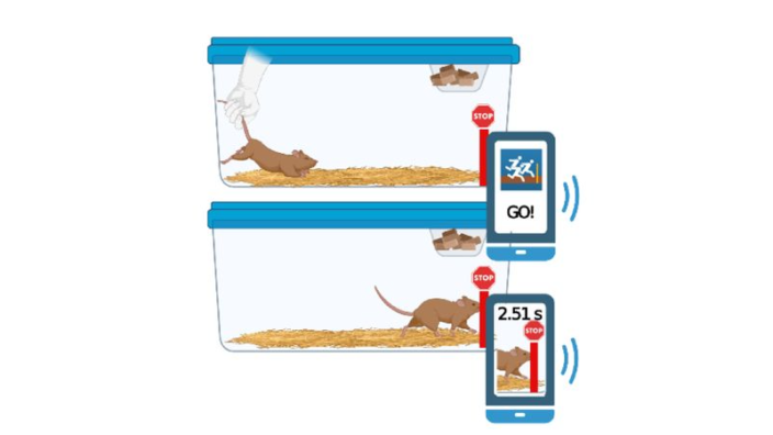
Let’s go back to your research. Do you think your results, particularly on the restoration of muscle mass, can be replicated in humans?

I like to think that the mouse is a miniature human, but I have colleagues who tell me the mouse is a terrible model. It depends on who you talk to. There are many success stories using mice for developing therapeutics, where what you find in the mouse does hold in human beings, but there are also many failures. It depends on the context and I’m not sure we can make a strong prediction one way or the other.

We like to think that the core processes which we’re studying in the mouse will hold over to humans, otherwise we wouldn’t be doing it. We’d be just sitting in our armchairs, talking about what we should do instead of actually doing stuff. I think the mouse is a good model for aging. No model is perfect, all models have downsides. Something I heard recently, “all models are wrong, it’s just a question of how wrong.” So, we hope to use the least wrong model in moving the field forward. You really want to hope that what you’re doing has some relevance to human biology, but ultimately, we don’t know until there is a successful translation of what you find at the mouse model level into a human therapy.

I’m hopeful, because of the dramatic success of speeding up of clinical trials with COVID, which would have been unheard of five years ago, that it is now possible to speed up the clinical trial process if there’s sufficient incentive. You can make the argument that that is the case with aging; there’s an incentive to speed it up just because of the whole ‘silver tsunami’ argument.

I think that’s already happening, but there’s a disconnect between the research, the epidemiology, and the political apparatus, which is weird, because the political apparatus is right in the middle of that tsunami. You would think that the basic biology of aging would be funded at least at the level of Alzheimer’s disease, but it is not. That’s because there’s an effective lobby group for Alzheimer’s disease, but there is not an effective lobby group for the functional decline associated with aging. I don’t understand why those dots are not being connected.

Such groups are beginning to emerge, for instance, an Alliance for Longevity Initiatives, a lobbying group here in the US. Were you personally involved in any conversations with politicians, decision makers about that?

I’m not involved with that at all, so I don’t really have an opinion on it beyond seeing a lack of funding. There has been a steady small increase in funding for the biology of aging, but when you think about it, basically all major medical problems stem from aging, except childhood infectious diseases.

There’s remarkably little funding, considering that basic fact, and it doesn’t make sense. Researchers in this area have bemoaned this situation for many years because to those of us who studied aging, it’s obvious where the results are going to come from in terms of overall improvement of human health – beyond things like basic nutrition, vitamins, and so on, which is a given, and even that’s not particularly well funded.

I know that in the States there’s still an appallingly high level of just basic poverty, which could be solved overnight if Congress got its act together. So, sometimes things are obvious, and it just doesn’t happen for whatever reason politically. I don’t know why aging falls into this category. For some reason, Alzheimer’s disease and cancer resonate with the politicians.

I’m not sure why it’s so hard for some people to understand that aging is a huge problem. I don’t know if it’s solvable, but we at least should try to solve it with all we have.

As someone who’s been in the field for quite a while, we’ve been talking about this for decades. It’s not new. We’ve had politicians at the Buck, we’ve talked to them about this, and there’s nodding of heads and an apparent understanding of the problem and then nothing ever happens. I’m not sure why, one of the reasons might be this inability to accept the fact that aging is a plastic biology. It’s malleable and we can muck around with it in the lab. It’s trivial for us to manipulate lifespan in simple organisms now. Still hard in mice, but in simple organisms, like drosophila and C. elegans, it’s really easy.

When I got started in the field in the early nineties, it was really hard. There were only a couple of labs in the world who were able to do it. My old boss Tom Johnson was one of the first to do it, discovering genes which extended the nematodes’ lifespan. That was unheard of at the time. Today, it’s something that a graduate student can do in a summer, it’s really trivial.

That’s different from what happened with the late politician Arlen Specter. He got cancer, I can’t recall which, and then he made it his mission to increase the budget of the NIH (National Institutes of Health) – as a function of him being ill – and he was dramatically successful in doing that.

We haven’t had the same sort of thing happening with Alzheimer’s; it’s been more of a sustained lobbying effort by multiple groups. As you said, there are groups trying to do this with aging, but it’s hard maybe in part because aging is so poorly defined. What does it mean to be old apart from the passage of years? There’s a lot of debate over it.

On the other hand, it’s something that every person is familiar with or will become familiar with.

But it’s also like accepting the weather. The weather just happens, and you don’t try to do anything about it. You might want to predict where it’s going (which in our case would be equivalent to, say, putting dollars into nursing homes), but you’re not going to try and fundamentally alter the weather. That is, unless it’s about climate change.

That’s an encouraging example. We had seen the same denial with climate change for decades, but then it changed. 

That’s true, but there are still large chunks of the population who don’t accept it. They just don’t believe science basically, that’s the bottom line.

Going back to your research again, what exactly is your target protein CRYAB? Like many other targets of senolytics, it seems to be protecting cells from apoptosis, right?

Yes, that’s correct. It’s a small heat shock protein. Interestingly enough, it has been reported previously to aggregate with age in human skeletal muscle. There is a report from more than a decade ago where they looked at the level of aggregated CRYAB represented by its insoluble form, which accumulates with age in human skeletal muscle quite dramatically.

We’re following up on that in multiple ways at the moment. We’re trying to understand if this target plays into the whole proteostasis argument around aging, which says that there’s a failure to maintain conformation with age and this somehow confers a benefit to senescent cells.

We’re investigating that at this point. We’re having a particular emphasis on skeletal muscle. It’s difficult to talk about apoptosis in skeletal muscle fibers, because they are long syncytial cells. This is a weird tissue to work on in many ways because there’s no single nucleus. They’ve got thousands of nuclei per fiber. They also have support cells and things like that, and maybe that’s where the action is. We are drilling into that at this point.

We know for certain that when you make cells senescent, multiple different cell types, everything from cardiac support cells through to liver cells, they will respond to the molecule (25HC), which we discovered, which somewhat bizarrely is also an anti-viral molecule discovered in the course of the COVID-19 pandemic. It kills senescent cells preferentially. We don’t really understand the interaction, but we’re working on the mechanism of that at the moment.

This brings me to the senolysis marker that was mentioned in your paper. How does it work; what exactly does it measure?

This marker was discovered by Judy Campisi and Chris Wiley early last year. They reported it in the context of a chemotherapeutic model, and we reported it now in the context of aging and treating aged animals with senolytics. It’s produced only in senescent cells, and when the senescent cell is lysed, this molecule is released into the plasma and the urine where it can be detected.

You get an increase in this marker when you’re killing senescent cells – increased signal is good, decreased signal is bad. Interestingly, we found an endogenous signal present in animals not treated with the senolytic, which might represent continuous turnover of senescent cells in aged animals. We were able to show that in old animals compared to young animals, there is a marked increase in the presence of this molecule. Then it goes up further when you treat them with a senolytic.

This marker is very useful because it’s really hard to enumerate senescence or senolysis in vivo. A lot of people are interested in that topic, and this is one way of doing it, which seems to work pretty well.

Since lifestyle changes are probably among the most potent anti-aging interventions that we have now…

The only anti-aging interventions we have now.

Do we have any indication that those lifestyle changes can help specifically with the accumulation of senescent cells or the rate of senolysis?

That’s a great question and one we’re actively interested in investigating. It’s likely going to be a major component in another initiative, which we have at the Buck in partnership with the Astera program.

This is this gigantic project to do single cell mapping on thousands of mice treated with different interventions that are reported to extend lifespan. I suspect some of these will not replicate when we test them. We will be looking at exercise as one of the modalities of improving function across multiple different domains of aging in mice in conjunction with senolytics like 25HC, and then we’re going to be able to answer that question.

It’s not something we have data on yet, but it’s certainly something there’s a lot of interest in, given the profound effects of exercise on slowing the functional decline associated with aging. You can’t stop aging, but exercise and a good diet appear to be the only way we have of slowing things down. You can actually make the philosophical argument that all we’re really doing is optimizing the system instead of fundamentally affecting aging.

It might be more appropriate to say that when you’re not exercising and you’re overeating, you’re just screwing the system up and you’re speeding things up. You’re making the system worse. So, when you exercise and you eat well, all you’re doing is putting the system back to what where it should be.

The question is whether many of the prospective anti-aging interventions work the same way, by simply optimizing the system?

We don’t know the answer to that yet. That’s something we are hoping to answer in the course of the Astera program over the next 5 to 10 years. It’s the largest project that Buck has ever launched. The whole thing is funded by Jed McCaleb, a crypto billionaire. I think it’s going to be tremendously useful to the community at large in the long run.

Do you have any plans or thoughts about human trials of 25HC? 

I have thoughts and plans, but I don’t know if they are realistic at this point. 25HC is being touted as a potential therapeutic for COVID. I don’t know whether that conversation is meaningful, but there’s been almost half a dozen papers published on 25HC and COVID in the last year alone. I think it might move forward in that route, not necessarily as a result of our work.

Still, there’s an intriguing possibility that the mechanism of action of endogenously produced 25HC is to kill senescent cells. In fact, it may be that the cytokine storm, which is very common in severely affected COVID patients, is linked to a high level of senescent cells. I haven’t seen anything on that, it’s pure speculation on my part, but if that’s true, then administration of 25HC might attenuate that storm by killing senescent cells.

Human trials are extremely hard to pull off. Do you think that the system might be broken, that things should be easier, less restrictive?

Yes, it’s just tremendously hard. It should be hard on one hand, I don’t want to say that it should be easy to do clinical trials: the possibility of bad things happening is high when you stick unknown substances into human beings. But there are things we can do.

For instance, I have a collaborator at Boulder (the University of Colorado), an investigator called Doug Seals. He’s really good at small-scale clinical trials, and he does them on an NIH budget, so it’s not true that you need billions of dollars to do a decent clinical trial. His trials are modest but well-designed; he’s assessing functional changes in the cardiovascular system, and he’s been very successful at running these little trials which involve just a couple of dozen people instead of hundreds.

I think it is possible to be creative within the constraints of the system without necessarily having to commit huge sums of money to large scale trials.

What are your thoughts on the situation in the aging field today? Are you more optimistic or more pessimistic? 

I’m optimistic, and it comes back to the point I made initially about technology. I do think the technology is here now to get fundamental insights at the level of a single cell, which we’ve lacked over the last three or four decades.

We can also make the readout at the level of the single cell for thousands, to hundreds of thousands, to maybe millions of cells. We’re going to have an instrument at the Buck fairly soon in conjunction with Astera which allows us to do spatial profiling at the level of the single cell.

It’s true single cell spatial profiling, and it yields enormous volumes of data because you get the positions of the transcripts within the cells, and you’re able to localize them to specific organelles, to quantitate them. It’s going to give tremendous insights into what’s happening at any given instant especially in conjunction with putative therapeutics.

Because we’re so good at measuring function in animal models (we can measure nearly any functional readout you care to name in human beings that we have a good proxy for in mice), we’ve got to be able to link function with therapeutics at the single cell level. That’s going to break down barriers like we’ve never seen before.

We would like to ask you a small favor. We are a non-profit foundation, and unlike some other organizations, we have no shareholders and no products to sell you. All our news and educational content is free for everyone to read, but it does mean that we rely on the help of people like you. Every contribution, no matter if it’s big or small, supports independent journalism and sustains our future.
Immune cell warrior

SENS Researchers Culture Immune Cells to Fight Senescence

Publishing in Aging, a team of researchers from SENS Research Foundation has described a new method of enriching natural killer (NK) cells to fight senescent cells.

Culturing NK cells to fight senescence

Previous experiments have shown that natural killer (NK) cells are partially responsible for the clearance of senescent cells from the human body [1]. While some senescent cells have ways of avoiding detection and clearance [2], NK cells are attracted to certain parts of the SASP, which trigger them to kill the cells expressing it. Techniques are being developed to use this senolytic ability of NK cells as a potential therapy.

The researchers lament two problems with the experiments used to develop some of these techniques: in many cases, the co-culturing lasted only 2 to 6 hours, and there was an extremely high ratio of NK (“effector”) cells to senescent (“target”) cells. Some of these experiments had 20 or more NK cells for every senescent cell [1]. The researchers hold that such experiments are not physiological: they do not match the conditions of the human body.

Enriching and using NK cells

After taking NK cells out of whole blood, the researchers sought to change the distribution of these cells. NK cells express different amounts of CD56 and CD16. NK cells that express high CD56 but low CD16 are immature and secrete interferon-γ; NK cells with low CD56 and high CD16 are responsible for cytotoxicity: the actual killing of other cells. The enrichment process, which involved activating the cells through the cytokine IL-2, substantially increased the percentages of both of these cell types.

These enriched cells were found to be very good at selectively eliminating senescent cells after 16 hours. In an experiment where there was only one NK cell for every senescent cell, 15% of normal fibroblasts and 43% of senescent fibroblasts died. These numbers remained largely the same regardless of how senescence was induced, and endothelial cells yielded similar results to fibroblasts.

Doubling or tripling the number of NK cells did kill more senescent cells; however, it also increased the number of normal cells being killed in the process. Therefore, instead of using more NK cells, the researchers increased the time in co-culture. This proved to be extremely helpful; while the number of normal fibroblasts dying remained low, only 10% of senescent cells survived after four days’ exposure to fresh, enriched NK cells. If the NK cells had been previously frozen (a common storage technique), 30% of senescent cells survived.

While the power of these cells, as measured by expression of cytotoxic factors, varied slightly by donor, all donor cells were significantly more cytotoxic towards senescent cells than normal cells.

Conclusion

The researchers describe the four-day experiment as follows:

a 4-day co-culture in which virtually all senescent cells were killed whereas viability of non-senescent cells with NK cell effectors was visually indistinguishable from negative control cells untouched by effector cells.

Even with this success, it’s easy to see potential ways in which this technique could be improved; it may be possible to adjust the protocol to increase specificity or attack different types of senescent cells. However, even in its current form, this technique could be explored in animal models to ascertain its potential therapeutic value.

Additionally, while this research is specific to this particular problem, the researchers are correct that lab conditions should match the conditions of the human body as closely as possible. Biology is an extremely complicated system, and if what cells experience in cell cultures (or animal models) do not match the experiences of cells in human beings, the results could be useless or misleading.

We would like to ask you a small favor. We are a non-profit foundation, and unlike some other organizations, we have no shareholders and no products to sell you. All our news and educational content is free for everyone to read, but it does mean that we rely on the help of people like you. Every contribution, no matter if it’s big or small, supports independent journalism and sustains our future.

Literature

[1] Sagiv, A., Biran, A., Yon, M., Simon, J., Lowe, S. W., & Krizhanovsky, V. (2013). Granule exocytosis mediates immune surveillance of senescent cells. Oncogene, 32(15), 1971-1977.

[2] Pereira, B. I., Devine, O. P., Vukmanovic-Stejic, M., Chambers, E. S., Subramanian, P., Patel, N., … & Akbar, A. N. (2019). Senescent cells evade immune clearance via HLA-E-mediated NK and CD8+ T cell inhibition. Nature communications, 10(1), 1-13.

Exercise Improves Cognition Depending on Type and Amount

In a new review paper, scientists show that exercise can alleviate age-related cognitive decline, but not all types of exercise are created equal [1].

Watch your head

Lifestyle choices, such as exercise and diet, are the most powerful anti-aging interventions currently available to us, and they might be the only effective ones. Exercise, in particular, has been proven to slow down various age-related changes [2].

One of these changes is cognitive decline. It spares virtually no one, and in some people, it grows into devastating dementias, such as the deadly Alzheimer’s disease. Geroscientists suspect that the reason not all old people become demented is that most die of other causes before age-related dementias can get to them. As life expectancy continues to rise, finding ways to maintain cognitive health becomes extremely important.

Numerous studies have tested the effect of exercise on age-related cognitive decline, mostly with positive results, but there are many confounding variables, such as types of exercise, sex, and BMI (body mass index). In this new review, which encompasses 44 studies with almost 5000 participants, the authors set out to determine  the minimum and optimal amounts of various types of exercise for influencing age-related cognitive decline.

Some exercise is better than nothing

The researchers begin with a brief overview of the current literature, noting that most of it still focuses on aerobic exercise, while other types of exercise have been shown to confer cognitive benefits as well – such as resistance training, dancing, and mind-body practices [3].

In their analysis, the scientists compare various types of exercise using their MET (metabolic equivalent), an established metric of energy expenditure. One MET-minute equals the amount of energy our body consumes during one minute of absolute rest. Brisk walking and vigorous weight training score at 5 MET, bicycling on flat terrain is equivalent to 9 MET, and running is one of the most energy-consuming activities at 11.5 MET. That means one minute of running uses up to 11.5 MET-mins worth of energy.

When measuring only MET equivalents and not type of exercise, the review shows that there is no minimum amount of exercise below which it does not promote cognition, however slightly. This sits well with the saying (officially endorsed the WHO) that some exercise is better than no exercise. However, according to the review, the difference is only perceptible at around 700 MET-minutes per week, which is on the smaller side of WHO recommendations (600-1200 MET-minutes per week).

Starting from 1200 MET-minutes per week, however, the effect becomes weaker, although it never plateaus, which led the researchers to conclude that beyond this point, the further benefits of exercise are unproven. This result aligns well with previous research showing that the benefits of exercise might peak at some point and even be reversed by too much exercise.

Exercise Recommendations

Obesity erases the effect

The differences between various types of exercise were quite significant. Resistance exercise shows a powerful effect, but it peaks at around 400 MET-minutes per week and then rapidly declines as the dose gets higher, producing an inverted U-shape on the chart. Walking and weightlifting had less pronounced effects on cognition than aerobic exercises. A mix of various aerobic exercises yielded good results, but a balanced combination of resistance training, weightlifting, and other exercise may be better. One previous meta-analysis indeed found that a mix of various types of exercise is most beneficial [4].

Finally, obesity seems to drastically change the picture. In people who are overweight, exercise improves cognition only slightly up to about 600 MET-minutes, and any further addition of MET-minutes diminishes the effect. Not only is obesity a major driver of aging, it can dampen the effect of exercise.

The researchers add the obvious observation, which can be taken as a warning, that it might be hard for aging people who had previously led sedentary lifestyles to drastically ramp up their exercise capacity to the recommended levels.

Conclusion

This review provides important insights into the effect that exercise can have on age-related cognitive decline, showing us that not all types of exercise are the same, and answering the question of how much exercise someone actually needs to start improving cognition in old age. It also reminds us that exercise is one of the few interventions available today that can put a dent in aging.

We would like to ask you a small favor. We are a non-profit foundation, and unlike some other organizations, we have no shareholders and no products to sell you. All our news and educational content is free for everyone to read, but it does mean that we rely on the help of people like you. Every contribution, no matter if it’s big or small, supports independent journalism and sustains our future.

Literature

[1] Gallardo-Gómez, D., del Pozo-Cruz, J., Noetel, M., Álvarez-Barbosa, F., Alfonso-Rosa, R. M., & del Pozo Cruz, B. (2022). Optimal Dose and Type of Exercise to Improve Cognitive Function in Older Adults: A Systematic Review and Bayesian Model-Based Network Meta-Analysis of RCTs. Ageing Research Reviews, 101591.

[2] Shephard, R. J., & Shek, P. N. (1995). Exercise, aging and immune function. International journal of sports medicine, 16(01), 1-6.

[3] Biazus-Sehn, L. F., Schuch, F. B., Firth, J., & de Souza Stigger, F. (2020). Effects of physical exercise on cognitive function of older adults with mild cognitive impairment: A systematic review and meta-analysis. Archives of Gerontology and Geriatrics, 89, 104048.

[4] Huang, X., Zhao, X., Li, B., Cai, Y., Zhang, S., Wan, Q., & Yu, F. (2021). Comparative efficacy of various exercise interventions on cognitive function in patients with mild cognitive impairment or dementia: a systematic review and network meta-analysis. Journal of Sport and Health Science.

Gut bacteria

Gut Viruses Found to Improve Cognition in Humans

A study published in Cell Host & Microbe has found that members of the Cuadovirales viral order positively affect cognition in multiple species, including humans.

Viruses in the human body, but not human viruses

Cuadovirales is an order of bacteriophages, which infect bacteria rather than animal cells. Previous research has noted that 80% of bacterial genomes contain viral DNA, which is linked to the diversity of gut bacteria, and therefore a healthy gut microbiome [1].

It has been previously discovered that viral abundance changes with age. Specifically, within the Cuadovirales order, Microviridae goes up and Siphoviridae goes down [2]. This change also occurs in mice fed a high-fat diet [3].

The taxonomy of these viruses is not fully eludicated, and, in fact, has been recently changed; because this change was done after the researchers finished their work, the researchers’ investigation into Siphoviridae now includes two other taxonomic familes as well; however, approximately 90% of the viruses in these three families are still Siphoridae. The researchers refer to members of these three families as “specific Cuadovirales“.

As the gut-brain axis is known to have effects on cognition, the researchers of this study sought to determine the extent of these viruses’ effects, and what they found was substantial.

Effects on bacterial metabolism

Viral infection, as expected, changes how the infected bacteria work. Surprisingly, the researchers found a strong negative association between specific Cuadovirales and multiple aspects of folate metabolism, including DNA repair mechanisms and the use of vitamins B2 and B6 in the folate cycle. However, the researchers also found that many of the bacterial pathways associated with folate metabolism were increased instead.

The researchers point to one specific bacterial gene upregulated by Siphoviridae: thyX, a gene that is known as TYMS in humans and is instrumental in neuroplasticity, neurological development, and memory retention [4]. Specific Cuadovirales also affected bacterial genes associated with multiple other aspects of central nervous system biochemistry, and they were shown to largely infect lactic acid-related bacteria, which are associated with dairy products [5].

Human cognition effects

Using multiple diagnostic tests and a careful examination of gut bacteria, the researchers investigated a discovery cohort of 114 people. Controlling for confounding factors such as age and BMI, the researchers found that specific Cuadovirales were associated with improved performance on the trail-making task B, a measurement of cognitive ability, in women, while Microviridae were associated with decreased performance in both sexes. On the other hand, specific Cuadovirales were associated with the backward digit span test, another measure of cognitive performance, in men but not women.

The researchers then examined a larger validation cohort of 942 people. The results in this cohort were even more pronounced in men; specific Cuadovirales were associated with improved short-term and long-term memory, executive function, and speed of information processing.

Animal results

The researchers also explored the effects of specific Cuadovirales in mice and fruit flies. Transplanting bacteria infected with these viruses into mice improved their scores on the novel object recognition test and increased gene expression associated with cognition; as expected, Microviridae were shown to negatively affect these genes.

In flies, the researchers chose to test specifically Lactococcal 936-type viruses, giving a control group whey without any viruses. Flies given live viruses were found to score better on learning and memory, which, in flies, is represented by an aversive taste test.

Conclusion

This study is extremely detailed with multiple layers of results, and it involves a great many individual viruses, bacteria, proteins, and genes. While some of the contradictory results are not fully elucidated, and the sex-based differences are not explained, this study offers very good insight into these topics, and it provides interesting potential for future development.

This study could be foundational for managing our fecal bacteria, even to the point of genetic manipulation, in order to keep our gut microbiomes functioning as they should to provide our brains with what they need.

We would like to ask you a small favor. We are a non-profit foundation, and unlike some other organizations, we have no shareholders and no products to sell you. All our news and educational content is free for everyone to read, but it does mean that we rely on the help of people like you. Every contribution, no matter if it’s big or small, supports independent journalism and sustains our future.

Literature

[1] Keen, E. C., & Dantas, G. (2018). Close encounters of three kinds: bacteriophages, commensal bacteria, and host immunity. Trends in microbiology, 26(11), 943-954.

[2] Gregory, A. C., Zablocki, O., Zayed, A. A., Howell, A., Bolduc, B., & Sullivan, M. B. (2020). The gut virome database reveals age-dependent patterns of virome diversity in the human gut. Cell host & microbe, 28(5), 724-740.

[3] Schulfer, A., Santiago-Rodriguez, T. M., Ly, M., Borin, J. M., Chopyk, J., Blaser, M. J., & Pride, D. T. (2020). Fecal viral community responses to high-fat diet in mice. Msphere, 5(1), e00833-19.

[4] Heyward, F. D., & Sweatt, J. D. (2015). DNA methylation in memory formation: emerging insights. The Neuroscientist, 21(5), 475-489.

[5] Mahony, J., & Van Sinderen, D. (2014). Current taxonomy of phages infecting lactic acid bacteria. Frontiers in microbiology, 5, 7.

Blood pressure

Carotid Bodies Play a Role in Glucose Regulation

An advance online preprint was recently released in Circulation Research examining carotid bodies’ role in hypertension and diabetes [1]. It will be published in the journal later this week.

Drugs that target the glucagon-like peptide 1 (GLP1) receptor are widely used to treat type 2 diabetes and have also been well-documented for their cardiovascular and antihypertensive benefits. However, the mechanism on how these medications achieve this is lacking.

The investigators of this study mention that despite established clinical protocols in Europe, less than 40% of patients with hypertension and diabetes achieve their treatment targets. Additionally, cardiovascular disease risk remains high for approximately 40% of patients whose treatment is controlled and within normal parameters [2]. Due to poor target achievement rates, the investigators set out to discover additional mechanisms at play that are impacting cardiovascular disease risk.

Background

The GLP1 receptor (GLP1R) stimulates insulin secretion and is found in the beta cells of the pancreas as well as in neurons in the brain.

The sympathetic nervous system (SNS) is responsible for the “fight or flight” response, which can be induced by stress. Carotid bodies (CBs) can be thought of as sensors that control peripheral reflexes. These reflexes are induced by chemical stimuli, which can be caused by environmental or bodily changes. These stimuli act by binding chemoreceptors on chemosensory cells, which can cause changes in the SNS.

The investigators of this study previously showed that hypertension in spontaneously hypertensive rats is dependent on carotid bodies promoting increased SNS activation [3,4]. These rats, which display diabetic traits, are used as a model for cardiometabolic disease, which describes a group of preventable diseases such as diabetes, insulin resistance, heart attack, stroke, and certain liver diseases.

GLP1R in CBs improves insulin signaling and blood glucose in rats

It was demonstrated in this study that the expression of the GLP1 receptor decreased expression of SNS activity in rats with cardiometabolic disease. When the researchers targeted CB with an agonist treatment for the GLP1 receptor, blood pressure was reduced. Additionally, when GLP1 receptors in CBs are activated, blood glucose is decreased. These results show that GLP1 receptors in CBs play a role in insulin signaling and blood glucose regulation.

The researchers then demonstrated that GLP1 receptors were localized in the CB chemosensory cells and a subset of blood vessels supplying the peripheral chemoreceptors.

GLP1 receptors are expressed in human CBs as well

Messenger RNA (mRNA) was isolated from CBs from five human cadavers. As in rats, gene expression of the GLP1 receptor was present in the glomus cells of these CBs. Additionally, with one human cadaver sample, they used an anti-GLP1 receptor antibody to confirm that GLP1 receptors are localized in the glomus cells of the CBs.

Additionally, as the authors provide in this figure, GLP1’s action on CBs is part of SNS regulation. Upon ingestion of food, the rise in blood glucose initiates CBs that cause the excitation of the SNS (sympathoexcitation), which stimulates the release of GLP1 from intestinal L-cells.

GLP1 Chart

Then, GLP1 mediates insulin secretion, which stimulates specific CB receptors. GLP1 can also inhibit chemosensory CB cells by counteracting sympathoexcitation, which tends to elevate levels of glucose and/or insulin, depending on physiological state. A GLP1 receptor agonist acts to reduce the sympathetic activity by suppressing sympathoexcitation activity from CBs.

Multiple receptors involved in energy metabolism

This robust study discussed multiple receptors involved in energy metabolism. Collectively, the results suggest that specific ligands that bind the GLP1 receptor suppress CB-driven SNS activation and that GLP1 may be released in the CBs to modulate its activity.

Conclusion

This novel study demonstrated a signaling circuit in which a GLP1 receptor agonist prevented SNS-induced elevated blood glucose. It does so by inhibiting specific chemoreceptors in response to the body’s physiological response to the increase in blood glucose.

These results suggest that CBs play a role in blood glucose regulation. Therefore, in individuals who have a hypertensive/diabetic condition, CBs could be a therapeutic target for decreasing excess SNS activity by using drugs that target GLP1 receptors.

Determining the mechanisms of action of these therapeutics opens the door for new therapeutics to be created. It will be exciting to see what other clinical research studies come of this work.

We would like to ask you a small favor. We are a non-profit foundation, and unlike some other organizations, we have no shareholders and no products to sell you. All our news and educational content is free for everyone to read, but it does mean that we rely on the help of people like you. Every contribution, no matter if it’s big or small, supports independent journalism and sustains our future.

Literature

[1] Pauza, A. G., Thakkar, P., Tasic, T., Felippe, I., Bishop, P., Greenwood, M. P., Rysevaite-Kyguoliene, K., Ast, J., Broichhagen, J., Hodson, D. J., Salgado, H. C., Pauza, D. H., Japundzic-Zigon, N., Paton, J., & Murphy, D. (2022). GLP1R Attenuates Sympathetic Response to High Glucose via Carotid Body Inhibition. Circulation research, CIRCRESAHA121319874. Advance online publication. https://doi.org/10.1161/CIRCRESAHA.121.319874

[2] Banegas, J. R., López-García, E., Dallongeville, J., Guallar, E., Halcox, J. P., Borghi, C., Massó-González, E. L., Jiménez, F. J., Perk, J., Steg, P. G., De Backer, G., & Rodríguez-Artalejo, F. (2011). Achievement of treatment goals for primary prevention of cardiovascular disease in clinical practice across Europe: the EURIKA study. European heart journal, 32(17), 2143–2152. https://doi.org/10.1093/eurheartj/ehr080

[3] McBryde, F. D., Abdala, A. P., Hendy, E. B., Pijacka, W., Marvar, P., Moraes, D. J., Sobotka, P. A., & Paton, J. F. (2013). The carotid body as a putative therapeutic target for the treatment of neurogenic hypertension. Nature communications, 4, 2395. https://doi.org/10.1038/ncomms3395

[4] Pijacka, W., Moraes, D. J., Ratcliffe, L. E., Nightingale, A. K., Hart, E. C., da Silva, M. P., Machado, B. H., McBryde, F. D., Abdala, A. P., Ford, A. P., & Paton, J. F. (2016). Purinergic receptors in the carotid body as a new drug target for controlling hypertension. Nature medicine, 22(10), 1151–1159. https://doi.org/10.1038/nm.4173

Chloroquine Increases Maximum Lifespan in Rats by 13%

Scientists have shown that chloroquine, a well-known anti-malarial drug and the cousin of hydroxychloroquine, attenuates inflammation and fibrosis while significantly extending median and maximum lifespan in naturally aged rats [1].

Fighting malaria since 1934

Chloroquine (CQ) was developed back in 1934 as an anti-malarial treatment and as such has enjoyed significant success. Since 2020, an analog of CQ – hydroxychloroquine – has attracted a lot of attention as a possible treatment for COVID-19, though its efficacy is heavily disputed [2], and the CDC currently does not recommend its use against the virus.

Over the years, more clinical uses for CQ have been found, including as an anti-inflammatory agent against rheumatoid arthritis. There were even some indications that CQ enhances the effects of chemotherapy and radiotherapy in cancer treatment [3]. More recently, CQ has been shown to reduce cellular senescence [4].

Can CQ extend lifespan?

Over the last few years, dozens of previously known compounds have been tested as life-prolonging interventions in animal models, and now CQ’s turn has arrived.

In this study, the scientists first conducted in vitro experiments to confirm the effect of CQ on cellular senescence. The researchers used human mesenchymal stem cells (hMSCs), multipotent cells present in many bodily tissues. The hMSCs were genetically engineered to recapitulate Werner’s syndrome, a type of progeria – in other words, they became a cellular model of accelerated aging.

Having treated the cells with various concentrations of CQ, the researchers found that in lower concentrations, CQ decreased markers of cellular senescence, but in higher doses, it actually increased them. This is consistent with CQ being known for rather severe side effects. Lower doses of CQ also increased markers of heterochromatin integrity, and heterochromatin destabilization is closely associated with aging and cellular senescence.

Next, the scientists treated naturally aged rats with CQ, sticking to a low and relatively safe dose of 0.1 mg/kg. 24-month-old male rats, approaching the end of their normal lifespan, were treated with CQ twice a week for five months. The treatment resulted in a 6% extension in median lifespan and a 13% extension in maximum lifespan, which is on par with some of the best anti-aging compounds ever tested in murine models.

CQ Chart

Various markers showed a significant decrease in systemic inflammation. Notably, the levels of tumor necrosis factor alpha (TNF-α) and white blood cell count fell to the levels observed in young healthy rats. CQ treatment also attenuated fibrosis in the kidney, liver, heart, small intestine, and lung.

Interestingly, after showing anti-senescence potential in hMSCs, CQ failed to lower the levels of ß-galactosidase, a popular senescence marker, in any of the tissues that were analyzed. On the bright side, although liver and cardiovascular damage are among the most well-documented side effects of CQ, none were detected in the experiments.

Good for the kidneys, not so for the heart

The researchers also conducted a transcriptomic analysis to determine how the CQ treatment changed the expression of genes that are either upregulated or downregulated with age. In all but one tissue, most changes reversed rather than exacerbated the aging phenotype, with the notable exception of the heart, where pro-aging changes were more pronounced. This is consistent with the adverse effect that CQ is known to have on vascular function and warrants caution, despite the lack of visible damage to the heart. Of all the organs, the kidney was the one whose age-related genes benefited the most from CQ.

Among the affected genes, those associated with inflammation and fibrosis were downregulated in most tissues, while those involved in protein homeostasis, glucose metabolism, and fatty acid metabolism were upregulated. Although the treatment did not lead to a noticeable decline in cellular senescence, it downregulated some genes involved in the production of the senescence associated secretory phenotype (SASP), the signature mix of mostly harmful molecules secreted by senescent cells. Moving to higher doses probably would not have helped, since in the in vitro experiments, higher doses were shown to be less effective against senescence.

Conclusion

With this study, CQ joins the growing list of molecules that prolong lifespan in animal models. The idea of repurposing existing drugs, which are usually relatively safe, cheap, and widely available, as anti-aging treatments, is not new, and it holds a lot of potential. Time will tell whether CQ can hold its own against anti-aging stars such as rapamycin and metformin that are already in human trials.

We would like to ask you a small favor. We are a non-profit foundation, and unlike some other organizations, we have no shareholders and no products to sell you. All our news and educational content is free for everyone to read, but it does mean that we rely on the help of people like you. Every contribution, no matter if it’s big or small, supports independent journalism and sustains our future.

Literature

[1] Li, W., Zou, Z., Cai, Y., Yang, K., Wang, S., Liu, Z., … & Zhang, W. (2022). Low-dose chloroquine treatment extends the lifespan of aged rats. Protein & Cell, 1-8.

[2] Chen, Y., Li, M. X., Lu, G. D., Shen, H. M., & Zhou, J. (2021). Hydroxychloroquine/chloroquine as therapeutics for COVID-19: truth under the mystery. International Journal of Biological Sciences, 17(6), 1538.

[3] Solomon, V. R., & Lee, H. (2009). Chloroquine and its analogs: a new promise of an old drug for effective and safe cancer therapies. European journal of pharmacology625(1-3), 220-233.

[4] Sargiacomo, C., Sotgia, F., & Lisanti, M. P. (2020). COVID-19 and chronological aging: senolytics and other anti-aging drugs for the treatment or prevention of corona virus infection?. Aging (Albany NY)12(8), 6511.

Rejuvenation Roundup February

Rejuvenation Roundup February 2022

February might be the shortest month, but it’s still full of exciting research on the path towards ending age-related diseases, including senolytics and new ways of looking at epigenetics. Let’s see what’s happened in these four weeks.

LEAF News

Life NogginLife Noggin Joins the lifespan.io Family: Today, we are delighted to officially announce that the popular pop-sci Youtube channel, Life Noggin, has joined the lifespan.io family. To celebrate the official launch, we are pleased to reveal a new episode of Life Noggin that focuses on some unexpected sources of life extension that we interact with every day.

EARD2021

Irina Conboy on Heterochronic Plasma at EARD2021: Irina Conboy discussed the effects of plasma dilution and heterochronic plasma exchange and their relationship to epigenetic alterations.

EARD2021 Greg FahyGrey Fahy on the TRIIM-X Trial at EARD2021: Greg Fahy discussed TRIIM-X, the ongoing continuation of his original TRIIM trial on reversing thymic involution, along with some surprising findings from it.

Lifespan News

WIRED and Rejuvenation: For this episode of Lifespan News, Ryan O’Shea focuses on Morgan Levine’s promotion of rejuvenation biotechnology on WIRED. WIRED, a media company historically associated with their coverage of the digital revolution, has released a video on the science of slowing down aging with Dr. Morgan Levine.

Resveratrol Controversy: On this episode of Lifespan News, Ryan O’Shea focuses on a recent controversy involving David Sinclair and his work regarding the well-known supplement resveratrol.

Rejuvenation Roundup Podcast

Ryan O’Shea of Future Grind hosts this month’s podcast, showcasing the events and research discussed here.

Journal Club

CRISPR activation of Yamanaka Factors in Human Cells: Journal Club returned on February 22nd at 12 PM Eastern time / 5 PM UK and was broadcast live to our Facebook page. This month, Dr. Oliver Medvedik took a look at a new method of partial cellular reprogramming using the gene editing tool CRISPR.

Interviews

Pankaj Kapahi InterviewDr. Pankaj Kapahi: “AGEs are a Trillion-Dollar Industry”: Dr. Pankaj Kapahi is a veteran geroscientist. His laboratory at the famous Buck Institute for Research on Aging is among the few that study advanced glycation end products (AGEs) and the many ways in which they affect aging. Dr. Kapahi’s company, Juvify, produces GLYLO, a supplement for detoxifying AGEs that also reduces food cravings.

Advocacy and Analysis

A Critique of Geroscience: Eric Le Bourg has published a thought-provoking paper in Biogerontology that offers a critique of geroscience. When people discuss the prospects of targeting the aging processes to prevent, halt, or even reverse age-related diseases, their beliefs often fall into two opposing categories: extreme optimism and extreme pessimism.

Research Roundup

Smart mouseSenolytic Navitoclax Rescues Neurogenesis and Memory in Mice: Scientists have shown that clearing senescent neural precursor cells with the senolytic drug navitoclax reverses the age-related decline in neurogenesis and improves spatial memory in mice.

Brain Cell Types Respond Differently to Aging Interventions: A new preprint on bioRxiv shows how researchers have developed transcriptomic, cell type-specific aging clocks from the regenerative zones of mouse brains. While chronological age is straightforward, it doesn’t necessarily capture the underlying biology.

Food DNAHigher Diet Quality Is Linked to Reduced Epigenetic Aging: A study published in the American Journal of Clinical Nutrition examined the relationship between diet quality and epigenetic aging clocks. This study used data from the Framingham Heart Study Offspring Cohort.

Naked Mole Rats Age, but Not Like Other Mammals: A research paper published in Nature by Dr. Vadim Gladyshev and his team has investigated the epigenetic aging of the naked mole rat, an animal whose mortality rate does not appear to increase with age.

ReprogrammingEffective New Method of Cellular Reprogramming Proposed: Scientists have proposed a novel method of cellular reprogramming that is more predictable and effective than traditional methods. Cellular reprogramming is a hot topic in geroscience that is being pursued by many academics and companies, including major players such as Google’s Calico along with Altos Labs.

Mapping the Gene Expression of Senescent Brain Cells: As explained in a paper published in GeroScience, researchers have used spatial transcriptomic analysis to show the inflammatory nodes that are caused by brain cells undergoing senescence.

Aging DNALongevity Allele Slows Immune and Cardiovascular Aging: In a new study, mice transduced with a longevity-associated variant (LAV) of the BPIFB4 gene showed less immunosenescence and healthier vasculature. Longevity-associated alleles prove that aging does not affect everyone equally. On average, centenarians and supercentenarians are more protected from age-related diseases than other people, and this protection is better explained by their genes than by their lifestyle choices.

Senolytics for Cardiac Regeneration in Diabetics: Research published in Diabetes has implicated senescent cardiac stem cells as the link between diabetes and cardiovascular disease. Aging is a major risk factor for diabetes, and individuals with diabetes exhibit several characteristics of aging.

MankaiA Polyphenol-Rich Diet Affects Age-Related Brain Atrophy: A preprint study originating from the DIRECT PLUS trial was recently released from the American Journal of Clinical Nutrition. This study has been accepted into the journal but has not yet been peer reviewed. It focused on a plant called Mankai, a strain of the Wolffia globosa duckweed.

Debunking Myths About Work Stress and Age Acceleration: A study published in Aging that studied the Northern Finland Birth Cohort of 1966 has found some interesting and counterintuitive results regarding work stress and accelerated epigenetic aging. Work stress is widely associated with premature death.

New BiotechNew Partial Reprogramming Drastically Lowers Cellular Age: In a pre-print paper that has not yet been peer reviewed, a group of researchers presented a new highly efficient method of partial cellular reprogramming. Our cells age, and old cells differ from young ones in multiple ways, including gene expression, telomere length, and protein milieu, but old cells turn back into young cells in the embryonic state.

Puerarin Shows Promise in Fighting COPD in Cells: Publishing in Aging, a team of researchers has discovered how puerarin, an extract of kudzu, ameliorates chronic obstructive pulmonary disorder (COPD) signs in cellular cultures by limiting mitophagy: the consumption of mitochondria by cells.

Hand arthritisNew Treatment for Osteoarthritis Proposed: In a paper published in Nature Aging, a group of scientists outlines a previously unknown pathway that leads to osteoarthritis and describes a promising treatment. There are plenty of deadly age-related diseases, but arthritis is one that shortens our healthspan rather than lifespan.

Muscular Oxidative Capacity Predicts Mobility Decline: Researchers publishing in Aging Cell have shown a relationship between mitochondrial dysfunction and mobility decline in older adults. Since 1958, a team of researchers from the National Institute on Aging Intramural Research program has been conducting the Baltimore Longitudinal Study of Aging (BLSA).

Rainbow ClocksA Fresh Examination of Epigenetic Clocks: In a preprint published in bioRxiv, Morgan Levine and colleagues have identified and grouped 5,717 epigenetic CpG sites into 12 different modules, conducting an in-depth examination into how epigenetic clocks work.

Caloric Restriction Promotes Rejuvenation in Human Trials: In a new paper, Yale scientists present encouraging data from an unprecedented human study of caloric restriction, a powerful anti-aging intervention. Despite all the might of today’s science, simple lifestyle choices are still the clearest option for living longer and healthier.

TelomeresA New Understanding of Telomere Attrition: In Nature Cell Biology, a team of researchers has presented a current review of telomeres and how they relate to aging, reflecting modern research into a decades-old topic.

NAD+ Levels Are Correlated with Physical Activity in Humans: In a study published in Nature Aging, a group of scientists has shown that NAD+ levels are correlated not only with age but with physical activity, with elder athletes rivaling normal young adults. Nicotinamide adenine dinucleotide (NAD) is as important as it is tiny.

Skeletal MuscleUrolithin A Affects Muscle and Mitochondria in Older Adults: A study published in the Journal of the American Medical Association has examined the dosage and safety of a urolithin A supplement. This study also examined urolithin A’s effect on muscle endurance and specific health biomarkers.

The Effects of Early Life Rapamycin Administration on Mice: A team of researchers, including Steve Horvath, Leonid Peshkin, and Vadim Gladyshev, has published a preprint on bioRxiv showing the effects of early administration of rapamycin over the lifespans of mice in a placebo-controlled experiment.

Rat LookResveratrol Ameliorates Erectile Dysfunction in Old Rats: Scientists have shown that resveratrol rescues erectile function in aged rats, most probably by activating SIRT1, and that it can work in synergy with the existing erectile dysfunction treatment tadalafil. Resveratrol, a plant-derived chemical of the polyphenol family, was once considered one of the most promising anti-aging molecules.

Combining Epigenetic and Imaging Biomarkers: Cardiologists, epidemiologists, and other researchers publishing in Aging have discovered that epigenetic aging and brain scans can be combined to accurately predict cognitive decline. The ongoing CARDIA study, from which this paper gets its data, was started in 1985 to monitor coronary artery risk development in young adults.

Optimal dose and type of exercise to improve cognitive function in older adults: There was a non-linear, dose-response association between overall exercise and cognition. The researchers found no minimal threshold for the beneficial effect of exercise on cognition.

Resistance exercise as a treatment for sarcopenia: prescription and delivery: The researchers state that two exercise sessions per week, with a combination of upper- and lower-body exercises performed with a relatively high degree of effort for 1-3 sets of 6-12 repetitions, is appropriate as a treatment for sarcopenia.

MIB-626, an Oral Formulation of NMN, Increases Circulating NAD in Middle-aged and Older Adults: MIB-626 1000-mg once daily or twice daily regimens were safe and associated with substantial dose-related increases in blood NAD levels and its metabolome.

Effect of 12-Week Intake of Nicotinamide Mononucleotide on Sleep Quality, Fatigue, and Physical Performance in Older Japanese Adults: Overall, NMN intake in the afternoon effectively improved lower limb function and reduced drowsiness in older adults. These findings suggest the potential of NMN in preventing loss of physical performance and improving fatigue in older adults.

Senolysis induced by 25-hydroxycholesterol targets CRYAB in multiple cell types: 25HC represents a potential class of senolytics, which may be useful in combating diseases or physiologies in which cellular senescence is a key driver.

Pharmacological Depletion of Microglia Leads to a Dose-Dependent Reduction in Inflammation and Senescence in the Aged Murine Brain: These results indicate that increased and detrimental brain inflammation in aged murine brain can be impaired by selectively reducing the microglial cell population with PLX5622.

Mycobacterium vaccae immunization in rats ameliorates features of age-associated microglia activation in the amygdala and hippocampus: Elevated neuroinflammatory priming, as is observed due to aging, is mediated in part by microglia, the primary immunocompetent cell in the CNS. Microglia in the amygdala and hippocampus appear most responsive to the anti-inflammatory effects of M. vaccae immunization, protecting against some age-associated microglia morphological changes.

Caudovirales bacteriophages are associated with improved executive function and memory in flies, mice, and humans: Supplementation of the Drosophila diet with the 936 group of lactococcal Siphoviridae bacteriophages resulted in increased memory scores and upregulation of memory-involved brain genes. Thus, bacteriophages warrant consideration as novel actors in the microbiome-brain axis.

Differences in Cognitive Functioning in Two Birth Cohorts Born 20 Years Apart: Data from the Interdisciplinary Longitudinal Study of Ageing: After correcting for age, a group born in the early 1950s significantly outperformed a group born in the early 1930s in all domains except concentration and verbal fluency.

Clinical Trials Targeting Aging: This review mentions caloric restriction, NAD+, senolytics, mTOR, and exercise. The future of clinical trials targeting aging may be phase 2 and 3 studies with larger populations if safety and tolerability of investigated medication continues not to be a hurdle for further investigations.

Cellular reprogramming and the rise of rejuvenation biotech: The researchers focus on an often-asked question: Can the ‘young’ science of rejuvenation, currently mostly based on in vitro studies, drive a new biotech field toward clinical applications?

The selection force weakens with age because ageing evolves and not vice versa: The researchers of this mathematical paper attempt to explain in evolutionary terms why aging exists.

Evidence that dog ownership protects against the onset of disability in an older community-dwelling Japanese population: Dog ownership appears to protect against incident disability among older Japanese adults. Additional benefits are gained from ownership combined with regular exercise. Daily dog care may have an important role to play in health promotion and successful aging.

Cannabis Use and Resting State Functional Connectivity in the Aging Brain: These findings suggest that future studies should examine both the potential risks of cannabinoids, as well as a potential benefits, on cognition and brain health for older adults.

We would like to ask you a small favor. We are a non-profit foundation, and unlike some other organizations, we have no shareholders and no products to sell you. All our news and educational content is free for everyone to read, but it does mean that we rely on the help of people like you. Every contribution, no matter if it’s big or small, supports independent journalism and sustains our future.
Brain Image

Combining Epigenetic and Imaging Biomarkers

Cardiologists, epidemiologists, and other researchers publishing in Aging have discovered that epigenetic aging and brain scans can be combined to accurately predict cognitive decline.

Two entirely different measurements

The ongoing CARDIA study was started in 1985 to monitor coronary artery risk development in young adults (who are no longer young). This large cohort has been repeatedly analyzed with multiple metrics, and this study took an interest in two measurements of epigenetic aging (taken in 2000 and 2005) along with two brain scans done in 2010 and 2015.

Fortunately, it is possible to use modern epigenetic analyses on older epigenetic data. While other clocks were also examined, the researchers focused most prominently on the well-known GrimAge, a predictor of all-cause mortality that has also been found to be associated with brain health in previous research [1].

The researchers also used an entirely different biomarker, Spatial Pattern of Atrophy for Recognition of Brain Age (SPARE-BA), which is a machine learning algorithm that uses imaging to detect brain deterioriation. As expected, this marker is associated with verbal fluency and attention problems [2].

When used to measure the acceleration of biological aging compared to chronological aging, these biomarkers are termed GrimAA and SPARE-BAA. Despite being associated with brain aging, these two biomarkers are not strongly correlated with each other, and this surprising finding led the researchers of this study to combine these two biomarkers into a unified analysis.

To measure cognition, the researchers chose the well-known Stroop test, which uses words in different colors than the words represent, the RAVLT long-delay recall test, and the Digit Symbol Substitution Test (DSST), which maps numbers to symbols.

Each marker is already powerful

Analyzing GrimAA findings compared to cognitive decline, the researchers found that the 2005 GrimAge results were strongly associated with the 2010 cognitive testing results and that the 2000 GrimAge results were, interestingly, even more strongly associated with the 2015 cognitive testing on both the Stroop and DSST cognitive biomarkers.

Similarly, the 2010 SPARE-BAA results were able to significantly predict 2015 measurements on Stroop and DSST, and cross-sectional analyses also significantly correlated SPARE-BAA to cognitive decline, particularly the 2015 results.

Combining the 2005 GrimAA results and the 2015 SPARE-BAA results yielded the best measurement that the researchers were able to find. Other combinations offered no benefit over GrimAge alone.

Conclusion

The researchers make a number of conclusions related to epigenetic aging and physical brain aging. Importantly, they point out that changes to brain synapses are governed by epigenetic alterations [3], which can explain a direct association between GrimAge and the cognitive results. Other potential correlations, such as inflammation, were also suspected.

While the CARDIA study used a large and wide-ranging cohort, the researchers note that epigenetic measurements were not taken at younger ages and that unmeasured cofactors might have biased the results, a common concern for longitudinal studies such as this one. Despite these potential issues, however, this study inspires confidence that it may be possible to develop combined biomarkers that examine both epigenetics and physical morphology in order to get a better picture of aging.

We would like to ask you a small favor. We are a non-profit foundation, and unlike some other organizations, we have no shareholders and no products to sell you. All our news and educational content is free for everyone to read, but it does mean that we rely on the help of people like you. Every contribution, no matter if it’s big or small, supports independent journalism and sustains our future.

Literature

[1] Hillary, R. F., Stevenson, A. J., Cox, S. R., McCartney, D. L., Harris, S. E., Seeboth, A., … & Marioni, R. E. (2021). An epigenetic predictor of death captures multi-modal measures of brain health. Molecular psychiatry, 26(8), 3806-3816.

[2] Eavani, H., Habes, M., Satterthwaite, T. D., An, Y., Hsieh, M. K., Honnorat, N., … & Davatzikos, C. (2018). Heterogeneity of structural and functional imaging patterns of advanced brain aging revealed via machine learning methods. Neurobiology of aging, 71, 41-50.

[3] Barter, J. D., & Foster, T. C. (2018). Aging in the brain: new roles of epigenetics in cognitive decline. The Neuroscientist, 24(5), 516-525.

Rat Look

Resveratrol Ameliorates Erectile Dysfunction in Old Rats

Scientists have shown that resveratrol rescues erectile function in aged rats, most probably by activating SIRT1, and that it can work in synergy with the existing erectile dysfunction treatment tadalafil [1].

A molecule of discord

Resveratrol, a plant-derived chemical of the polyphenol family, was once considered one of the most promising anti-aging molecules. David Sinclair’s lab at Harvard University showed that resveratrol works by activating SIRT1 [2], a protein thought to be protective against various diseases of aging [3].

Sinclair’s research eventually led to a huge 720-million-dollar deal with GlaxoSmithKline, but a few years later, GSK reluctantly shut the program down due to various problems. Currently, there is an open controversy about resveratrol, involving several prominent figures in the field, the gist of which you can get from this Lifespan News video.

Yet, resveratrol was not abandoned, and numerous studies have documented its various beneficial effects, some of which may be unrelated to SIRT1 [4]. This should not come as a surprise, since many other polyphenols, such as quercetin and curcumin, have shown strong anti-aging potential.

It would be safe to say that the question of whether resveratrol indeed activates SIRT1, and whether such activation is beneficial for lifespan and healthspan, is currently debated. In this new study, a group of Chinese scientists contributes to the discussion in an interesting way.

Rats have the same problem

The researchers decided to analyze the effect of SIRT1 activation by resveratrol on erectile dysfunction in aged rats (apparently, humans are not the only mammals suffering from age-related loss of erection). Sixty-eight 20-month-old rats were divided into four groups: a control group, a resveratrol group, a group that was given the SIRT1 inhibitor nicotinamide (NAM), and a group that was given a combination of resveratrol and tadalafil, a known treatment for erectile dysfunction.

In this study, 8-week-long resveratrol supplementation led to a significant amelioration of erectile dysfunction and coincided with an increase in SIRT1. In the NAM group, SIRT1 levels were lower, and erectile dysfunction was more severe than in the controls. Finally, the fourth group showed the best results, which led the researchers to believe that resveratrol and tadalafil work in unison against erectile dysfunction.

The researchers also analyzed histological changes in the rats’ penises. Resveratrol supplementation led to a significant increase in smooth muscle and endothelial tissue in the corpus cavernosum, which is indicative of better function. Yet again, NAM supplementation delivered the opposite results, while tadalafil reinforced the resveratrol-related gains.

Nitric oxide (NO) levels are negatively correlated with erectile dysfunction. In this study, resveratrol supplementation increased NO levels. This might be the mechanism behind the synergy between resveratrol and tadalafil, since drugs like tadalafil need a minimum amount of NO in order to work. One study has shown that SIRT1 promotes vascular relaxation (hence, erectile function) by activating the NO-producing enzyme nitric oxide synthase, which might also have contributed to the overall effect [5].

Erectile dysfunction has also been linked to oxidative stress. The researchers checked for two markers of oxidative stress: the antioxidant enzyme SOD, which eliminates free radicals, and MDA, which is indicative of tissue damage, and found that resveratrol supplementation had a positive effect on the levels of both (that is, it increased SOD levels and decreased MDA levels). In the context of oxidative stress, the advantage of the combination treatment was less pronounced than in other experiments but still significant. Overall, the benefits of resveratrol were shown to be statistically significant across all the experiments.

A safer ED drug?

This is not the first study of its kind. A previous one found that resveratrol reduces erectile dysfunction in rats that was induced by diabetes [6], another problem that these rodents seem to share with humans. According to the researchers, those previous findings, as well as their own, point at resveratrol as a potentially effective treatment for erectile dysfunction that is also safer than the current ones.

This study has some limitations. First, although the researchers did employ a SIRT1 inhibitor, it would be interesting to also see SIRT1 activation by other means, in order to be sure that resveratrol indeed works via this pathway. Second, a group treated with tadalafil alone could have provided a deeper insight into the supposed synergy between tadalafil and resveratrol.

Conclusion

This interesting study appears at a time when resveratrol’s beneficial effects and its ability to activate SIRT1 are under discussion. Although this cannot be considered an ironclad proof, and more research is needed, this study shows that in a rodent model, resveratrol does activate SIRT1, which leads to the amelioration of erectile dysfunction.

We would like to ask you a small favor. We are a non-profit foundation, and unlike some other organizations, we have no shareholders and no products to sell you. All our news and educational content is free for everyone to read, but it does mean that we rely on the help of people like you. Every contribution, no matter if it’s big or small, supports independent journalism and sustains our future.

Literature

[1] Yu, W., Wang, J., Dai, Y. T., Wang, B., Xu, Y., Gao, Q. Q., & Xu, Z. P. (2022). Modulation of SIRT1 expression improves erectile function in aged rats.

[2] Baur, J. A., Pearson, K. J., Price, N. L., Jamieson, H. A., Lerin, C., Kalra, A., … & Sinclair, D. A. (2006). Resveratrol improves health and survival of mice on a high-calorie diet. Nature444(7117), 337-342.

[3] Elibol, B., & Kilic, U. (2018). High levels of SIRT1 expression as a protective mechanism against disease-related conditions. Frontiers in endocrinology, 614.

[4] Pollack, R. M., Barzilai, N., Anghel, V., Kulkarni, A. S., Golden, A., O’Broin, P., … & Crandall, J. P. (2017). Resveratrol improves vascular function and mitochondrial number but not glucose metabolism in older adults. Journals of Gerontology Series A: Biomedical Sciences and Medical Sciences72(12), 1703-1709.

[5] Mattagajasingh, I., Kim, C. S., Naqvi, A., Yamamori, T., Hoffman, T. A., Jung, S. B., … & Irani, K. (2007). SIRT1 promotes endothelium-dependent vascular relaxation by activating endothelial nitric oxide synthase. Proceedings of the National Academy of Sciences104(37), 14855-14860.

[6] Fukuhara, S., Tsujimura, A., Okuda, H., Yamamoto, K., Takao, T., Miyagawa, Y., … & Okuyama, A. (2011). Vardenafil and resveratrol synergistically enhance the nitric oxide/cyclic guanosine monophosphate pathway in corpus cavernosal smooth muscle cells and its therapeutic potential for erectile dysfunction in the streptozotocin-induced diabetic rat: Preliminary findings. The journal of sexual medicine, 8(4), 1061-1071.

Running Mice

The Effects of Early Life Rapamycin Administration on Mice

A team of researchers, including Steve Horvath, Leonid Peshkin, and Vadim Gladyshev, has published a preprint on bioRxiv showing the effects of early administration of rapamycin over the lifespans of mice.

Rapamycin before and after weaning

In this placebo-controlled experiment, the researchers selected the UMHET3 strain of mice, as it is a crossbreed that does not suffer the negative effects of inbreeding. 130 newborn mice were selected for a lifespan study, and another 40 were selected for a healthspan study. The mothers of the newborns selected for treatment were administered rapamycin, which was shown to be transmitted to the newborns through their milk. This continued until the mice were weaned at three weeks, after which they were administered rapamycin until they were 45 days old.

In line with previous research [1], the pups administered rapamycin were substantially smaller than their untreated counterparts, even within a few days. Protein content and milk consumption were the same between the groups, showing that the difference was caused by rapamycin rather than a nutritional deficit. Rapamycin was also shown to delay the onset of sexual maturity, and it was shown to significantly reduce the size of the spleen, liver, heart, kidney, and brain.

Effects on murine lifespan

Early life rapamycin was shown to significantly increase lifespan only in male mice. As this research shows, male mice do not normally live as long as female mice, but rapamycin administration brought their lifespan nearly to that of females, who were only slightly affected by rapamycin. The reasons why the mice died were the same between both groups; rapamycin simply delayed the mortality of age-related diseases.

Gait disorders, body condition, distended abdomen, and a loss of forelimb grip strength, all of which are regularly chosen aging biomarkers for mice, were shown to be delayed with early life administration of rapamycin. Rapamycin significantly improved glucose and insulin tolerance only in males.

The effects on epigenetics largely matched the effects on lifespan. Male mice maintained a comparatively youthful transcriptome, but this did not hold true for females, particularly in the liver. Many of the effects brought about by early life rapamycin were attenuated with age in both sexes.

To confirm their findings, the researchers examined the effects of early life rapamycin on the commonly studied Daphnia species, receiving similar results to their murine experiment. Daphnia given a very small dose of rapamycin eventually reached the body size of the control group while living significantly longer.

Conclusion

The researchers offer several hypotheses and explanations for their findings. The somatic mutations that drive cancer later in life may be suppressed by early life rapamycin [2]. Other research has shown that rapamycin has lifelong benefits for the chromatin in the mouse intestine [3].

The researchers also note that they used only one dose of rapamycin in this study, and, given the Daphnia results, it may have been too large. There may be a rapamycin dose that has substantial effects on murine lifespan while having reduced effects on body weight.

However, it must be noted that this preprint study is simply a scientific curiosity that may lead to other research, not a proposed intervention. Using such a treatment on human newborns is obviously a very bad idea, not least because this research shows that rapamycin shrinks the organs, including the brains, of baby mice.

We would like to ask you a small favor. We are a non-profit foundation, and unlike some other organizations, we have no shareholders and no products to sell you. All our news and educational content is free for everyone to read, but it does mean that we rely on the help of people like you. Every contribution, no matter if it’s big or small, supports independent journalism and sustains our future.

Literature

[1] Siegmund, S. E., Yang, H., Sharma, R., Javors, M., Skinner, O., Mootha, V., … & Schon, E. A. (2017). Low-dose rapamycin extends lifespan in a mouse model of mtDNA depletion syndrome. Human molecular genetics, 26(23), 4588-4605.

[2] Williams, N., Lee, J., Mitchell, E., Moore, L., Baxter, E. J., Hewinson, J., … & Nangalia, J. (2022). Life histories of myeloproliferative neoplasms inferred from phylogenies. Nature, 1-7.

[3] Lu, Y. X., Regan, J. C., Eßer, J., Drews, L. F., Weinseis, T., Stinn, J., … & Partridge, L. (2021). A TORC1-histone axis regulates chromatin organisation and non-canonical induction of autophagy to ameliorate ageing. Elife, 10, e62233.

Skeletal Muscle

Urolithin A Affects Muscle and Mitochondria in Older Adults

A study published in the Journal of the American Medical Association has examined the dosage and safety of a urolithin A supplement [1]. This study also examined urolithin A‘s effect on muscle endurance and specific health biomarkers.

A randomized, controlled clinical trial 

This study is a follow-up to prior work done by the Amazentis company, with collaborators at the University of Washington Medical center and the Red Hutchinson Cancer Research center in Seattle [2]. This placebo-controlled, double-blinded study examined 50 women and 16 men ranging in age from 65 to 90. All of the participants identified as White, and all of them could perform daily activities without assistance.

Participants took four gel caps, totaling 1000 milligrams of urolithin A (Mitopure®), in the morning on an empty stomach with 1 cup of water between March 2018 to July 2020. 15 individuals in the placebo group and 16 in the urolithin A group reported adverse events, but none were medically serious.

Improvements in function

120 days into this study, the placebo and urolithin A groups walked the same distance on a flat and hard surface in six minutes; however, both groups had significantly improved in this regard during that time. At month two, the urolithin A group enjoyed significant improvements in hand and leg skeletal muscles compared to placebo, and this improvement in the urolithin A group was further increased at month four.

Biomarker improvements

The researchers investigated whether taking urolithin A improved mitochondrial capacity in aged skeletal muscle. They did this by measuring maximal ATP production via magnetic resonance spectroscopy (MRS) and by examining the recovery rate of phosphocreatine. From baseline to 4 months, there was no significant difference in the maximal ATP production of skeletal muscles in the hand and leg.

In the urolithin A group, higher levels of urolithin A and its conjugate, urolithin A glucuronide, were detected 4 months after supplementation began. At two and four months, the urolithin A group also had significant reductions in several acylcarnitines and ceramides, which, in this study, represented improved mitochondrial function and decreased inflammation, respectively. Inflammation was directly examined by measuring the ratio of plasma levels of C-reactive protein from the start of the study to 4 months. The urolithin A group had significant reductions in plasma C-reactive protein during this time, but the placebo group did not.

There were no other significant differences between the placebo and urolithin A groups regarding blood chemistry parameters, vital signs, urinalysis, and hematology.

Conclusion 

This study was limited in several ways. Daily physical activity was not monitored in this study, the participants were demographically homogenous, and the 6-minute walking distance did not differ between the two groups. The researchers suspect that this may be due to the study motivating some individuals to exercise. They suggest that future studies should have longer intervention times than 6 months, track daily physical activity, and select participants with reduced physical performance.

This study was funded by Amazentis, and the authors disclosed many conflicts of interest. Nevertheless, these results still provide pioneering insight on urolithin A supplement tolerability, and the results of this study suggest that it may be a promising approach to combat age-associated muscle decline. More research studies with larger sample sizes and a wider variety of participants are needed to confirm these results.

Disclaimer

This article is only a very brief summary. It is not intended as an exhaustive guide and is based on the interpretation of research data, which is speculative by nature. This article is not a substitute for consulting your physician about which supplements may or may not be right for you. We do not endorse supplement use or any product or supplement vendor, and all discussion here is for scientific interest.

We would like to ask you a small favor. We are a non-profit foundation, and unlike some other organizations, we have no shareholders and no products to sell you. All our news and educational content is free for everyone to read, but it does mean that we rely on the help of people like you. Every contribution, no matter if it’s big or small, supports independent journalism and sustains our future.

Literature

[1] Liu, S., D’Amico, D., Shankland, E., Bhayana, S., Garcia, J. M., Aebischer, P., Rinsch, C., Singh, A., & Marcinek, D. J. (2022). Effect of Urolithin A Supplementation on Muscle Endurance and Mitochondrial Health in Older Adults: A Randomized Clinical Trial. JAMA network open, 5(1), e2144279. https://doi.org/10.1001/jamanetworkopen.2021.44279

[2] Singh, Anurag, D’Amico, D., Andreux, P. A., Dunngalvin, G., Kern, T., Blanco-Bose, W., … Rinsch, C. (2021). Direct supplementation with Urolithin A overcomes limitations of dietary exposure and gut microbiome variability in healthy adults to achieve consistent levels across the population. European Journal of Clinical Nutrition. https://doi.org/10.1038/s41430-021-00950-1

Exercising man

NAD+ Levels Are Correlated with Physical Activity in Humans

In a study published in Nature Aging, a group of scientists has shown that NAD+ levels are correlated not only with age but with physical activity, with elder athletes rivaling normal young adults [1].

NAD: small, crucial, age-related

Nicotinamide adenine dinucleotide (NAD) is as important as it is tiny. As its name suggests, this molecule consists of just two nucleotides joined via their phosphate groups. Despite that, NAD performs many functions in the cell, the most important among them being carrying electrons around to facilitate redox (reduction/oxidation) reactions. Therefore, NAD exists in two forms: NAD+ acts as an oxidizing agent that takes electrons away from molecules, becoming NADH – the reducing agent that can donate electrons. With this “shuttle diplomacy”, NAD plays a crucial role in mitochondrial energy production.

NAD+ levels are known to decline with age, and this decline has been linked to multiple age-related diseases [2]. In animal models, NAD+ supplementation has been shown to provide health benefits, although the science is less clear regarding its effect on lifespan [3-5]. Yet, human data is hard to come by. To complicate things further, various tissues display different age-related NAD+ dynamics.

Aging incurs many metabolic changes to the body, with skeletal muscles taking a heavy toll. Age-induced loss of skeletal muscle mass and function, called sarcopenia in its most extreme form, limits a person’s mobility and ability to exercise, and it can cause frailty and falls. Despite mitochondrial dysfunction being linked to sarcopenia, the age-related dynamics of NAD+ in muscle have not been well-understood.

Regular exercise keeps your NAD+ up

This new important study analyzes what happens with NAD+ in our muscles as we age. The researchers recruited 52 people, dividing them into four groups: 12 young people (20-30), 17 old people (65-80) reporting normal levels of physical activity, 17 “trained” old people with above-average levels of physical activity, and 12 old people categorized as physically impaired with abnormally low physical activity levels.

“Trained” older adults reported having completed at least 3 one-hour exercise sessions a week for the last year, “normal” adults complete one session a week, and the physically impaired were people who scored less than 9 on the SPPB (Short Physical Performance Battery) test, a usual metric of fitness in such trials.

In addition to self-reporting, the researchers measured physical activity levels for five days after the recruitment. During that period, both the young adults and the “normal” older adults averaged around 10 thousand steps daily, while “trained” older adults were considerably more active, clocking more than 13 thousand daily steps on average.

The scientists then conducted a deep metabolomic analysis, recording the levels of more than a hundred various metabolites. They found that NAD+ was among the metabolites most downregulated with aging. Not only were NAD+ levels correlated with aging, they also depended on the amount of physical activity. While there was a considerable difference in NAD+ levels between the young adults and the normally active older adults, trained older adults were almost on par with the young group. The physically impaired older adults fared the worst, with the lowest levels of NAD+ recorded. The researchers also plotted NAD+ levels against physical activity in individual participants and found a strong correlation between the two.

Correlation or causation?

NAD+ was not the only metabolite correlated with physical activity. In fact, according to the researchers, their results imply that “most metabolic changes that occur with age in muscle can be reversed with regular exercise training.”

While NAD+ was negatively correlated with aging and positively correlated with physical activity, many metabolites exhibited an opposite behavior, including markers of oxidative stress. This led the researchers to believe that the decline in NAD+ occurs in parallel with increased reactive oxygen species (ROS) production.

Despite the results strongly suggesting that physical activity can rescue NAD+ levels, this is still not hard proof. As the authors note, the cross-sectional design of their study (that is, the participants were asked about their recent physical activity levels, and NAD+ was measured once) makes it difficult to establish a causal relationship. For this, multiple measurements across a period of time are required.

Conclusion

It has been known for a while that NAD+ levels decline with aging, and scientists have been searching for ways to replenish it. By showing the correlation between NAD+ levels and physical activity, this study suggests that exercise could be an effective way to boost your NAD+, at least in skeletal muscle. Still, more research is needed to establish a causal relationship between physical activity and NAD+ levels.

We would like to ask you a small favor. We are a non-profit foundation, and unlike some other organizations, we have no shareholders and no products to sell you. All our news and educational content is free for everyone to read, but it does mean that we rely on the help of people like you. Every contribution, no matter if it’s big or small, supports independent journalism and sustains our future.

Literature

[1] Janssens, G. E., Grevendonk, L., Perez, R. Z., Schomakers, B. V., de Vogel-van den Bosch, J., Geurts, J. M., … & Hoeks, J. (2022). Healthy aging and muscle function are positively associated with NAD+ abundance in humans. Nature Aging, 1-10.

[2] Garrido, Amanda, and Nabil Djouder. “NAD+ deficits in age-related diseases and cancer.” Trends in cancer 3, no. 8 (2017): 593-610.

[3] Mitchell, S. J., Bernier, M., Aon, M. A., Cortassa, S., Kim, E. Y., Fang, E. F., … & de Cabo, R. (2018). Nicotinamide improves aspects of healthspan, but not lifespan, in mice. Cell metabolism27(3), 667-676.

[4] Miao, Y., Cui, Z., Gao, Q., Rui, R., & Xiong, B. (2020). Nicotinamide mononucleotide supplementation reverses the declining quality of maternally aged oocytes. Cell reports32(5), 107987.

[5] Hou, Y., Wei, Y., Lautrup, S., Yang, B., Wang, Y., Cordonnier, S., … & Bohr, V. A. (2021). NAD+ supplementation reduces neuroinflammation and cell senescence in a transgenic mouse model of Alzheimer’s disease via cGAS–STING. Proceedings of the National Academy of Sciences118(37).