Exosomes From Stem Cells Fight Liver Disease in Mice
- Autophagy was increased while senescence declined.

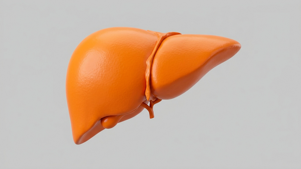
- Exosomes derived from human umbilical cord stem cells improve autophagy and decrease senescence in liver cells.
- This restores key biomarkers in older mice, restoring them to be more like those of young mice.
- This is due to the protein THBS1, which activates PPARα in the target cells.
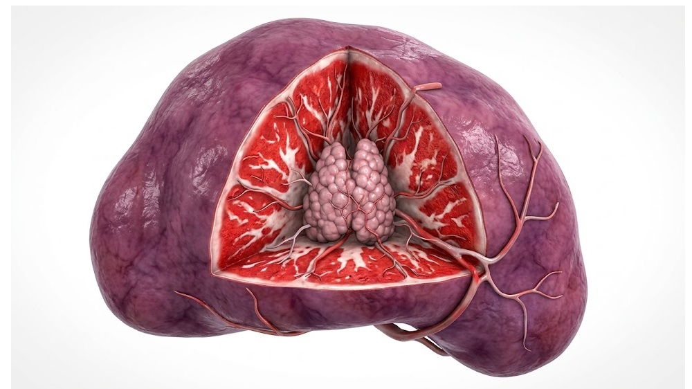
Researchers have described a method of using exosomes derived from mesenchymal stem cells (MSCs) to fight harmful metabolic changes in the liver.
Another look at exosomes
This is far from the first attempt at using MSC-derived exosomes, particularly exosomes derived from MSCs originally taken from human umbilical cord tissue (HucMDEs derived from HucMSCs), to treat an age-related disease. For example, we have previously reported on such exosomes being used to fight sarcopenia in mice, and the researchers note previous work suggesting that they can be used to fight liver disease [1].
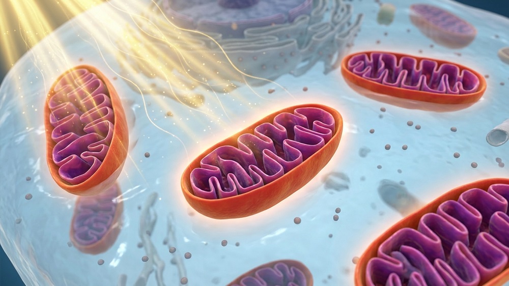
This research focuses specifically on autophagy, the maintenance process in which cells consume their own malfunctioning organelles. Autophagy is responsible for removing fatty droplets within liver (hepatic) cells [2], and a lack of autophagy leads to liver diseases such as non-alcoholic fatty liver disease (NAFLD) [3]. Previous research has found that increasing autophagy fights liver injury in a mouse model [4].
The authors began by ascertaining whether the human umbilical cord cells could be differentiated into bone cells (osteoblasts) and fat cells (adipocytes), then beginning to derive exosomes from them. These exosomes were labeled with a marker before being injected into the tail veins of mice, and then the livers were examined for the presence of this marker, confirming that the exosomes were successfully taken up into liver tissue.
Fighting unwanted liver fat
The next experiment involved three groups of animals: 8-week-old (young) mice, a control group of 18-month-old (old) mice, and an experimental group of old mice given HucMDEs. These mice were all male. While the control group of old mice was substantially heavier in body weight than the young mice, the treatment group was only slightly heavier. The control group had a substantial increase in blood glucose, while the treatment group was nearly indistinguishable from the young mice.
Four key markers of fatty liver disease, alanine aminotransferase (ALT), aspartate aminotransferase (AST), triglyceride (TG), and total cholesterol (TC), were tested. ALT had the strongest spike in the untreated old group, which was reduced to the levels of young mice. AST was similarly upregulated in the control group, and its levels were significantly leveled as well, although not quite to the levels seen in young mice. The TC results were similar to the ALT results, and the TG results were similar to the AST results.
“The above results demonstrate that aging induces liver dysfunction and dyslipidemia in mice, and that HucMDE administration can effectively improve liver function and normalize serum lipid levels in aging mice.”
Confirming this, the researchers also found substantial decreases in lipid deposition in the livers of the treatment group compared to the old group. SREBP1, a protein that encourages fat deposition, was increased with age but diminished with treatment, while PPARα, a protein that is involved in the destruction of such fat deposits, was substantially diminished with age and somewhat recovered with treatment. An in vitro analysis found that HucMDEs, but not exosomes derived from lung fibroblasts (HEDEs), had similar results in liver cells that had been driven senescent by palmitic acid (PA).
Effects against senescence
Similar results were found with two key senescence-related proteins, p16 and p21. In the old controls, both of these proteins were elevated in the liver; however, in the treatment group, the levels were similar to those of young mice. These results were likewise confirmed with in vitro testing, which found that HucMDEs but not HEDEs reduce a key senescence biomarker, SA-β-Gal, in PA-treated liver cells.
Autophagy was similarly increased by HucMDEs in these cells, with one key related protein, LC3, being increased by HucMDE treatment. Mouse experiments found similar results, with LC3 levels becoming more similar to those of young animals after treatment, although the reversal was not complete. Further experiments involving RNA silencing confirmed that HucMDEs reliably increase autophagic flux.
These effects were found to be due to THBS1, a protein that is highly expressed in HucMDEs but not HEDEs. Knocking down THBS1 in HucMSCs, and then deriving HucMDEs from them, created exosomes that were powerless to affect autophagy or senescence in liver cells. PPARα is responsible for THBS1’s effects within these cells; knocking down PPARα in the target cells similarly blunted the effects of useful HucMDEs.
The researchers note that there is no current drug therapy that is specific for NAFLD, so finding potential targets and therapies is a priority for the field. While these results are only, so far, in male mice, exosomes offer promise as a treatment method for this and other disorders involving aging and senescence.
Literature
[1] Lou, G., Chen, Z., Zheng, M., & Liu, Y. (2017). Mesenchymal stem cell-derived exosomes as a new therapeutic strategy for liver diseases. Experimental & molecular medicine, 49(6), e346-e346.
[2] Sakane, S., Hikita, H., Shirai, K., Myojin, Y., Sasaki, Y., Kudo, S., … & Takehara, T. (2021). White adipose tissue autophagy and adipose-liver crosstalk exacerbate nonalcoholic fatty liver disease in mice. Cellular and molecular gastroenterology and hepatology, 12(5), 1683-1699.
[3] Qian, H., Chao, X., Williams, J., Fulte, S., Li, T., Yang, L., & Ding, W. X. (2021). Autophagy in liver diseases: a review. Molecular aspects of medicine, 82, 100973.
[4] Li, X., Gong, S., Chen, W., Zhao, Y., Fu, K., Zheng, Y., & Chen, J. (2023). Schisandrol A, a bioactive constituent from Schisandrae Chinensis Fructus, alleviates drug-induced liver injury by autophagy activation via exosomes. Bioorganic Chemistry, 139, 106751.
About the author
Josh Conway