Physical Activity Delays Ovarian Aging in Mice
- This effect can be recreated by activating a hormone receptor.

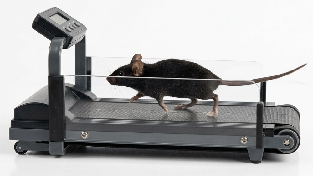
- In mice, ovarian aging is delayed by exercise.
- This effect is facilitated through adiponectin in the ovaries.
- Directly activating the adiponectin receptor yields similar benefits.
Results from a recent study suggest a delay in ovarian aging in mice following treadmill training; this effect was mediated by adiponectin. Pharmacologically activating adiponectin receptors extended reproductive span in mice. [1]
Beyond reproduction
The female reproductive system strongly influences the female aging trajectory, and its impact goes well beyond the cessation of reproduction. While ovarian aging is accompanied by a decline in the ability to produce egg cells capable of fertilization and a healthy pregnancy (ovarian reserve), it also spurs hormonal changes that affect the brain, bones, muscles, cardiovascular system, mental health, and more [2, 3].
Given that about half of the population undergoes ovarian aging, interventions are needed to slow it down. So far, the only available option, hormone replacement therapy, can be used to alleviate symptoms of menopausal transition, but it’s unable to prevent ovarian aging.
Exercise and ovarian reserve
One intervention known to slow aging in other organs, such as the brain or skeletal muscle, is physical activity [4]; however, whether physical activity affects ovarian aging is still debated, as studies so far have produced mixed results [5-8].
The authors of this study analyzed data from over 150,000 women across two studies and found that postmenopausal women have lower physical activity levels than premenopausal women. While these differences might stem from other factors, the authors suggested that ovarian aging might be implicated.
To test the impact of physical activity on ovarian aging, they used adult female mice that underwent 1 month of treadmill exercise training. Comparing this group with mice that didn’t exercise showed that exercise can help preserve ovarian reserve in adult mice and reduce primordial follicle loss. Primordial follicles contain an immature oocyte arrested in early development. Each female has a set number of follicles at birth, and, over time, those reserves are depleted because, in each menstrual cycle, several are recruited and one develops into a mature oocyte ready for fertilization.
Mice in the exercise group had more primordial follicles than controls. The exercise group also had higher AMH, an ovarian reserve marker, and increased levels of growth factors produced by oocytes.
The mediator of the effect
One molecule previously reported to increase with exercise is adiponectin [9], a hormone mainly produced by fat tissue that positively affects metabolism (e.g., insulin sensitivity) and reduces inflammation. It has also been reported to extend lifespan in mice [9] and slow muscle and brain aging [10, 11]. Adiponectin expression is also reduced in aging ovaries of model animals [12].
Those researchers hypothesize that adiponectin mediates exercise’s protective impact on ovarian health and delays ovarian aging. Their results supported this hypothesis and showed higher adiponectin levels in the exercise group than in the control group.
Adiponectin has previously been shown to regulate the mTOR signaling pathway [13]. Moreover, one mTOR pathway component was suggested as the primary activator of primordial follicle development [14]. In this study, the researchers observed lower levels of phosphorylated mTOR (p-mTOR) and phosphorylated rpS6 (p-rpS6) in the exercise group than in the control group, suggesting reduced mTOR signaling.
To investigate this further, they tested cultured newborn mouse ovaries exposed to various adiponectin levels. Higher adiponectin significantly reduced the proportion of growing follicles and p-mTOR levels compared with the control group. Decreased adiponectin levels resulted in more growing follicles and higher mTOR levels. These data suggest that “adiponectin regulates follicle activation through suppression of the mTOR signaling pathway.”
Further investigation found that adiponectin-deficient mice have fewer follicles than wild-type mice. After exercise, the number of follicles in wild-type and adiponectin-deficient mice was higher than in the non-exercised groups, but the protective effect of exercise was significantly less pronounced in adiponectin-deficient mice.
While adiponectin is mainly produced by adipose tissue, some adiponectin is also produced by ovaries. The protective role of exercise was significantly reduced against primordial follicle loss in mice with ovary-specific adiponectin knockout (76% reduction compared to wild-type). Additionally, the ovary-specific adiponectin-knockdown exercise group had reduced mTOR protein phosphorylation in the ovaries compared with controls, suggesting that ovarian adiponectin plays a significant role in how exercise delays ovarian aging.
An exercise substitute
After identifying the molecules mediating exercise’s effect on ovarian aging, the researchers looked for molecules that could affect those pathways. They identified AdipoRon, an adiponectin receptor agonist that binds to and activates the adiponectin receptor, as a good candidate to substitute for exercise to delay ovarian aging.
Adult mice received AdipoRon for one month. The treatment group had significantly more primordial follicles, more total follicles, higher AMH protein levels, and higher adiponectin receptor 1 protein levels within the ovaries than the control group, but adiponectin levels did not differ significantly between groups. Additionally, p-mTOR and p-rpS6 protein levels in the ovaries were lower in the AdipoRon-treated group. Similar results were obtained when mice were treated at their mid-reproductive age. These effects persisted for a few months, which is already an older age in mice.
The reproductive capacity of female mice also improved following AdipoRon treatment. While AdipoRon-treated females at mid-reproductive age had a similar number of offspring as control mice, as mice aged, AdipoRon-treated females had significantly more offspring than the untreated group. At an age when control animals had almost stopped producing offspring, most AdipoRon-treated mice were still producing offspring.
The authors conclude that both physical activity “and adiponectin may serve as promising targets for delaying ovarian aging, with substantial implications for both reproductive health and longevity.”
Literature
[1] Li, B., Zheng, N., Luo, T., Chen, Y., Mei, X., Huang, Q., Wang, W., Li, L., Han, L., Wang, W., Lin, B., Fan, Y., Liu, Y., Liu, Z., Ai, S., Liang, Y. Y., Wang, H., Tan, X., Zhang, M., Feng, H., … Zhang, J. (2026). Physical activity delays ovarian aging in part through adiponectin-related signaling pathways. Nature aging, 6(9), 1886–1900.
[2] Coughlan, G. T., Betthauser, T. J., Boyle, R., Koscik, R. L., Klinger, H. M., Chibnik, L. B., Jonaitis, E. M., Yau, W. W., Wenzel, A., Christian, B. T., Gleason, C. E., Saelzler, U. G., Properzi, M. J., Schultz, A. P., Hanseeuw, B. J., Manson, J. E., Rentz, D. M., Johnson, K. A., Sperling, R., Johnson, S. C., … Buckley, R. F. (2023). Association of Age at Menopause and Hormone Therapy Use With Tau and β-Amyloid Positron Emission Tomography. JAMA neurology, 80(5), 462–473.
[3] Mehta, J. M., & Manson, J. E. (2024). The menopausal transition period and cardiovascular risk. Nature reviews. Cardiology, 21(3), 203–211.
[4] Sun, S., Ma, S., Cai, Y., Wang, S., Ren, J., Yang, Y., Ping, J., Wang, X., Zhang, Y., Yan, H., Li, W., Esteban, C. R., Yu, Y., Liu, F., Izpisua Belmonte, J. C., Zhang, W., Qu, J., & Liu, G. H. (2023). A single-cell transcriptomic atlas of exercise-induced anti-inflammatory and geroprotective effects across the body. Innovation (Cambridge (Mass.)), 4(1), 100380.
[5] Zhao, M., Whitcomb, B. W., Purdue-Smithe, A. C., Manson, J. E., Hankinson, S. E., Rosner, B. A., & Bertone-Johnson, E. R. (2018). Physical activity is not related to risk of early menopause in a large prospective study. Human reproduction (Oxford, England), 33(10), 1960–1967.
[6] Kiranmayee, D., Praveena, T., Himabindu, Y., Sriharibabu, M., Kavya, K., & Mahalakshmi, M. (2017). The Effect of Moderate Physical Activity on Ovarian Reserve Markers in Reproductive Age Women Below and Above 30 Years. Journal of human reproductive sciences, 10(1), 44–48.
[7] Gudmundsdottir, S. L., Flanders, W. D., & Augestad, L. B. (2013). Physical activity and age at menopause: the Nord-Trøndelag population-based health study. Climacteric : the journal of the International Menopause Society, 16(1), 78–87.
[8] Dorjgochoo, T., Kallianpur, A., Gao, Y. T., Cai, H., Yang, G., Li, H., Zheng, W., & Shu, X. O. (2008). Dietary and lifestyle predictors of age at natural menopause and reproductive span in the Shanghai Women’s Health Study. Menopause (New York, N.Y.), 15(5), 924–933.
[9] Liu, L., Tang, J., Liang, X., Li, Y., Zhu, P., Zhou, M., Qin, L., Deng, Y., Li, J., Wang, Y., Jiang, L., Huang, D., Zhou, Y., Wang, S., Xiao, Q., Luo, Y., & Tang, Y. (2024). Running exercise alleviates hippocampal neuroinflammation and shifts the balance of microglial M1/M2 polarization through adiponectin/AdipoR1 pathway activation in mice exposed to chronic unpredictable stress. Molecular psychiatry, 29(7), 2031–2042.
[10] Selvais, C. M., Davis-López de Carrizosa, M. A., Nachit, M., Versele, R., Dubuisson, N., Noel, L., Gillard, J., Leclercq, I. A., Brichard, S. M., & Abou-Samra, M. (2023). AdipoRon enhances healthspan in middle-aged obese mice: striking alleviation of myosteatosis and muscle degenerative markers. Journal of cachexia, sarcopenia and muscle, 14(1), 464–478.
[11] He, K., Nie, L., Ali, T., Liu, Z., Li, W., Gao, R., Zhang, Z., Liu, J., Dai, Z., Xie, Y., Zhang, Z., Liu, G., Dong, M., Yu, Z. J., Li, S., & Yang, X. (2023). Adiponectin deficiency accelerates brain aging via mitochondria-associated neuroinflammation. Immunity & ageing : I & A, 20(1), 15.
[12] Wu, M., Huang, Y., Zhu, Q., Zhu, X., Xue, L., Xiong, J., Chen, Y., Wu, C., Guo, Y., Li, Y., Wu, M., & Wang, S. (2022). Adipose tissue and ovarian aging: Potential mechanism and protective strategies. Ageing research reviews, 80, 101683.
[13] Park, J. S., Choe, K., Lee, H. J., Park, T. J., & Kim, M. O. (2023). Neuroprotective effects of osmotin in Parkinson’s disease-associated pathology via the AdipoR1/MAPK/AMPK/mTOR signaling pathways. Journal of biomedical science, 30(1), 66.
[14] Zhang, H., Risal, S., Gorre, N., Busayavalasa, K., Li, X., Shen, Y., Bosbach, B., Brännström, M., & Liu, K. (2014). Somatic cells initiate primordial follicle activation and govern the development of dormant oocytes in mice. Current biology : CB, 24(21), 2501–2508.
About the author

Anna Drangowska-Way