
A recent study showed that a treatment with the short peptide elamipretide reversed multiple signs of age-related decline in oocyte quality in mice and in human oocytes in vitro [1].
An early decline
In females, the reproductive system ages relatively early in life, with fertility declining as early as the mid-thirties; combined with the recent trend to delay motherhood, this often makes it difficult for women to have healthy pregnancies when they choose to become mothers. While researchers are investigating multiple compounds for their ability to delay or stop ovarian aging, no effective treatment is currently available on the market.
In a recent study published in Aging Cell, the researchers investigated a four-amino-acid peptide called elamipretide for its ovarian anti-aging properties. Elamipretide was previously described as selectively targeting and accumulating in the inner mitochondrial membrane, where it helps improve mitochondrial function and reduce oxidative stress damage in different contexts [2]. Since these outcomes are linked to decreased ovarian aging, testing elamipretide in the context of female fertility was a logical next step.
Molecular players
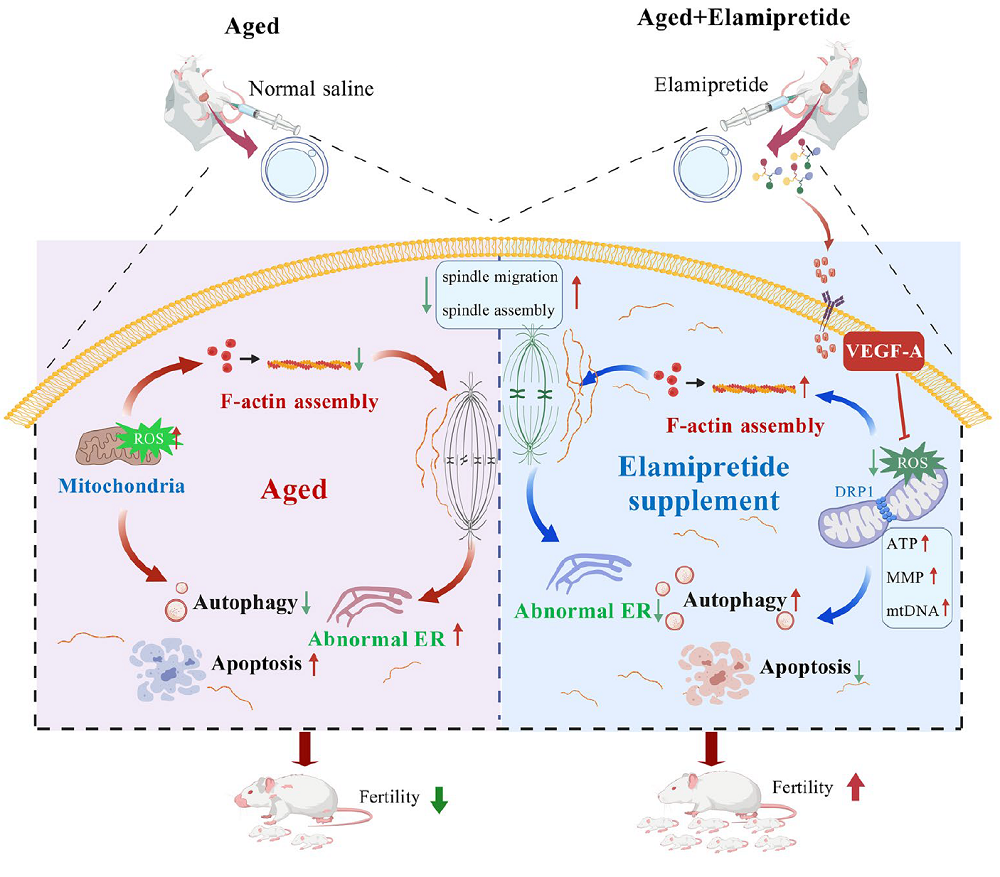
The researchers began by identifying optimal doses and treatment regimens in aged mice. A 7-day elamipretide treatment regimen was optimal for rejuvenating the aged mouse ovary, improving fertilization rates in aged oocytes, and increasing litter size in aged mice for 1 month after treatment.
A molecular analysis found that 250 metabolites accumulated in different amounts in aged mouse ovaries compared with young ones. Elamipretide treatment partially reversed those changes.
One affected molecule was pyruvate, a compound related to mitochondrial energy metabolism. The researchers described an aging-related decrease in pyruvate levels and suggested that declining mitochondrial energy metabolism contributes to declining female fertility.
Elamipretide significantly affected Vitamin B6 metabolism in aged mice, and Vitamin B6 supplementation improved oocyte quality in these animals. This suggests that “elamipretide may enhance the maturational quality of aged oocytes by modulating Vitamin B6 metabolism.”
Other studies suggested a link between Vitamin B6 and pyruvate; for example, Vitamin B6 deficiency was shown to lead to pyruvate accumulation along with other compounds, which, in turn, impairs mitochondrial oxidative metabolism [3].
Gene expression analysis in oocytes confirmed some metabolic findings and identified the vascular endothelial growth factor (VEGF) signaling pathway as an important player in modulating the maturation quality of aged oocytes, since this pathway was significantly affected in oocytes from aged mice and elamipretide treatment reversed those changes. Inhibiting VEGF-A improved oocyte quality, suggesting that elamipretide acts through VEGF-A to restore oocyte quality in aged oocytes.
Maturing oocytes
A key factor in ovarian aging is the age-related decline in oocyte quality, which is largely determined by the complex process of oocyte maturation. This consists of two parts: nuclear maturation and cytoplasmic maturation [4]. The most important part of nuclear maturation is the separation of chromosomes, strings of DNA and proteins, which, when done properly, allows the cells to divide. Problems with proper separation of chromosomes during egg production can lead to such diseases as Down Syndrome in the resulting children.

“Cytoplasmic maturation of the oocytes involves the accumulation of mRNAs, proteins, and substrates required for subsequent fertilization/development.” Age-associated cytoplasmic maturation problems are linked to mitochondrial dysfunction, which can lead to oxidative damage and other issues. Mitochondrial dysfunction has been linked to reduced oocyte quality in both humans and mice [5-7]. Because mitochondria are the cell’s powerhouses, they provide essential energy for oocyte maturation, fertilization, and embryonic development.
These researchers analyzed several processes linked to oocyte maturation and the impact of elamipretide on those processes. They observed that elamipretide treatment helped to rejuvenate cytoskeleton-related biological processes in aged oocytes. For example, investigations into spindle assembly (the formation of a structure that enables proper chromosome segregation during cell division and egg production), chromosome alignment, and the stability and assembly of other cellular structures showed that these processes were dysregulated in aged oocytes, and elamipretide ameliorated these changes.
Mitochondrial rearrangement plays an essential role in cytoplasmic maturation, and these researchers identified key mitochondria-related genes whose levels changed in aged oocytes. Elamipretide treatment reversed those age-associated changes in gene expression. Similarly, elamipretide restored the aging-related displacement of mitochondria in oocytes and restored (in full or in part) other physiological processes related to mitochondrial metabolism. This included the partial reversal of age-related overexpression of genes related to oxidative stress, which led to reduced levels of reactive oxygen species (ROS).
Another important component of cytoplasmic maturation of oocytes is remodeling and functional maturation of the cell’s organelles. Elamipretide helped to reverse age-related perturbations in processes involving the endoplasmic reticulum, Golgi apparatus, and lysosomes.
Beyond mice
The researchers examined oocytes from human donors. Supplementing in vitro cultured oocytes from women aged 35 and up with elamipretide improved oocyte maturation, suggesting its potential to improve developmental competence in humans.
They also analyzed oocytes derived from pigs. They induced oxidative stress in those oocytes, which had a detrimental effect on oocyte maturation, while elamipretide supplementation alleviated the oxidative stress-induced cellular changes and restored the maturation rate.
These positive results, combined with properties that make elamipretide easily applicable in the clinic, such as “small molecular size, easy synthesis, good water solubility, not easily degraded by peptidase, and high stability in solution,” make it a good candidate for future research into its ability to extend the fertility window in women.
Literature
[1] Zhang, H. L., Wang, Y., Wang, C., Guo, X., Chen, H., Hou, Y. X., Wu, X., Wu, Z. J., Pan, W. L., Ma, R. J., Lu, P. S., Shu, J., & Sun, S. C. (2026). Elamipretide Rejuvenates Oocyte Quality and Restores Female Fertility During Reproductive Aging. Aging cell, 25(10), e70749.
[2] Sabbah, H. N., Gupta, R. C., Kohli, S., Wang, M., Hachem, S., & Zhang, K. (2016). Chronic Therapy With Elamipretide (MTP-131), a Novel Mitochondria-Targeting Peptide, Improves Left Ventricular and Mitochondrial Function in Dogs With Advanced Heart Failure. Circulation. Heart failure, 9(2), e002206.
[3] Ciapaite, J., van Roermund, C. W. T., Bosma, M., Gerrits, J., Houten, S. M., IJlst, L., Waterham, H. R., van Karnebeek, C. D. M., Wanders, R. J. A., Zwartkruis, F. J. T., Jans, J. J., & Verhoeven-Duif, N. M. (2023). Maintenance of cellular vitamin B6 levels and mitochondrial oxidative function depend on pyridoxal 5′-phosphate homeostasis protein. The Journal of biological chemistry, 299(9), 105047.
[4] Jiang, Y., He, Y., Pan, X., Wang, P., Yuan, X., & Ma, B. (2023). Advances in Oocyte Maturation In Vivo and In Vitro in Mammals. International journal of molecular sciences, 24(10), 9059.
[5] Sun, Q. Y., & Nagai, T. (2003). Molecular mechanisms underlying pig oocyte maturation and fertilization. The Journal of reproduction and development, 49(5), 347–359.
[6] Watson A. J. (2007). Oocyte cytoplasmic maturation: a key mediator of oocyte and embryo developmental competence. Journal of animal science, 85(13 Suppl), E1–E3.
[7] Kujoth, G. C., Hiona, A., Pugh, T. D., Someya, S., Panzer, K., Wohlgemuth, S. E., Hofer, T., Seo, A. Y., Sullivan, R., Jobling, W. A., Morrow, J. D., Van Remmen, H., Sedivy, J. M., Yamasoba, T., Tanokura, M., Weindruch, R., Leeuwenburgh, C., & Prolla, T. A. (2005). Mitochondrial DNA mutations, oxidative stress, and apoptosis in mammalian aging. Science (New York, N.Y.), 309(5733), 481–484.