
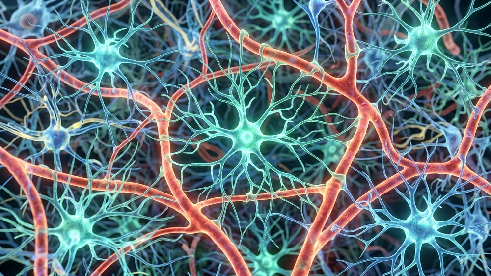
In Aging Cell, researchers have described how increasing fibroblast growth factor 13 (FGF13) in astrocytes reduces aging throughout the bodies of mice.
A factor from a well-known family
FGFs are signaling molecules that share structural and functional commonalities and play crucial roles in biology, including neural function [1]. Some of these molecules are secreted, and others are not; one of the secreted molecules, FGF21, has been thoroughly researched as a regulator of aging processes.
However, the subject of this study, FGF13, is a non-secreted molecule with functions that are not related to the FGF receptor [2]. Previous work has found that it has beneficial effects in a mouse model of Parkinson’s [3], while its expression in endothelial cells is detrimental in a mouse model of diabetes [4]. Unlike FGF21, its role in age-related diseases has not been thoroughly explored.
Brain inflammation may spread to the body
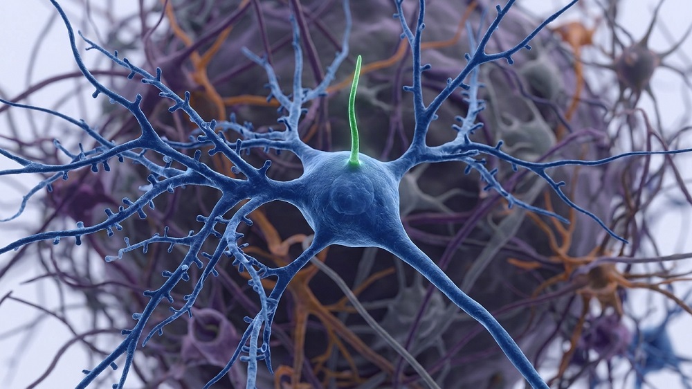
The researchers first examined public mouse datasets and confirmed that FGF13 indeed decreases in the brains of mice; while it does not decrease in neurons, it decreases in other cells, including oligodendrocytes, astrocytes, and microglia. Interestingly, while FGF13 smoothly declined with age in the hippocampus, it increased in the cortex at 6 months of age before decreasing. While macaque data was less comprehensive, it was similar to that of mice, with a particularly strong effect in the astrocytes. Therefore, the researchers chose those cells for further experimentation.
Knocking out FGF13 in the astrocytes of aging mice exacerbated their aging. The affected mice performed even more poorly on the Morris water maze test than the aged control group did. Additionally, the affected mice had more cellular senescence in the heart, skin, liver, spleen, and kidney, even though the knockout was targeted to astrocytes, and they also had higher levels of circulating inflammatory cytokines. This led the researchers to conclude that these mice’s brains were “amplifying and transmitting aging signals to other tissues via the blood to accelerate systemic aging.”
The researchers then performed the opposite experiment, using a similar technique to increase FGF13 in the astrocytes of older mice instead. In the Morris water maze test, the treated mice performed similarly to young mice, having a substantial advantage over the untreated mice. In related spatial reasoning tests, the elevated plus maze test and the open field test, the treated mice also performed significantly better than the untreated mice, although not quite to the level of young mice.
Similar benefits were found in other tissues as well, despite this still being an astrocyte-specific intervention. Inflammatory cytokines in the bloodstream were substantially decreased, particularly IFN-γ and IL-6. Most other benefits were significant but modest, with decreases in skin, liver, spleen, and kidney senescence (although not in the heart). FGF13 overexpression in astrocytes also decreased age-related weight gain in mice.
The researchers then performed cellular experiments. They found that complement protein C3, which marks the presence of neurodegenerative diseases, is significantly increased in FGF13-deficient astrocytes. Cx43, which marks age-related neuronal injury, is increased with FGF13 deficiency. Co-culturing these deficient astrocytes with neurons found that they impaired the neurons’ synapses, and co-culturing them with microglia revealed that they induced the microglia to release inflammatory factors. FGF13 overexpression in neurons revealed benefits in all of these areas, restoring neuronal synaptic proteins, reducing microglial expression of cytokines, and reducing levels of proteins associated with injury and degeneration.
Autophagy may be the crux
RNA sequencing revealed some of the mechanisms of action behind these effects. 625 genes were upregulated and 195 were downregulated in astrocytes with the overexpression of FGF13. These genes affected multiple pathways, including lysosomal transport, inflammatory factor production, metabolism, and various signals.
FGF13 was specifically found to have a physical interaction with the protein Phafin2, a protein that is related to the cellular maintenance process of autophagy [5]. Like FGF13, Phafin2 decreases with age in mice, while increasing FGF13 also increases Phafin2. An analysis of the mice’s brain tissue found that the FGF13-overexpressing animals had increases in autophagy, as evidenced by increases in autophagosomes and autolysosomes along with the autophagy markers P62 and LC3. Similar results were found in cellular experiments.

These researchers have provided evidence that an upstream regulator in one particular brain cell has downstream ramifications throughout the body, suggesting that a treatment to increase FGF13 in the astrocytes of older people may improve multiple age-related conditions. However, this paper does not suggest any potential methods of doing so, and further work will need to be done to confirm these results and determine whether this approach is valid in non-human primates and then in people.
Literature
[1] Bollenbecker, S., Barnes, J. W., & Krick, S. (2023). Fibroblast growth factor signaling in development and disease. International Journal of Molecular Sciences, 24(11), 9734.
[2] Naderi, J., Johnson, A. K., Thakkar, H., Chandravanshi, B., Ksiazek, A., Anand, A., … & Chaurasia, B. (2025). Ceramide-induced FGF13 impairs systemic metabolic health. Cell metabolism, 37(5), 1206-1222.
[3] Song, N., Wang, X., Ha, L., Hu, L., Mei, S., Liang, Y., … & Lu, M. (2025). Neuronal FGF13 Inhibits Mitochondria‐Derived Damage Signals to Prevent Neuroinflammation and Neurodegeneration in a Mouse Model of Parkinson’s Disease. Advanced science, 12(28), 2503579.
[4] Sun, J., Guan, X., Niu, C., Chen, P., Li, Y., Wang, X., … & Cong, W. (2023). FGF13-Sensitive alteration of parkin safeguards mitochondrial homeostasis in endothelium of diabetic nephropathy. Diabetes, 72(1), 97-111.
[5] Tang, T. X., Jo, A., Deng, J., Ellena, J. F., Lazar, I. M., Davis, R. M., & Capelluto, D. G. (2017). Structural, thermodynamic, and phosphatidylinositol 3‐phosphate binding properties of Phafin2. Protein Science, 26(4), 814-823.