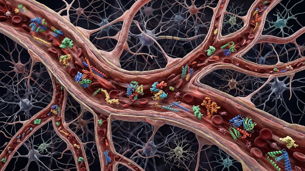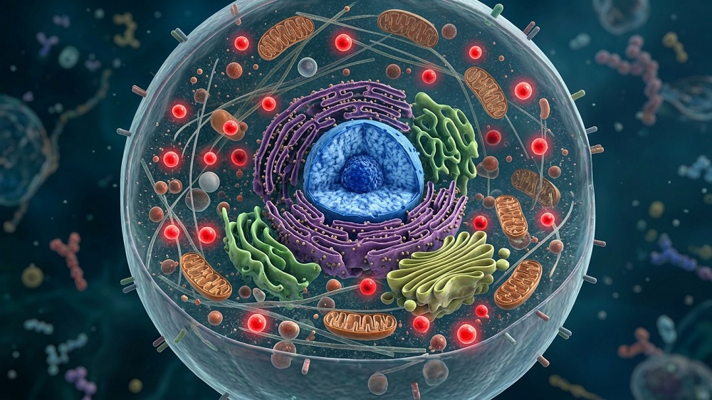


Reviewers publishing in Biology have elucidated SIRT1 functional reserve as an aspect of aging, describing how its depletion is an upstream cause of age-related disorders at the cellular level and explaining why many prospective therapies that increase SIRT1 and NAD+ have not had promising results.
Managing a dangerous byproduct
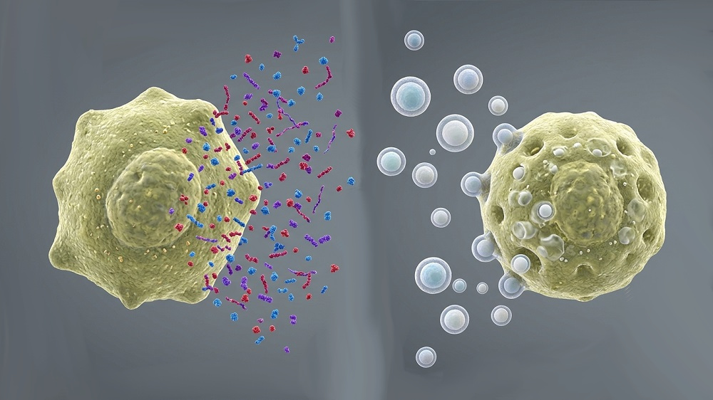
Reactive oxygen species (ROS) are necessarily produced as part of basic cellular biology, and under normal conditions, they serve signaling functions [1]. With aging, however, ROS handling becomes dysregulated, leading to their accumulation and thus to age-related cellular dysfunction [2].
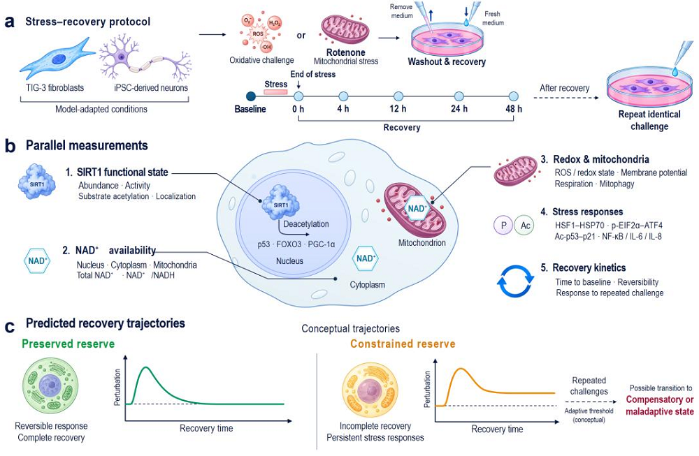
The sirtuin SIRT1, which depends on NAD+ to function [3], is very well-known in its utility in managing ROS. However, the simple presence of SIRT1 within a cell does not necessarily mean that it is active, and even if it is active, it may not be meeting the cell’s fluctuating needs. These reviewers, therefore, introduce the concept of SIRT1 functional reserve, a metric intended to “describe the remaining operational flexibility of SIRT1-dependent regulatory processes under a defined stress demand” and thus measure how well a cell can “engage, coordinate, and recover SIRT1-dependent responses”.
As this is intended to be a measurable and experimentally testable metric rather than simply a broad concept, the reviewers largely focused on SIRT1’s relationship to redox reactions and mitochondria; while SIRT1’s use throughout cells should be measured in its analysis, SIRT1 functional reserve does not reflect every use of SIRT1, nor does it measure cellular aging as a whole.
Functional reserve is not availability
The reviewers note that measuring total cellular NAD+ may not reflect its complete relationship to SIRT1 functional reserve. NAD+ is spatially located within various parts of a cell and is used and consumed by a variety of processes and molecules such as CD38 [4]. While the various pools that hold NAD+ may be connected, and mitochondria also hold NAD+ [5], there is no guarantee that a cell that is presumably abundant in total NAD+ is delivering enough of it to SIRT1 in a way that properly handles ROS. Similiarly, NAD+ availability does not necessarily reflect SIRT1 prevalence.
SIRT1 also appears as various isoforms, and the v2 and v3 varieties, which accumulate with age, are less functional than the full-length v1 version [6]. Measuring total SIRT1 in a way that does not account for these isoforms, therefore, lacks usefulness in estimating its functional reserve. Additionally, SIRT1 can be modified as a protein by oxidative and other stresses, some of which degrade its capabilities [7] while others enhance them [8].
SIRT1 is used throughout cells
The reviewers note that aging often impairs proper responses and the recovery from such [9]. SIRT1 is a crucial component of these responses, as it directly interacts with energy-sensing pathways [10]. Measuring this interaction, however, can be counterintuitive; for example, during high-intensity interval exercise, SIRT1 activity increased while its total level decreased [11]. Furthermore, the strength of a stress response may or may not correspond to its resolution; a strong response does not signify full recovery, and a weaker response may signify that the cell has handled the situation with less effort [12].
The reviewers also add that repeated stress, a well-known precursor of cellular decline, may alter SIRT1 dynamics [13] but does not necessarily result in a loss of SIRT1 function. Additionally, SIRT1 helps to regulate the heat shock protein HSF1 [14], which aid in proteostasis, while other heat shock proteins regulate SIRT1 [15]. Disruption of this, therefore, leads to a harmful feedback loop: the very machinery that is supposed to regulate proteostasis is harmed by the lack of proteostasis regulation. SIRT1 also regulates proteostasis in mitochondria [16].
SIRT1 is often part of a greater regulatory framework rather than an independent factor. Therefore, the reviewers warn that “enhancement of SIRT1 activity may yield progressively smaller benefits if other determinants of adaptation become limiting,” which may explain some weaker results seen in studies aimed at SIRT1 enhancement [17]. Such other causes need to be separated in order to properly measure SIRT1 functional reserve.
From function to dysfunction
This review discusses the adaptive-to-maladaptive transition within cells, which is a gradual process that does not have a clear line. During chronic stress, cells compensate in ways that involve either the constant use of compensatory mechanisms or altering their structure. SIRT1 is involved in both cases, although in different ways; it can be engaged in ways that involve stress tolerance, and its function can be affected by cellular changes, particularly senescence. According to the reviewers, therefore, “the same level of SIRT1 protein or activity may not produce equivalent functional output under different cellular conditions.” They propose including stress history as part of estimating the functional reserve of SIRT1.
The species and cell involved also determine the role of SIRT1. Some experiments show a straightforward relationship: increasing oxidative stress in various human cells depletes SIRT1. Others, however, such as caloric restriction and exercise in mice, change SIRT1 in a tissue-specific way. Neural stem cells are particularly vulnerable to perturbations in SIRT1 [18]. Therefore, systemic measurements of SIRT1 and/or NAD+ may not properly reflect its use or influence in cells, and its functional reserve must be measured with cell type in mind.
Towards the clinic
This review provides heavily substantiated explanations of why commonly tested methods, such as increasing SIRT1 and NAD+, along with directly targeting oxidative stress at the mitochondrial level through MitoQ [19], have had mixed results. However, this paper also acknowledges that its own framework requires experimental validation. They propose experiments in which SIRT1 functional reserve is measured by analyzing its forms and locations within the cell along with the locations of NAD+, ROS measurements, and various biomarkers of cellular health and inflammation.
Of course, the “main translational question is whether changes in SIRT1-dependent adaptive capacity can be linked to meaningful functional outcomes in humans.” The amount of work needed to verify and validate this approach, and so move towards SIRT1 and NAD+ therapies with a higher chance of working in people, will be substantial.
Literature
[1] Sies, H., Mailloux, R. J., & Jakob, U. (2024). Fundamentals of redox regulation in biology. Nature Reviews Molecular Cell Biology, 25(9), 701-719.
[2] Atayik, M. C., & Çakatay, U. (2023). Redox signaling and modulation in ageing. Biogerontology, 24(5), 603-608.
[3] Covarrubias, A. J., Perrone, R., Grozio, A., & Verdin, E. (2021). NAD+ metabolism and its roles in cellular processes during ageing. Nature reviews Molecular cell biology, 22(2), 119-141.
[4] Chini, C. C., Peclat, T. R., Warner, G. M., Kashyap, S., Espindola-Netto, J. M., de Oliveira, G. C., … & Chini, E. N. (2020). CD38 ecto-enzyme in immune cells is induced during aging and regulates NAD+ and NMN levels. Nature metabolism, 2(11), 1284-1304.
[5] Høyland, L. E., VanLinden, M. R., Niere, M., Strømland, Ø., Sharma, S., Dietze, J., … & Ziegler, M. (2024). Subcellular NAD+ pools are interconnected and buffered by mitochondrial NAD+. Nature Metabolism, 6(12), 2319-2337.
[6] Patyal, P., Ameer, F. S., Verma, A., Zhang, X., Azhar, G., Shrivastava, J., … & Wei, J. Y. (2024). The role of Sirtuin-1 isoforms in regulating mitochondrial function. Current Issues in Molecular Biology, 46(8), 8835-8851.
[7] Caito, S., Rajendrasozhan, S., Cook, S., Chung, S., Yao, H., Friedman, A. E., … & Rahman, I. (2010). SIRT1 is a redox-sensitive deacetylase that is post-translationally modified by oxidants and carbonyl stress. The FASEB Journal, 24(9), 3145.
[8] Han, C., Gu, Y., Shan, H., Mi, W., Sun, J., Shi, M., … & Yu, W. (2017). O-GlcNAcylation of SIRT1 enhances its deacetylase activity and promotes cytoprotection under stress. Nature communications, 8(1), 1491.
[9] Lomeli, N., Bota, D. A., & Davies, K. J. (2017). Diminished stress resistance and defective adaptive homeostasis in age-related diseases. Clinical Science, 131(21), 2573-2599.
[10] Cantó, C., Jiang, L. Q., Deshmukh, A. S., Mataki, C., Coste, A., Lagouge, M., … & Auwerx, J. (2010). Interdependence of AMPK and SIRT1 for metabolic adaptation to fasting and exercise in skeletal muscle. Cell metabolism, 11(3), 213-219.
[11] Gurd, B. J., Perry, C. G., Heigenhauser, G. J., Spriet, L. L., & Bonen, A. (2010). High-intensity interval training increases SIRT1 activity in human skeletal muscle. Applied Physiology, Nutrition, and Metabolism, 35(3), 350-357.
[12] Lamech, L. T., & Haynes, C. M. (2015). The unpredictability of prolonged activation of stress response pathways. The Journal of cell biology, 209(6), 781
[13] Zhang, J., Wang, H., Slotabec, L., Cheng, F., Tan, Y., & Li, J. (2023). Alterations of SIRT1/SIRT3 subcellular distribution in aging undermine cardiometabolic homeostasis during ischemia and reperfusion. Aging Cell, 22(9), e13930.
[14] Westerheide, S. D., Anckar, J., Stevens Jr, S. M., Sistonen, L., & Morimoto, R. I. (2009). Stress-inducible regulation of heat shock factor 1 by the deacetylase SIRT1. Science, 323(5917), 1063-1066.
[15] Białopiotrowicz-Data, E., Noyszewska-Kania, M., Jabłońska, E., Sewastianik, T., Komar, D., Dębek, S., … & Juszczyński, P. (2023). SIRT1 and HSP90α feed-forward circuit safeguards chromosome segregation integrity in diffuse large B cell lymphomas. Cell Death & Disease, 14(10), 667.
[16] Chen, Q., Chang, Y. L., Hung, M. W., Lee, P. L., Hsin, C., & Lin, Y. F. (2026). Sirt1 coordinates the mitochondrial UPR and myocellular proteostasis to preserve muscle integrity during muscle atrophy in zebrafish. Frontiers in Cell and Developmental Biology, 14, 1761278.
[17] Alcendor, R. R., Gao, S., Zhai, P., Zablocki, D., Holle, E., Yu, X., … & Sadoshima, J. (2007). Sirt1 regulates aging and resistance to oxidative stress in the heart. Circulation research, 100(10), 1512-1521.
[18] Wang, G., Wang, F., Ren, J., Qiu, Y., Zhang, W., Gao, S., … & Xu, J. (2018). SIRT1 involved in the regulation of alternative splicing affects the DNA damage response in neural stem cells. Cellular Physiology and Biochemistry, 48(2), 657-669.
[19] Broome, S. C., Whitfield, J., Janssens, K., & Hawley, J. A. (2025). MitoQ supplementation does not impact redox responses to acute exercise in skeletal muscle of older individuals. Redox Biology, 103927.