A recent study suggests that hyperglycosylation in brain tissue can be a hallmark of Alzheimer’s disease [1].
Lesser-known Alzheimer’s molecular features
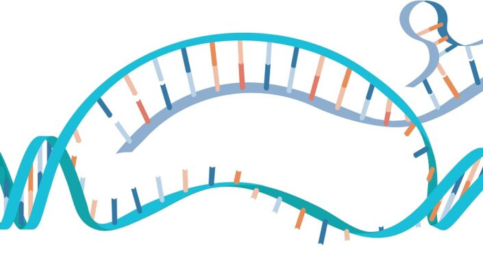
Pathological deposits of amyloid-β plaques and tau tangles in the brain are probably the most well-known features of Alzheimer’s disease. However, Alzheimer’s also involves various metabolism-related pathologies, such as alterations in glucose metabolism [2], mitochondrial function [3], lipid homeostasis [4], and N-linked glycosylation, a reaction in which a complex carbohydrate (a glycan) is attached to a protein.
N-linked glycans have been found to contribute to protein stability, intercellular communication, and immune signaling [5] and to regulate the blood–brain barrier [6]. Previous studies have reported a link between alterations in glycosylation patterns and neurological disorders [7] and suggested changes in glycosylation in Alzheimer’s disease [8]. Glycosylation has also been found to impact tau post-translational modifications and aggregation [9] and might alter amyloid-β plaque composition and clearance [10].
The authors of this study delved deeper into this topic and leveraged advances in -omics technologies to elucidate the role of glycosylation in Alzheimer’s disease.
Hyperglycosylation appears in Alzheimer’s
Advancements in laboratory techniques enabled researchers to conduct spatial metabolomics, lipidomics, and glycomics (techniques that allow comprehensive examination of metabolites, lipids, and glycans) on frontal cortex samples from deceased patients with Alzheimer’s disease and from healthy donors. “This technology allows us to examine thousands and thousands of molecules created when the body breaks down food or drugs and to uncover intricate pathways that otherwise would stay hidden,” said senior author Ramon Sun, Ph.D., director of the Center for Advanced Spatial Biomolecule Research and associate director for innovation of UF’s McKnight Brain Institute.
While they observed Alzheimer’s disease-specific changes in metabolomics and lipidomics analyses, the differences that caught their attention most were in glycan abundance, which was significantly increased across both white and grey matter regions in samples from patients with Alzheimer’s disease.
The hyperglycosylation observed in human brains was also observed in two mouse models of Alzheimer’s disease. These experiments discovered that those modifications were brain region-specific and preferentially affected brain regions associated with memory, cognitive processing, and neuroinflammation.
Subsequent experiments in human and mouse models found that the increase in glycosylation was the result of increased glycan biosynthesis rather than reduced degradation and recycling, and that the hyperglycosylation in Alzheimer’s disease “predominantly involves an increase in glycosylation modifications on pre-existing glycoproteins rather than the appearance of novel glycosylated proteins.”
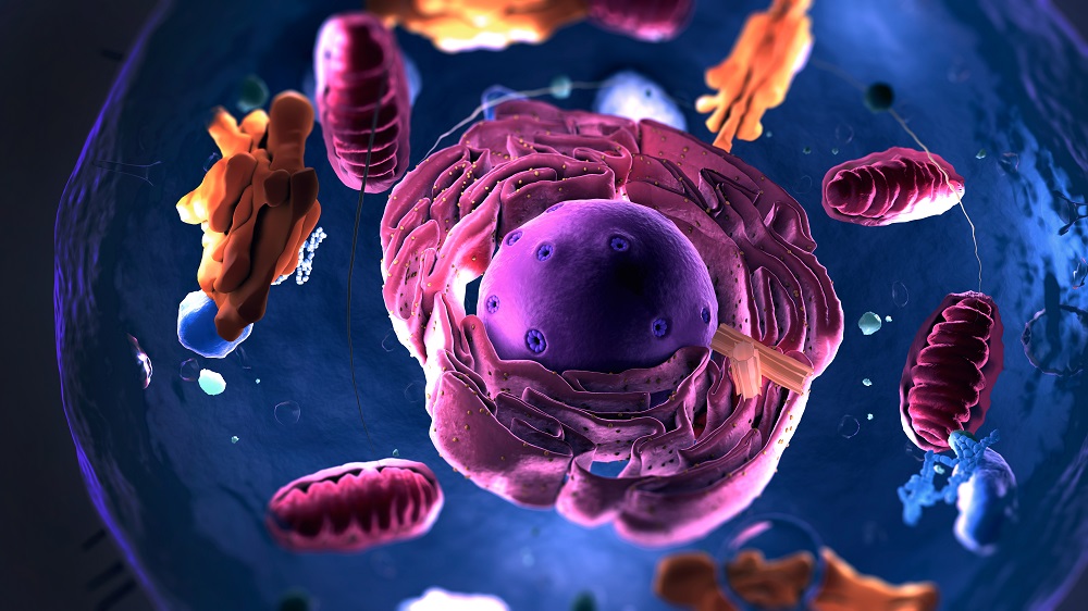
Additionally, the researchers identified neurons as the predominant cell population affected by increased glycosylation, supporting the role of glycosylation in Alzheimer’s disease pathogenesis.
A cause or a consequence?
The observed changes in glycosylation can be either a driver of neurodegeneration or a consequence of it. To determine this relationship, the authors experimentally decreased or increased N-glycan levels in the brain.
First, they used two approaches to block glycosylation: genetic engineering tools to reduce the levels of a key enzyme involved in the production of glycans, and a small molecule to block transfer of glycans to proteins. Mouse models of Alzheimer’s disease treated this way showed improvement on a social memory test (which measures “social interaction time across repeated exposures”) compared to controls. Results from those experiments suggest that reducing excessive N-glycosylation results in behavioral benefits.
Increasing glycosylation in mice with Alzheimer’s had the opposite effect. The authors used glucosamine, a molecule that can be incorporated into brain glycans, and supplemented the mice with Alzheimer’s disease with it. Glucosamine supplementation increased brain glycosylation and further exacerbated social memory deficits in those mice. Supplementing glucosamine to healthy mice didn’t result in hyperglycosylation in the brain nor impaired social recognition memory, suggesting that normal brains have mechanisms that can protect them against glucosamine supplementation-induced perturbation.
Together, these results show a causal role for hyperglycosylation in Alzheimer’s disease-associated cognitive dysfunction and suggest that glycosylation can be used as a therapeutic target for Alzheimer’s disease-related neurocognitive deficits. This finding is independent of other Alzheimer’s features such as neuroinflammation or amyloid deposits, which were not affected when glycosylation was experimentally blocked. The authors suggest future studies into small-molecule inhibitors with the ability to selectively decrease pathological glycosylation levels in Alzheimer’s patients’ brains.
“Our results suggest that altered metabolism is a significant contributor to Alzheimer’s progression and, in addition, addressing the metabolic defect could be an important complement to approaches focused on Alzheimer’s plaques and tangles,” Sun said.
Glucosamine supplementation drawbacks
While the results of these experiments can be used for future therapy development, the authors found that the knowledge they gathered here has immediate implications. They demonstrated this by examining glucosamine use in Alzheimer’s disease patients. Glucosamine, which they used in mouse supplementation experiments, is an over-the-counter supplement used for joint health.
From the University of Florida Health system health records, the authors identified 24,481 patients with Alzheimer’s disease-related dementias; among them, 1,896 patients had at least 1 year of documented glucosamine usage following a dementia diagnosis. 41,884 people with mild cognitive impairment were used as controls; 2,750 of them received glucosamine. The analysis indicated that “glucosamine usage was associated with a 25% increase in mortality risk” among patients with Alzheimer’s disease-related dementias, but no significant increase was observed among people with mild cognitive impairment.
When the rate of mild cognitive impairment-to-Alzheimer’s disease-related dementias conversion rate (indicating disease progression) was quantified, the conversion rate was increased by 25% among people who took glucosamine, suggesting that it might accelerate disease progression.
“The electronic health record data are very provocative,” said Matt Gentry, Ph.D., chair of UF’s Department of Biochemistry and Molecular Biology and a study co-author. “While it’s an association and not proof of causality, it does raise an important clinical question that now deserves much more attention.”
“In the United States, there are about seven million people living with Alzheimer’s and millions more with related dementias such as Lewy body or frontotemporal dementia,” Sun adds. “A lot of these people actively take an over-the-counter supplement that could be making their disease progression worse.”
The authors emphasize the need for a well-designed clinical trial to evaluate glucosamine’s impact on cognitive health in people with dementia and to identify which groups are at risk for worsening their dementia symptoms.
We would like to ask you a small favor. We are a non-profit foundation, and unlike some other organizations, we have no shareholders and no products to sell you. All our news and educational content is free for everyone to read, but it does mean that we rely on the help of people like you. Every contribution, no matter if it’s big or small, supports independent journalism and sustains our future.
Literature
[1] Hawkinson, T. R., Liu, Z., Ribas, R. A., Medina, T., Nielsen, R. S., Clarke, H. A., Ma, X., Mueller, A. C., Plasencia, A. F., Sheer, A. L., Simpson, S. T., Soto, C. M., Sudderth, J., Cai, F., Cantrell, A. R., Colpaert, M. G., Shedlock, C. J., Wu, L., Young, L. E. A., Kooser, D. D., … Sun, R. C. (2026). Hyperglycosylation is a metabolic driver of Alzheimer’s disease. Nature metabolism, 10.1038/s42255-026-01538-4. Advance online publication.
[2] Butterfield, D. A., & Halliwell, B. (2019). Oxidative stress, dysfunctional glucose metabolism and Alzheimer disease. Nature reviews. Neuroscience, 20(3), 148–160.
[3] Swerdlow R. H. (2018). Mitochondria and Mitochondrial Cascades in Alzheimer’s Disease. Journal of Alzheimer’s disease : JAD, 62(3), 1403–1416.
[4] Di Paolo, G., & Kim, T. W. (2011). Linking lipids to Alzheimer’s disease: cholesterol and beyond. Nature reviews. Neuroscience, 12(5), 284–296.
[5] Conroy, L. R., Hawkinson, T. R., Young, L. E. A., Gentry, M. S., & Sun, R. C. (2021). Emerging roles of N-linked glycosylation in brain physiology and disorders. Trends in endocrinology and metabolism: TEM, 32(12), 980–993.
[6] Nesselbush, M. C., Luca, B. A., Jeon, Y. J., Jabara, I., Meador, C. B., Garofalo, A., Binkley, M. S., Hui, A. B., van ‘t Erve, I., Xu, N., Shi, W. Y., Liu, K. J., Sugio, T., Kastelowitz, N., Hamilton, E. G., Liu, C. L., Olsen, M., Bonilla, R. F., Wang, Y. P., Jiang, A., … Diehn, M. (2025). An ultrasensitive method for detection of cell-free RNA. Nature, 641(8063), 759–768.
[7] Freeze, H. H., Eklund, E. A., Ng, B. G., & Patterson, M. C. (2015). Neurological aspects of human glycosylation disorders. Annual review of neuroscience, 38, 105–125.
[8] Hawkinson, T. R., Clarke, H. A., Young, L. E. A., Conroy, L. R., Markussen, K. H., Kerch, K. M., Johnson, L. A., Nelson, P. T., Wang, C., Allison, D. B., Gentry, M. S., & Sun, R. C. (2022). In situ spatial glycomic imaging of mouse and human Alzheimer’s disease brains. Alzheimer’s & dementia : the journal of the Alzheimer’s Association, 18(10), 1721–1735.
[9] Losev, Y., Frenkel-Pinter, M., Abu-Hussien, M., Viswanathan, G. K., Elyashiv-Revivo, D., Geries, R., Khalaila, I., Gazit, E., & Segal, D. (2021). Differential effects of putative N-glycosylation sites in human Tau on Alzheimer’s disease-related neurodegeneration. Cellular and molecular life sciences : CMLS, 78(5), 2231–2245.
[10] Perdivara, I., Petrovich, R., Allinquant, B., Deterding, L. J., Tomer, K. B., & Przybylski, M. (2009). Elucidation of O-glycosylation structures of the beta-amyloid precursor protein by liquid chromatography-mass spectrometry using electron transfer dissociation and collision induced dissociation. Journal of proteome research, 8(2), 631–642.