The Blog

Building a Future Free of Age-Related Disease

Public Longevity Group

Lifespan Research Institute Launches Public Longevity Group

[Mountain View, September 17, 2025]Lifespan Research Institute (LRI) today announced the launch of the Public Longevity Group (PLG), a new initiative focused on bridging the cultural gap between scientific breakthroughs in aging and their public acceptance. To kickstart its work, PLG has opened a crowdfunding campaign to develop tools that measure and strengthen public trust in longevity science.

While the science of longevity biotechnology continues to advance, skepticism and cultural resistance limit progress, with some studies showing that more than half of Americans would reject a safe, proven therapy to extend life. This hesitation poses risks of raising costs, delaying health-promoting regulation, and slowing the delivery of treatments that could combat age-related diseases and extend healthy lifespan.

“The breakthrough that unlocks all other breakthroughs is public trust,” said Sho Joseph Ozaki Tan, Founder of PLG. “Without it, even the most promising therapies may never reach the people they’re meant to help. PLG exists to change that.”

“Persuasion is a science too,” said Keith Comito, CEO of Lifespan Research Institute. “To bring health-extending technologies to the public as quickly as possible, we must approach advocacy with the same rigor as our research. With PLG, we’ll be able to systematically measure and increase social receptivity, making the public’s appetite for credible longevity therapies unmistakable to policymakers, investors, and the public itself.”

PLG is developing the first data-driven cultural intelligence system for longevity—a platform designed to track real-time sentiment, test narratives, and identify which messages resonate and which backfire. Early tools include:

  • The Longevity Cultural Clock: a cultural barometer mapping readiness and resistance across demographics and regions.
  • Sentiment Dashboards: real-time monitoring of public, investor, and policymaker perceptions.
  • Narrative Testing Tools: data-driven analysis that will enable robust pathways to public support.

The crowdfunding campaign will provide the initial $100,000 needed to launch these tools, creating the cultural foundation required for healthier, longer lives.

With a lean, data-driven team, the group aims to provide open-access cultural insights for advocates and policymakers while offering advanced analytics to mission-aligned partners.

Campaign Timeline:

  • Campaign completion: November 2, 2025
  • Dashboard development: Dec 2025 – Feb 2026
  • First survey deployment: Feb – Apr 2026
  • Beta dashboard launch: May 2026
  • First public insight report: June 2026

Supporters can contribute directly at: https://lifespan.io/campaigns/public-longevity-group/

The PLG campaign is sponsored by the members of LRI’s Lifespan Alliance, a consortium of mission-aligned organizations that believe in the promise of extending healthy human lifespan. Newly-joined members include OpenCures, AgelessRx, and Lento Bio.

About Lifespan Research Institute

Lifespan Research Institute accelerates the science and systems needed for longer, healthier lives by uniting researchers, investors, and the public to drive lasting impact. LRI advances breakthrough science, builds high-impact ecosystems, and connects the global longevity community.

Media Contact:

Christie Sacco

Marketing Director

Lifespan Research Institute

christie.sacco@lifespan.io

(650) 336-1780

We would like to ask you a small favor. We are a non-profit foundation, and unlike some other organizations, we have no shareholders and no products to sell you. All our news and educational content is free for everyone to read, but it does mean that we rely on the help of people like you. Every contribution, no matter if it’s big or small, supports independent ethical journalism and sustains our future.
Longevity Science Foundation

Invested in Her Campaign Extends Fundraising Deadline

Driven by ongoing support from their community, The Longevity Science Foundation is extending the deadline for the Invested in Her campaign to October 1, 2026, as they work toward a $250,000 fundraising target to support early-stage research into ovarian aging and menopause.

Hundreds of millions of women live longer than men yet spend more years in poor health. The biology driving this disparity is rooted in ovarian aging and the menopausal transition, and it remains one of the most underfunded areas in medicine. Invested in Her is raising $250,000 to fund the science that closes that gap, for mothers, daughters, and women everywhere.

Only 8.8% of NIH funding is directed to women’s health research, and only a fraction of that studies ovarian aging and the menopausal transition. The result is that a critical biological transition remains insufficiently studied, limiting prevention, early intervention, and targeted treatment.

The implications extend across women’s long-term health. Women live longer than men on average but spend more years in poor health, while the hormonal cascade associated with ovarian decline and menopause reverberates across major systems, including cardiometabolic, brain, and skeletal health. Closing the research gap is how we begin to close the health gap.

Funding the Full Picture

Through its scientific review process, the LSF has identified a portfolio of high-impact projects ready for immediate funding. Rather than funding isolated therapies alone, the LSF is targeting the full arc of women’s reproductive aging through three coordinated pillars: measurement, biological understanding, and early-stage intervention.

The work will support:

  • Measurement: developing better ways to measure ovarian aging and the menopausal transition, because better measurement is the first step to better medicine.
  • Biology: answering why women’s aging unfolds the way it does and improving our understanding of the mechanisms connecting reproductive aging to broader systemic decline.
  • Intervention: advancing early-stage approaches that treat reproductive aging as modifiable, not inevitable.

Building Momentum Around Women’s Health

The campaign recently brought its community together at 7 World Trade Center in New York City for the Invested in Her event, uniting investors, clinicians, researchers, founders, philanthropists, advocates, and other leaders across healthcare and industry. Through personal stories, presentations, panel discussions, and networking, participants explored the challenges facing women’s health and the opportunities to create meaningful change.

Attendee feedback reinforced the need for action. When asked about the biggest barriers to progress in women’s health, healthcare system design and lack of research funding led the room. For the LSF, the answer is clear: it needs to continue doing its part in funding holistic women’s health research to create equitable solutions for every woman.

Invested in Her will continue building on this momentum through October 1, directing attention and funding toward research that can improve how women’s reproductive aging is measured, understood, and ultimately addressed. 100% of donations go directly to research, with no overhead deducted, and all eligible gifts are tax-deductible. Support Invested in Her and help fund the science that closes the gap in women’s health, menopause, and ovarian aging research.

We would like to ask you a small favor. We are a non-profit foundation, and unlike some other organizations, we have no shareholders and no products to sell you. All our news and educational content is free for everyone to read, but it does mean that we rely on the help of people like you. Every contribution, no matter if it’s big or small, supports independent ethical journalism and sustains our future.
Rejuvenation Roundup August 2026

Rejuvenation Roundup August 2026

Properly untangling aging means untangling its various counterbalances, sometimes on the individual level. Here’s what’s been investigated this month.

Interviews

Wei-Wu HeWei-Wu He: People Should Become the CEOs of Their Own Health: Wei-Wu He is an unusual combination of scientist and businessman, and his current company focuses on using precision medicine to lengthen people’s lives.

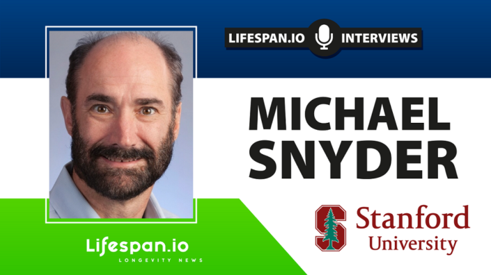
Michael Snyder: We Need to Track Health, Not Disease: Michael Snyder, Director of the Center for Genomics and Personalized Medicine at Stanford University, is convinced that wearables will help drive the transition from “sickcare” to healthcare.

Advocacy and Analysis

Ships togetherWhy Democratizing Rejuvenation Is an Economic Imperative: Worldwide healthcare faces an unsustainable situation. We spend trillions managing the late-stage effects of biological aging, yet routinely overlook the underlying causes that drive them. Driven by an aging society, the financial burdens of treating diabetes, dementia, and heart disease are pushing Social Security and Medicare towards increasing and potentially catastrophic budget deficits.

Research Roundup

Valine Restriction Increases Male Mouse Lifespan by 23%: A new study has found that restricting dietary valine extends both median and maximum lifespan in male mice while improving healthspan in both sexes.

PastaA New Transcriptomic Clock for Intervention Analysis: A team of researchers has developed Pasta, a transcriptomic clock that accurately predicts the age-related effects of various compounds and gene expressions.

A New Target Against High Blood Pressure: Researchers have discovered why the protein AGGF1 has significant effects on blood pressure and published their findings in Aging Cell.

Organism complexityWhy Affecting Aging in Complex Organisms Is So Hard: A new study proposes a theoretical framework that explains why the more complex an animal is, the harder it is to move the needle on its rate of aging.

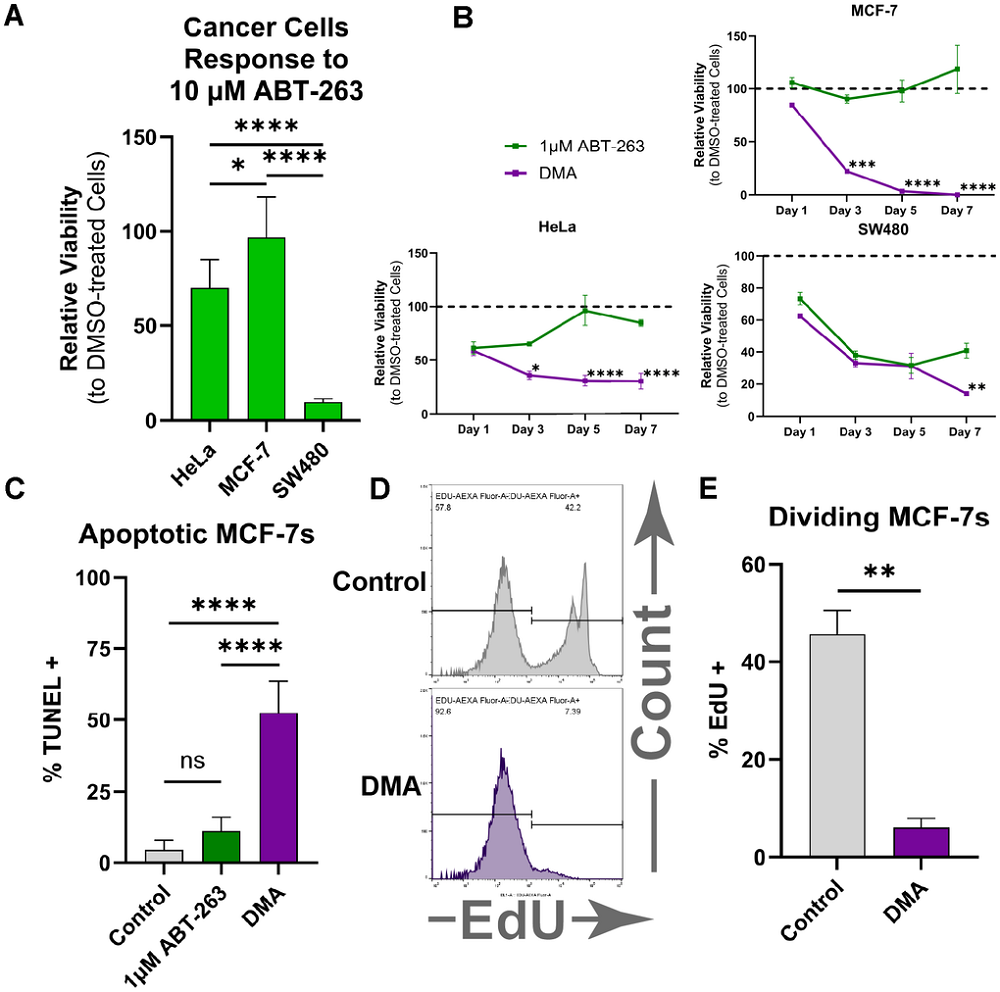
A Drug Combination Fights Both Senescence and Cancer: In Aging, the Conboys and their team have described how a combination treatment kills both senescent and cancer cells and lengthens the lives of old mice.

No getting fatA Million-Person Study Finds a Protective Metabolic Gene: A massive human genetics study identified rare folliculin-interacting protein 1 (FNIP1) mutations associated with favorable metabolism and much lower cardiometabolic disease risk. Experiments in human liver cells and mice suggest a therapeutic route.

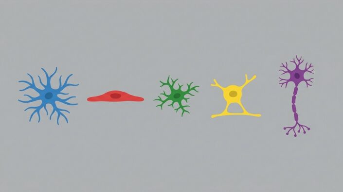
How Senescence Spreads Between Brain Cells: Researchers have gone into deep detail regarding how each of five senescent brain cell types expresses and receives factors that encourage other cells to become senescent.

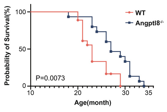
Fat cellsHow Old Fat Cells Trigger Inflammation and Raise Risks: Using cohort studies and a mouse model, researchers have found that the circulating factor ANGPTL8, which is produced by senescent fat cells, is related to age-related diseases and mortality in mice and people.

In ALS, Microglia Eat Living Neurons, Mistaking Them for Dead: This study suggests that in amyotrophic lateral sclerosis – and possibly other neurodegenerative diseases – the brain’s immune cells devour stressed but still living neurons due to altered signaling.

Rat tendonsA Tougher Extracellular Matrix Strengthens Tendons in Rats: Researchers have found that an upstream promoter of two extracellular matrix proteins increases healing and strength capabilities in the tendons of rats.

Short, Intense Exercise Elicits Beneficial Metabolic Changes: Compared to moderate-intensity exercise, high-intensity sprint-interval exercise produced larger changes in circulating protein and metabolite levels and stimulated proteins associated with cardiometabolic health benefits.

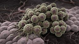
Aggressive cancerWhy Turning Off Cancer Genes Doesn’t Always Work: A recent study has examined whether senescence might allow cancer cells to survive oncogene withdrawal and become even more dangerous.

A Mechanistic Explanation for SIRT1’s Effects: Researchers have found that the sirtuin SIRT1 stabilizes the human genome by suppressing retrotransposition, which occurs when parts of the genome transcribe themselves onto other parts.

Impact of N-PEP-12 Supplementation on Attentional Performance and Mental Wellbeing in Healthy Adults with Subjective Cognitive Complaints: These findings support further investigation of N-PEP-12 as a nutritional intervention for early subjective cognitive changes associated with aging.

Diet-dependent, beneficial and adverse effects of rapamycin on life span of Drosophila melanogaster: These results confirm the adverse effects of rapamycin during development and demonstrate its potential to switch between beneficial and harmful effects on adult life span depending on other components of the diet.

Natural Bioactive Compounds Targeting Key Hallmarks of Aging: Functional Food Potential of Spermidine, Fisetin, Berberine, and Urolithin A: Together, the four compounds primarily target distinct but complementary aging-associated pathways (autophagy, senolysis, metabolic regulation, mitophagy), suggesting rational potential for combined functional food formulations.

A Systems Pharmacology Model of Aging Identifies Optimal Combination Therapies With Secondary Benefits on Weight Loss and Metabolic Health: Metabolic optimization and aging optimisation are distinct objectives that do not converge on the same drug combination.

A machine learning-derived dietary pattern for aging: The MYTH Diet may offer a biologically informed, scalable framework for developing personalized nutrition strategies aimed at supporting healthy aging and longevity.

Social media use duration and epigenetic aging among U.S. adults in the MIDUS refresher study: These findings provide preliminary evidence that social media use duration is associated with the pace of biological aging as measured by DunedinPACE.

Living Beyond Our Evolutionary Warranty: Why Non-Communicable Diseases May Be The Inevitable Costs of an Extended Lifespan: Accepting NCDs as partially intrinsic to extended lifespan while pursuing interventions delaying onset may yield more realistic health goals than assuming complete preventability.

Intrasplenic Thymus Organogenesis from Injectable Tissue Fragments Restores Functional T-Cell Immunity: These results identify the spleen as an optimal ectopic niche for thymus regeneration and provide a promising strategy for clinical immune reconstitution and regenerative immunology.

Beyond Diabetes: Continuous Glucose Monitoring as a Candidate Precision Tool for Cardiovascular Prevention and Healthy Longevity: The shared longevity genes identified in this work offer potential targets to promote healthy aging and decrease age-onset disease.

News Nuggets

Forever Healthy FoundationForever Healthy Launches the Evipedia Browser Extension: The extension currently recognizes 3,700+ terms across 630+ evidence reviews. It is free to use and available for Chrome, Firefox & Safari. Installing the extension is a simple one-click process from Evipedia’s extension page.

GenBio Launches a “Virtual Cell” AI Model: This AI startup, which lists Nobel laureate David Baker among its co-founders, has announced a “world model” of a cell that can simulate both its natural state and responses to successive perturbations, potentially transforming biological research.

We would like to ask you a small favor. We are a non-profit foundation, and unlike some other organizations, we have no shareholders and no products to sell you. All our news and educational content is free for everyone to read, but it does mean that we rely on the help of people like you. Every contribution, no matter if it’s big or small, supports independent ethical journalism and sustains our future.
Chromatin

A Mechanistic Explanation for SIRT1’s Effects

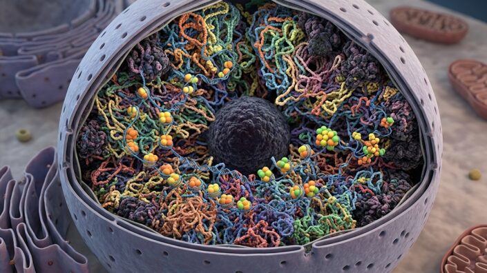
Researchers have found that the sirtuin SIRT1 stabilizes the human genome by suppressing retrotransposition, which occurs when parts of the genome transcribe themselves onto other parts.

When DNA modifies itself

Long interspersed elements-1 (LINE-1) have existed in organisms’ genomes, including our own, for well over a billion years [1]. They are the only autonomous elements that transpose themselves within the human genome, and they occupy a full sixth of it [2]. Two proteins generated by LINE-1 elements, ORF1 and ORF2, are responsible for a third of the human genome [1].

As catalysts of genomic instability, LINE-1 elements have aided in human evolution, and they are directly responsible for the complexity of the human brain [3]. However, this comes at a high cost: the same instability that allowed us to evolve is itself an aspect of aging, and it is directly responsible for multiple other aspects, including senescence [4] and cancer [5].

Heterochromatin is the packed non-coding DNA that functions as a transcriptional regulator. While it is well-known in aging research as a key part of epigenetic alterations [6], newer research has found that its age-related diminishment allows LINE-1 elements to proliferate [7]. H3K9me3, a fundamental marker of heterochromatin, has been found to be directly responsible for keeping LINE-1 elements in check [8].

A possible explanation for sirtuins’ effects

Sirtuins have been heavily researched in the context of aging. SIRT6 has been specifically identified as a suppressor of LINE-1 activity [9], and SIRT1 is known to have benefits against age-related disorders in several organisms, including in the lungs of mice [10]. However, before this study, no one had yet investigated whether or not SIRT1 suppresses LINE-1 as well.

In their first experiment, the researchers used HeLa cells, an established line of human cancer cells. Using fluorescent reporter proteins to identify LINE-1 activity, the researchers found that the overexpression of SIRT1 in these cells minimized this activity, and silencing SIRT1 increased it. Similar results were found in IMR90 human cells and mouse embryonic fibroblasts.

These results were due to direct effects on a LINE-1 internal promoter. Silencing SIRT1 increased the activity of this promoter, increasing the production of ORF2, which led to increased DNA damage within cells. Similarly, overexpressing SIRT1 decreased this damage. These results were confirmed with the DNA damage marker γH2AX.

Protection on multiple fronts

LINE-1 is also known to trigger the cGAS-STING inflammatory pathway [11], which often leads to cellular senescence. After a high dose of radiation exposure, the researchers found that 30% of a control group of HeLa cells became senescent; however, this dose was only sufficient to drive 13% of a SIRT1-overexpressing group into senescence. In HCA2-hTERT, another cell line, SIRT1 overexpression dropped senescence from 38% to 20% after a high radiation dose. Similarly, SIRT1 overexpression reduced the SASP factors secreted by senescent cells, while silencing SIRT1 increased them.

Quiescence is a state in which cells do not divide; however, unlike senescence, these cells are not incapable of division but are simply waiting for a trigger. SIRT1 was found to have exceptionally strong effects on LINE-1 in quiescent cells, being enriched at its loci and preventing it from harming these reserve cells.

The team then investigated how SIRT1 interacts with two well-known regulators of genomic stability, Lamin B1 and KAP1. Interestingly, while SIRT1 did not have any effects on the levels of these proteins, it improved their ability to interact, aiding in heterochromatin stability. SIRT1 was also found to be positively associated with H3K9me3 in chromatin.

While this research only involved cells and not mice or people, it offers plausible explanations for why SIRT1 has the effects reported in other studies. The researchers state that their “findings establish a new mechanistic framework for SIRT1-mediated senescence intervention, with profound implications for delaying aging and mitigating age-related diseases.” Further work will need to be done to confirm if this framework is correct and that these results hold true in vivo.

We would like to ask you a small favor. We are a non-profit foundation, and unlike some other organizations, we have no shareholders and no products to sell you. All our news and educational content is free for everyone to read, but it does mean that we rely on the help of people like you. Every contribution, no matter if it’s big or small, supports independent ethical journalism and sustains our future.

Literature

[1] Baldwin, E. T., van Eeuwen, T., Hoyos, D., Zalevsky, A., Tchesnokov, E. P., Sánchez, R., … & Taylor, M. S. (2024). Structures, functions and adaptations of the human LINE-1 ORF2 protein. Nature, 626(7997), 194-206.

[2] Beck, C. R., Collier, P., Macfarlane, C., Malig, M., Kidd, J. M., Eichler, E. E., … & Moran, J. V. (2010). LINE-1 retrotransposition activity in human genomes. Cell, 141(7), 1159-1170.

[3] Garza, R., Atacho, D. A., Adami, A., Gerdes, P., Vinod, M., Hsieh, P., … & Jakobsson, J. (2023). LINE-1 retrotransposons drive human neuronal transcriptome complexity and functional diversification. Science Advances, 9(44), eadh9543.

[4] De Cecco, M., Criscione, S. W., Peckham, E. J., Hillenmeyer, S., Hamm, E. A., Manivannan, J., … & Sedivy, J. M. (2013). Genomes of replicatively senescent cells undergo global epigenetic changes leading to gene silencing and activation of transposable elements. Aging cell, 12(2), 247-256.

[5] Rodić, N., & Burns, K. H. (2013). Long interspersed element–1 (LINE-1): passenger or driver in human neoplasms?. PLoS genetics, 9(3), e1003402.

[6] Lee, J. H., Kim, E. W., Croteau, D. L., & Bohr, V. A. (2020). Heterochromatin: an epigenetic point of view in aging. Experimental & molecular medicine, 52(9), 1466-1474.

[7] Li, X., Yu, H., Li, D., & Liu, N. (2024). LINE-1 transposable element renaissance in aging and age-related diseases. Ageing Research Reviews, 100, 102440.

[8] Guerra, M. V., Cáceres, M. I., Herrera-Soto, A., Arredondo, S. B., Varas-Godoy, M., van Zundert, B., & Varela-Nallar, L. (2022). H3K9 methyltransferases Suv39h1 and Suv39h2 control the differentiation of neural progenitor cells in the adult hippocampus. Frontiers in Cell and Developmental Biology, 9, 778345.

[9] Van Meter, M., Kashyap, M., Rezazadeh, S., Geneva, A. J., Morello, T. D., Seluanov, A., & Gorbunova, V. (2014). SIRT6 represses LINE1 retrotransposons by ribosylating KAP1 but this repression fails with stress and age. Nature communications, 5(1), 5011.

[10] Zhou, J., Chen, H., Wang, Q., Chen, S., Wang, R., Wang, Z., … & Jin, J. (2022). Sirt1 overexpression improves senescence‐associated pulmonary fibrosis induced by vitamin D deficiency through downregulating IL‐11 transcription. Aging Cell, 21(8), e13680.

[11] Mathavarajah, S., & Dellaire, G. (2023). LINE-1: an emerging initiator of cGAS-STING signalling and inflammation that is dysregulated in disease. Biochemistry and Cell Biology, 102(1), 38-46.

Michael Snyder Interview

Michael Snyder: We Need to Track Health, Not Disease

Michael Snyder, Director of the Center for Genomics and Personalized Medicine at Stanford University, is obsessed with health-tracking wearables. During our interview, he wore four wrist-worn devices – watches and bands – and two rings. He is convinced that these small but powerful gadgets will help drive the transition from today’s “sickcare” to true healthcare, which, as its name suggests, should start with proactively monitoring health rather than waiting for disease to develop. They can also provide the longitudinal health data we need for personalized medicine – and to feed data-hungry AI models.

However, Snyder’s scope of interests is much wider than that. He made waves with a study that reported two distinct periods of pronounced age-related change in people’s 40s and 60s. He also studies the deeply individual patterns of aging, which he calls “ageotypes,” and is developing the concept of intrinsic capacity as a quantitative measure of health to be used in longevity research and drug trials.

I just saw you wearing all those wearables, and I think that’s a great place to start: how do we work with all the data from these devices?

In my case, I’m collecting a lot of data, but that’s more on the research side. The average person will normally wear just one, possibly two of these things – not six or seven or eight like I do.

Wearables are powerful because they measure 24/7, as long as you keep them charged. That means they’re tracking your health, they know your healthy baseline, and you can look for shifts from it. We got involved when they first came out as fitness trackers and thought they could be powerful health monitors because they measured resting heart rate and a few other things. Now they measure even more – heart rate variability, which is super important for health monitoring, blood oxygen. There’s medical value in that information.

We have strong circadian patterns throughout the day. Heart rate goes up during the day, blood pressure shifts – but if you track all that, you can look for shifts from the norm.

We started putting wearables on our cohort and discovered pretty much right away that we could tell when someone was getting ill. In my case, it started with Lyme disease. I picked it up presymptomatically because my blood oxygen dropped.

Then we showed that we could detect respiratory viral infections. It really hit home when the pandemic arrived in 2020. We partnered with Fitbit and showed that you could tell when people were getting COVID in advance of symptoms.

It works particularly well for COVID because it has a long presymptomatic period. Influenza and some other respiratory viruses have more like a 36- to 48-hour incubation before symptoms appear. We can still pick those up before symptoms, but with COVID you have more time.

Especially with COVID, I guess that’s important in two ways: preventing transmission and starting treatment as early as possible, such as with Paxlovid.

Yes, definitely. Paxlovid is thought to be especially effective when you use it right away. We’re running studies where family members of transplant patients have devices. If a family member gets ill, they’re alerted and can self-isolate so the transplant patient doesn’t get infected, which could be very serious.

We’ve had family members get red alerts. They quickly test for COVID or influenza, and often we can tell it’s one or the other. Sometimes, we know they’re ill but aren’t quite sure with what. The treatment differs depending on what they have, as you point out, and they can self-isolate. They’ve been pretty grateful for the alerting system. I think that’s the first sort of clinically actionable demonstration.

You’ve been working with wearables for years, and both their capabilities and the accuracy of the measurements have been increasing. Are they really coming of age now? Can they meaningfully change how we do research? There’s a distinction to be made here between tracking an individual trajectory and getting population-level insights.

They’re definitely embedded in research now. Everybody uses them to track activity and basic physiological parameters. They’re not yet embedded in the clinic. Some concierge services are bringing in these data – but not most standard health plans – partly because the existing system hasn’t been set up for it and isn’t incentivized that way.

We tend to practice sick care rather than healthcare because the financial incentives aren’t aligned. We need to fix that. If we actually practice healthcare, people will routinely wear these devices because tracking is so integral to keeping people healthy.

Other groups have shown that you can pick up atrial fibrillation with a smartwatch, and it works reasonably well – in the 30-50 percent range, I think. None of these devices is perfect, although they keep getting better.

Interestingly, for our alerting system, the number one trigger of red alerts is workplace stress, not a respiratory viral infection. That makes sense because it’s mostly built around heart rate and related measures. Now that we have other data types, I’m quite confident we can distinguish respiratory infections from mental stress. That’s something we’re working on.

Knowing when people are mentally stressed is a big deal. If something is mentally or physically stressing you, you should take care of yourself. We don’t have good biomarkers for mental health. There’s a lot more to do, but I think wearables will be excellent indicators of both mental and physical stress.

We haven’t published this yet, but we had a case of someone who died of a heart attack, and his wife shared his data. He had an Apple Watch and an Oura Ring, and it’s pretty clear he had a step-function change about four and a half months before the event – resting heart rate and a lot of other parameters shifted.

We need real-time systems that pull in the data, track people, and alert them when something is off. It comes back to the car-dashboard analogy. If a light goes on, you may not know exactly what it is, and it may be a false alarm in terms of something serious. But usually if an alert goes off, something is happening, and then you can follow up.

On the population side, wearable companies are sitting on these huge mountains of data. Do you think it will ever be possible to anonymize and use it? Are there efforts in that direction?

I don’t think companies are incentivized to share their data, and I expect they generally won’t. Some are forced to share if they want to publish. So a lot of this is going to come from academic researchers.

Many of us share our smartwatch data, but continuous glucose monitoring is a good example of the problem. There are many CGM studies, but it’s very hard, if not impossible, to get the underlying data. With smartwatches, some big biobanks are now putting wearables on people. All of Us has Fitbits on many participants. UK Biobank has done more with ActiGraph, but they’re discussing wearables as well.

I’d like to think the cohort we’ve followed pioneered some of this too. It’s relatively small, but we did deep -omics profiling, put wearables on people early, and learned that these things are powerful.

One of the more important things we learned was with continuous glucose monitoring. CGMs were being used a lot for insulin-dependent type 1 and type 2 diabetics. When we got involved, they weren’t really being used in so-called normal people and prediabetics. We put them on those groups and discovered right away that a lot of people thought to be normal weren’t so normal. They were spiking pretty badly – sometimes as badly as diabetics.

That was actually one of my questions. I’ve worn a CGM a few times for a few weeks.

Very powerful, right? You’ll never eat the same again. I mean that in a good way. You see what spikes you, and that’s very personal. What spikes your glucose can be very different from what spikes mine. We’re all very different.

Do you think the question about the importance of glucose spikes is settled? Are they really that crucial for health?

I think it’s settled in certain ways. There are at least two obvious lines of evidence. Time in range is strongly related to diabetes, and many studies show that diabetes strongly affects health, especially cardiovascular disease. There are also studies showing that postprandial spikes – spikes after meals – are associated with cardiovascular disease independently of diabetes. So, in my mind, the data on spiking are pretty clear. There are nuances, of course. If you lift weights, for example, you can break down glycogen into glucose, so not every rise has the same meaning.

Constant monitoring obviously is amazing: it gives you important insights into your health and can flag many things early on. But what about overdiagnosis, overtreatment, false positives, or just the constant background anxiety you can get when you measure yourself all the time?

I think it’s an education issue. You’re right that some people get overly anxious, and you want to be careful about how information is returned. It’s up to the person to decide how they want that information, in my opinion, but most people are quite capable of handling it.

When people first get these devices, everybody gets very absorbed in them – what foods spike you and all that. Then you settle into a pattern where they’re fairly useful. They can alert you to some pretty serious health issues, so I think it’s better to know. I like the car analogy. Your car has lots of sensors. You wouldn’t dream of driving it without a dashboard.

I could even argue that things like whole-body MRI can reduce anxiety in some cases. I know someone who was very worried because their family had a history of ovarian cancer. They got a whole-body MRI and were very pleased that everything looked good.

The mismatch with whole-body MRI is that people assume, ‘If you have nodules, you may have cancer.’ That’s the wrong way to think about it. You want to know what nodules you have, but the real issue is whether any are growing. Everybody has nodules. We need longitudinal data.

I have nine nodules. I get whole-body MRIs every three months. That may be overkill – I’m trying to see how often we really should measure people. If you have an aggressive cancer, it can take off pretty quickly.

Yes, we’re believers in whole-body MRI. You’re going to have nodules, and the key is knowing where they are so that if you ever get cancer, have it operated on, and then get follow-up imaging, you know what your background looked like. Without that baseline, you may be in a difficult position.

In my opinion, everybody should get a whole-body MRI so they know their baseline. I think you should get a discount on your health plan if you do these sorts of things – whole-body MRI, wearables, genome sequencing – because you’ll be better able to manage your health.

Hopefully we’ll eventually see some involvement from insurance companies. But for that, we need hard evidence that continuous monitoring actually works. Do we have it now? Are we close?

For wearables, I’d argue there are plenty of cases where they’ve been useful, but have the proper trials the medical establishment wants to see been done? Not really. It would be nice to have them so we can show that this keeps people healthier and maybe saves lives in some cases. They’ll have to be large because we’re doing health tracking, not disease tracking, and that’s a big difference.

Let’s move to your ARPA-H project, which I think is the biggest recent news. It studies intrinsic capacity. What is intrinsic capacity, why is it important, and how could it affect the way we do longevity research?

Intrinsic capacity is sort of a wellness score – a functional health state, if you will. We have lots of measurements for disease, and that’s what’s embedded in our health system. We have ICD codes: if you have a disease, it can be coded, with reimbursement mechanisms for the diagnostic tests and therapeutics associated with it. We don’t have comparable measures for wellness. That’s where intrinsic capacity comes in.

It’s built around functional areas. The WHO came up with five categories: cognition, locomotion, psychological well-being (things like depression and anxiety), sensory function (such as hearing and vision), and vitality. Vitality is kind of a giant bucket and probably should be broken into subtypes because it involves heart aging, blood, and many other things.

There are validated tests already, many of them surveys, and other measurements are coming from wearables. You can measure gait, heart rate, heart rate variability. Grip strength is a good one. Locomotion in general relates to mobility and strength. We’re funded to do two things. One is to build an intrinsic-capacity score that gets FDA approved – that’s the mission – and, if possible, divide it into subdomains. I like that because we’re big on something called ageotypes, which we can come back to. The idea is to have a quantitative score for someone’s health, not their disease.

That could also be a big deal for drug trials. Imagine you have a good drug – the GLP-1s, for instance, are now thought to have many important health benefits. How do you know whether someone’s health is actually improving? Or, you discover a new drug and think, ‘This is the solution.’ How do you prove it? You need a quantitative measurement that tracks improvement. That’s what intrinsic capacity is about.

The other part of this ARPA-H PROSPR grant is to build a home test. We think the final score will probably combine blood, wearables, and surveys. The idea is to make something simple that you can do frequently to see your health state. We’re supposed to get the home test down to around $100.

We’ve invented microsampling in the lab, where you collect small drops of blood, mail them in, and we can measure thousands of analytes from that tiny sample.

Is it fair to say that you basically did what Theranos tried to do?

In a sense, yes, but they could have done what we did, and they didn’t. They tried to reproduce a lot of conventional clinical tests. Some of our measurements are clinical-grade, but we don’t try to do every standard clinical assay – we don’t do LDL, for example – because that’s not how the technology is set up. What we do are scientifically validated measurements that are in the literature and are valuable markers.

You collect these small blood samples – from a fingertip, or there are methods that collect them from the upper arm – mail them in, and we make all these measurements. We spun out a company called Iollo that does this. You send in the sample, they make about 650 measurements, combine your data with information from the literature, and use AI to make very specific recommendations.

Most people improve their markers. We think this is the future: health tracking through wearables, facial and voice recognition, and biochemical measurements you can make at home. Instead of going to a physician every two years and waiting until you’re sick, you could do this routinely while you’re healthy.

If it’s easy, people will do it often. Going to a doctor’s office is inconvenient – you have to take time off work. It’s a pain. The key is making this easy and convenient so people get measured often and keep themselves healthy. That’s the mission.

We’re not doing every biochemical measurement you’d do in a physician’s office. We do quite a few, but some are surrogates, and they may even be better for certain purposes.

Physiology measured in a physician’s office can also be quite off. There’s white-coat syndrome: people get nervous, their heart rate and blood pressure may be high. If you pull someone’s resting heart rate from a smartwatch first thing in the morning, that’s often a much better reflection of what’s going on.

That’s a good point. When I go to the doctor, my blood pressure is always high for no apparent reason. It’s also one measurement a year or a few months, so unless it’s extremely off, it doesn’t reveal very much.

Exactly. What do you do with a measurement you know is flawed? Same with heart rate. Longitudinal measurements can be useful for infectious disease, mental health, and, we believe, heart issues and other things as well.

Another area we’re getting involved in is supplements. Supplements have a bad reputation, somewhat deservedly. You walk into CVS and there’s a whole row of them, and for most, the data aren’t very strong.

The data around foods containing many of those compounds are often better. Diets rich in antioxidant-containing foods, for example, are generally associated with better health outcomes, fewer events, and lower all-cause mortality.

But, critics will say, ‘You haven’t shown that the same thing works as a supplement.’ And that’s fair. We need more studies. There are some supplements, such as vitamin D in particular settings, where there’s evidence of benefit, but broadly, we need much better data.

So we launched a website called MySuppleHub. It’s a community-driven supplement encyclopedia. You can look up supplements you use or are curious about, get information about them, and share your experiences.

That actually sounds like it could be a game changer.

I hope so. We’ll see if it works. We just launched it and several thousand people have already signed up. We’d love to get millions. Then we want to take the supplements that are most widely used or look most interesting and run studies around them to see how they really affect health. I think that could be super cool.

I can see how an intrinsic-capacity score works for health monitoring and early detection. But, if we’re talking about aging biomarkers for use in aging research, what are its advantages over something like an epigenetic clock, which may be less explainable but perhaps easier to measure?

I think the epigenetic clocks from Steve Horvath and others are the prototype for all of this. They work. The data are pretty strong that they’re associated with all-cause mortality and other outcomes when the clock is accelerated. The limitation is that they don’t give you as much actionable information. What do you do with a methylation clock per se? There are methylation markers that are surrogates for particular things, but the overall number doesn’t necessarily tell you what to act on.

Through our deep profiling – metabolomics, proteomics, transcriptomics, and other measurements – we track people over time and see how they change. Everybody changes differently. Some are cardiovascular agers, some metabolic agers, some show more oxidative-stress aging. Some are all of the above; you can have combinations of things going off. Back to the car analogy: your car ages as a whole, but certain parts may age faster.

The entire car doesn’t break at the same time.

Exactly. You want to know what the weak link is. We call these aging patterns ageotypes. I like that name because it covers organ-specific aging – heart age, kidney age – but also more systemic things like oxidative stress and inflammation. So we can track how you’re aging.

Iollo, the company I mentioned, uses microsampling to measure your metabolic profile. They estimate biological age and your ageotype. You can see things like heart age and oxidative stress. Then AI can see what’s off and make very specific recommendations – not just ‘exercise more’ or ‘eat better,’ but specific dietary and lifestyle changes. People who follow the recommendations improve their markers about 95% of the time.

I think this is the future. Between wearables and microsampling for biochemical measurements, we’ll be able to measure people much more often, track their trajectories, and follow how they progress.

That brings up an important concept. The healthcare system focuses on population averages: are you inside the normal range or outside it? We think the individual trajectory is much more important. You can sit at the low end of normal as your healthy baseline, then double a value – a liver enzyme, for example – and still technically be within the normal range. Your physician may say nothing. But if a marker suddenly doubles, something may be off.

We’ve seen this in our research. In one case, a person reached out after a liver marker shifted and said, ‘Mike, what’s going on here?’ I said, ‘I don’t know – why don’t you get another measurement?’ He did, and the next measurement was outside the normal range. Under the traditional system, he might not have gone back until a routine checkup two years later, if at all. Who knows what damage could have occurred by then?

So, we think the individual trajectory is much more important than comparing one measurement with a population reference range.

That’s a paradigm shift that could eventually require redoing our entire healthcare system.

It is a paradigm shift, but I don’t think it’s that hard, technologically. You can have algorithms tracking you. We’re all going to have agents tracking our health in the future. There’s going to be a lot of information around you, and the system can alert you when things are off.

What about the incentives in the US healthcare system? Are they likely to help or impede this transition, especially compared with other countries?

The US is at a huge disadvantage because the financial incentives aren’t aligned. Our health system is fragmented. The average time someone stays in a health plan is about 18 months. Why would an insurer put a lot of money into prevention if, 18 months from now, you’re probably going to move to another plan?

Other countries often have single-payer systems. Once you get your genome sequenced, for example, that information stays in the system and can be used over time to help manage your health.

I think health plans should give people incentives for having a smartwatch, getting their genome sequenced, getting checkups – things that help people keep themselves healthy. Ideally, those people will have less chronic disease and cost the healthcare system less, although you could argue that maybe you’re just delaying some costs until the last year of life.

Do you think we’re too obsessed as a society with keeping our health data private?

Way too obsessed, in my view, because almost nothing is private anymore. There are cameras on every street corner. We all use credit cards, which generate a lot of personal data, and nobody panics about that because nobody wants to walk around with bags of cash.

With health data, what people are really worried about is abuse. I believe that in a wealthy society there should be some minimum level of healthcare, and you shouldn’t be discriminated against because of your health information. If you can protect people from misuse, everybody should be able to benefit from health tracking. To me, the privacy issue is overemphasized relative to the potential value.

Healthcare organizations have vast troves of data that we can barely touch because of all kinds of restrictions.

And they haven’t figured out the analytics. That will change with AI. The physician of the future is going to be an AI agent. In the immediate future, there’ll still be a human in the loop, and humans will remain very important for a while. Down the road, we’ll see.

But, we all need AI agents because there’s simply too much information. An agent can pull together all the information collected about you and make recommendations. And you can interface with it 24/7, which is kind of nice.

If people are shown clear benefits from sharing their data, you think they’ll become more open to it?

I hope so, because not sharing is a disaster. We have eight billion people on the planet. Even detailed information from just 0.1% of them is about eight million people. That would be an enormous amount of data. We need detailed data if we want to track all the elements of health.

Can we touch on your famous paper about the transitions around ages 44 and 60 and nonlinear aging? It became very widely discussed and probably misunderstood in some places. It also wasn’t a huge study, so I’d like you to explain what it actually showed.

It was a small number of people, but they were densely tracked, and that’s the key. The main point is that aging is nonlinear – certain things change more at certain times.

Some of what happens in the 60s was already well known before our study. Your immune system declines, you lose muscle mass, and so on. But we saw other things too. Oxidative stress changes throughout life and tends to increase sharply as you hit your 60s. We also saw a lot of changes in the 40s.

Some of the statistical methods we used have since been questioned, and those criticisms are correct – we did make a mistake there. But the broader conclusion that aging is nonlinear is correct, and we still see waves of change in the 40s and 60s.

Then the question is what underlies those changes. For the wave in the 40s, we think lifestyle is probably part of it. In your teens and 20s, you’re often very active – at least I was. In your 30s, you’re developing your career, you may have a family, and however hard you try, you’re probably not quite as active. I think some of that catches up with you.

We also shift our preferences as we go through life. But, if you look at people who live long, healthy lives, there are some basic ingredients. They’re very active. They tend to avoid ultra-processed foods. They generally have good social and community networks, and many have strong family networks. That’s understudied and underappreciated. My prediction – also not studied nearly enough – is that they probably have good sleep patterns too. Most people don’t sleep enough. Sleep is my weak point, by the way. I’m pretty good on the other things, but not sleep. I’ve been working on it.

It’s hard to maintain all these things throughout your lifespan. In fact, we train people improperly from the start. We put kids in school where they sit all day, and prolonged uninterrupted sitting is bad. Studies show that getting people to move every half hour is beneficial. That’s hard in many settings, but at least moving once an hour would help. We need to ingrain healthy habits earlier, and maybe then we can make aging a little more linear.

Circling back to wearables, if young people start adopting them en masse, maybe that will help move them toward the idea that they need to adopt a healthy lifestyle earlier, while they still feel fine, or to continue being active in their 30s, just like you said.

Exactly. Don’t wait and try to fix a broken system. Keep people healthy rather than fixing something after it breaks.

One more question about that study. It included a little over a hundred participants, if I remember correctly. Did you see people who didn’t show that two-wave pattern or didn’t show it nearly as clearly? There’s probably something to be learned from such outliers.

That’s a good question. We’ve tended to look more at people aging unusually rapidly than at the straight-liners you’re talking about. In our earlier work on ageotypes, we see people with very different aging patterns. Some are metabolic agers, for example, and some are quite obese.

But your question about people with relatively straight trajectories is a great one. We should go back and look more carefully. We’ve enlarged the study somewhat and are almost finished collecting about 12 years of data. What’s powerful is that the dataset is both dense and long.

But the follow-up in the original study was pretty short.

It was – around three and a half years for that analysis. Now, for some data types, we have much longer follow-up. That means we can correlate very specific lifestyle patterns with aging phenotypes and biochemical and physiological changes. I think that’s going to make a huge difference.

We run all kinds of studies – fiber supplementation, for example – and people in the cohort also go on and off things like statins or GLP-1 drugs as part of their normal lives. A lot of that is embedded in the longitudinal data. The number of people is still small, but the information is extremely dense. It may not generalize to a million people, but it can give us strong hints.

We already know that GLP-1 drugs have many effects, but I predict we’ll find some new ones that aren’t as well known because of how densely we sample and follow people. We’ve seen the same thing when people become sedentary or, on the flip side, start exercising – dramatic changes.

Correlating those changes with lifestyle will be very valuable. At the end of the day, we want actionable information that lets people improve their ageotype and their metabolic and other health phenotypes.

I want to end with AI. Like many researchers in aging and longevity, you seem to see AI as essential for deciphering the extraordinary complexity of aging – especially once we start collecting huge amounts of wearable, biochemical, and other data on each person. But AI has also become very controversial in society. How do you think about its advance? Should people in our field be ambassadors for the beneficial side of AI?

Some people feel threatened about job security and things like that, I guess. But to me, AI is the future. It’s going to be integrated into our lives and we’ll use it. Smartphones are integrated into our lives now. There are downsides – as a society, we probably have too much screen time – but it’s still an enormously useful tool.

Information is incredibly valuable for managing health. Medicine and health are information sciences, and there’s more information than any human can handle. We need AI agents that can collect the information about you, pull it together, and help determine what’s best for your health.

The caution is that AI builds on existing information. You need data to make these recommendations. AI doesn’t inherently know where the blank spots are. It can try to project into them, but that’s not the same as actually having the information. We need to fill those gaps – first so everyone is represented and can benefit, and second so we can give the best medical and health advice possible.

AI is already capable of a lot more than many people realize. In some circumstances, it clearly outperforms physicians.

I don’t think that message is out there enough. What people keep hearing is, ‘AI will help us develop new drugs.’ That’s important, but it’s not the whole story.

Not at all. The health-management side is already emerging. For certain tasks, like diagnosis from images, AI can be much more powerful than a human. We’re going to use things like retinal scans for health in the future, and much of that will be AI-driven.


Aggressive cancer

Why Turning Off Cancer Genes Doesn’t Always Work

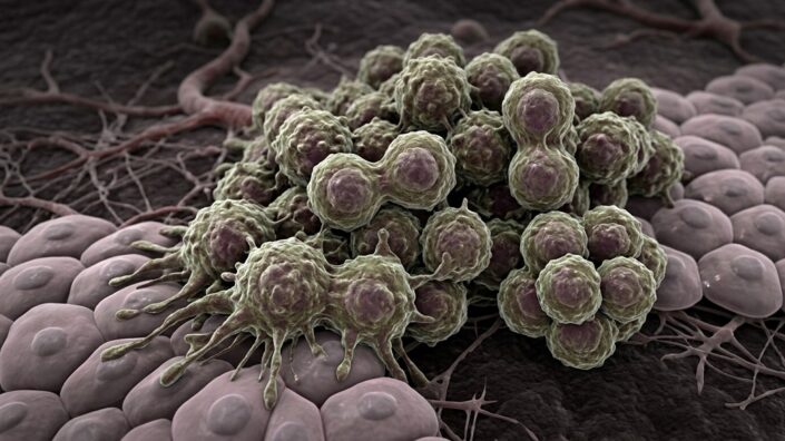
A recent study has examined whether senescence might allow cancer cells to survive oncogene withdrawal and become even more dangerous [1].

Some blocking techniques are temporary

Many cancers depend heavily on a particular oncogenic signal to maintain their growth. Drugs that block these drivers can, therefore, produce dramatic tumor shrinkage. A major clinical problem, however, is that a small population of cancer cells may survive treatment and eventually regenerate the tumor, which no longer responds to the same treatment and often grows in a more aggressive, invasive manner. The biological changes that allow these surviving cells to persist during prolonged suppression of the oncogene are not fully understood.

Therefore, these investigators used a genetically controllable system in which cancer cell growth is driven by the SV40 large T antigen (Tag). Expression of this oncogenic protein could be switched on or off by adding or removing doxycycline, respectively.

Doxycycline can be used either by directly adding it to a cell culture or by adding it to the drinking water given to animals who carry such modified cells. This provides a way to reproduce, experimentally, the situation in which a tumor suddenly loses the oncogenic signal on which it has become dependent. The researchers followed the cells after oncogene withdrawal both in culture and in tumors grown in mice.

They also examined whether similar phenomena occur in a human cancer model. For this purpose, they used A375 melanoma cells carrying the common BRAFV600E mutation and treated them with vemurafenib, a drug that inhibits mutant BRAF. This second model tested whether the observations from the engineered mouse system might also apply to a clinically relevant form of targeted therapy.

Oncogene loss drives cellular senescence

When the oncogenic driver was removed, tumor cells rapidly stopped dividing and developed morphological and molecular characteristics associated with senescence. They became larger and flatter and accumulated senescence-associated β-galactosidase, a well-known biomarker. At the same time, cell-cycle regulator expression changed in a way that signified durable proliferation arrest: the cells were no longer able to divide.

Interestingly, this process did not follow the classic pattern in which p16 is strongly induced. Instead, senescence induction relied largely on a p21-associated mechanism. The authors explained this unusual pattern in terms of the interaction between SV40 Tag and the tumor-suppressor proteins p53 and Rb. Once Tag was removed, the regulatory relationship between these proteins changed, producing a form of senescence that differs from the canonical pathway.

Senescent cells remain biologically active

Although the cells stopped proliferating, they did not become metabolically or functionally inert. They altered their gene-expression programs and began producing a range of secreted molecules associated with inflammatory signaling and tissue remodeling.

These cells also underwent substantial metabolic adaptation. Rather than simply reducing energy production, they increased activity in both glycolysis and mitochondrial respiration. This suggests that the surviving population enters an energetically demanding state in which multiple metabolic pathways are running simultaneously. Such flexibility may help cells remain viable while they are unable to divide.

This finding changes our understanding of residual senescent cancer cells. They may appear dormant because they are no longer proliferating, but they remain metabolically active and capable of substantially influencing their surroundings.

Senescence can be followed by tumor regrowth

The most important observation of the study was that tumor cells that experienced oncogene withdrawal were more capable of producing recurrent tumors than cells that had not undergone this state. Tumors initially regressed when the oncogenic driver was switched off, but a subset subsequently began growing again.

In some animals, tumors eventually emerged even though the original oncogene remained suppressed. Nine of twelve animals in one experimental group developed tumors after a long delay despite continued inhibition of Tag. This demonstrates that recurrence did not necessarily require restoration of the original oncogenic stimulus. Instead, some cells acquired new ways of sustaining proliferation.

Thus, this study suggests a two-stage process: oncogene removal initially forces cancer cells into a non-proliferative state, but the prolonged survival of these cells creates an opportunity for genetic and functional adaptations that can eventually restore tumor growth.

Genetic changes accompany this escape from senescence

The cells isolated from recurrent tumors were substantially different from the original tumor population. They displayed extensive chromosomal abnormalities and increases in chromosome number, indicating that genome instability had developed during or after the senescent period. Such abnormalities can generate genetic diversity, potentially allowing a subset of cells to find alternative routes around the growth restriction imposed by oncogene loss.

The recurrent cells also displayed altered metabolic programs. Pathways involved in nucleotide production, amino-acid metabolism, and folate metabolism became more active, consistent with the increased biosynthetic requirements of cells that had returned to proliferation.

One of the most notable molecular changes was the increased expression of Mdm2, a protein that suppresses p53 activity. This provided a plausible mechanism for overcoming the growth arrest that had followed loss of SV40 Tag. Importantly, cells from recurrent tumors were particularly sensitive to an Mdm2 inhibitor, whereas the original tumor cells were not. This suggests that Mdm2 became a new dependency during the transition from oncogene dependence to oncogene-independent growth.

Changes in the tumor microenvironment

This study also indicates that recurrence cannot be explained entirely by alterations within the cancer cells. The investigators compared the immune and stromal composition of tumors before oncogene withdrawal, during tumor regression, and after recurrence.

The composition of the tumor environment changed substantially during this process. Recurrent tumors contained more endothelial cells, consistent with renewed blood-vessel formation. They also had fewer conventional dendritic cells and more regulatory macrophages. Together, these changes suggest that the environment surrounding the recurrent tumor becomes less favorable for effective immune surveillance and more supportive of tumor growth.

The secretory activity of senescent cells may contribute to this remodeling. Molecules released by these cells can affect neighboring immune, stromal, and vascular cells. Consequently, a population that initially suppresses tumor expansion by ceasing to divide may simultaneously create conditions that make later tumor growth easier.

Relevance to BRAF-targeted therapy

The experiments with human A375 melanoma cells provide evidence that the phenomenon is not restricted to the engineered mouse model. Inhibition of BRAFV600E with vemurafenib generated cells with several features of senescence, including prolonged growth arrest and changes in cellular morphology and secretory activity. The authors therefore suggest that a similar response could occur when human tumors are treated with drugs that remove a major oncogenic growth signal.

However, the authors also emphasize that the molecular mechanism is likely to depend on the genetic background of the cancer. For example, Mdm2-based escape may be particularly relevant to tumors in which p53 remains functional. Tumors carrying TP53 mutations would be expected to use different mechanisms to bypass senescence, so the therapeutic implications cannot simply be generalized to all cancers.

These findings have potentially important implications for targeted therapy. A treatment that efficiently suppresses an oncogenic driver may nevertheless leave behind a population of viable cells with a capacity to adapt. Consequently, preventing relapse may require strategies that eliminate these surviving cells or block the mechanisms they use to escape growth arrest.

We would like to ask you a small favor. We are a non-profit foundation, and unlike some other organizations, we have no shareholders and no products to sell you. All our news and educational content is free for everyone to read, but it does mean that we rely on the help of people like you. Every contribution, no matter if it’s big or small, supports independent ethical journalism and sustains our future.

Literature

[1] Schmitt, P., Hönig, K., Norcia, M. T., Nogueira, M. F., Flore, V., Vesperinas, I. S., … & Blankenstein, T. (2026). Oncogene inactivation-induced senescence facilitates tumor relapse. Nature Communications, 17(1), 6244.

Bicycle racers

Short, Intense Exercise Elicits Beneficial Metabolic Changes

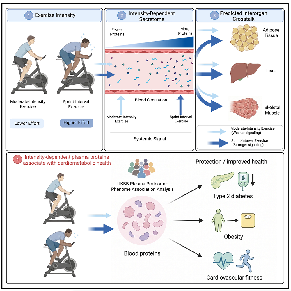
A new study compared high-intensity sprint-interval exercise with moderate-intensity exercise. The former produced larger changes in circulating protein and metabolite levels than the latter and stimulated proteins associated with cardiometabolic health benefits [1].

Short and intense vs. long and moderate

Exercise is a well-known lifestyle factor that can positively impact health. Exercise benefits multiple cells, tissues, and organs through factors that are secreted by various cells and circulate in the body [2]. However, the particular type of exercise that offers the greatest return on investment remains debated. Similarly, the circulating factors that mediate exercise benefits are still not fully defined.

Since intensity affects the composition of secreted molecules, the researchers employed “young, active, metabolically healthy males” in order to understand how exercise intensity affects these molcules and their crosstalk between organs. One group performed sprint-interval exercise (SIE), a routine that consists of short bursts of physical activity: here, it was 6 sets of 30-second all-out cycling with 4-minute rests between sets. The other group did 90 minutes of continuous cycling as moderate-intensity exercise (MIE), with individually adjusted intensity.

Intensity-dependent changes

Initial analysis of all proteins (the proteome) in plasma showed intensity-dependent changes, encompassing almost a quarter of the total detected proteins, including factors involved in the formation of new blood vessels (angiogenesis), extracellular matrix remodeling, gut signaling, and potential neuroregulation, immediately after SIE. Most of these proteins returned to resting levels three hours after exercise, showing that SIE drives rapid changes in the plasma proteome. MIE shows only modest time-dependent changes.

The researchers also noted intensity-dependent and time-dependent changes in secreted metabolites following both SIE and MIE. For SIE, significant changes were observed immediately after exercise and again three hours later, while MIE showed a delayed response: only a few molecules changed immediately after exercise, but that number increased three hours later. The metabolites that changed following SIE are associated with high energetic demands, while those that changed following MIE reflect the sustained energetic demands that are characteristic of continuous exercise.

The researchers repeated the experiment on a subset of participants who underwent 8 weeks of training; similar results emerged. Similar observations also emerged when the researchers tested runners, suggesting these changes are specific to exercise intensity rather than training level or exercise modality.

“What’s exciting here is that just a few minutes of intense exercise can trigger a significant molecular response,” says Paul Cohen, Associate Professor at The Rockefeller University and the corresponding author of the study. “And we still see it after eight weeks of training, which tells us this response isn’t simply a product of the body struggling to keep up with unfamiliar stress. It may be that the responses we observed are intrinsic to intense exercise.”

Multi-organ crosstalk

Because those exercise-responsive proteins and metabolites were in plasma, the question is which organs released them and which organs they affect. Investigating this question suggested crosstalk among many organs and the systemic effects of the studied metabolites.

First, experiments suggested that multiple organs can be a potential source for plasma proteins released following both SIE and MIE, with immune-system proteins most represented. However, later experiments focused on skeletal muscle, a tissue known to be affected by physical exercise. Experiments using human and mouse skeletal muscle cell cultures and skeletal muscle samples collected from study participants before and three hours after SIE and MIE suggested a role for skeletal muscle in intensity-dependent release of organ-specific proteins, specifically muscle fiber-derived proteins, which was greater following SIE.

Next, the researchers focused on the tissues affected by those circulating proteins, since secreted proteins can affect organs if they bind to receptors on their surfaces. There were multiple ligand-receptor pairs that were differentially regulated following SIE but not MIE.

Using primary human adipocyte cell cultures as an example, the authors showed that exercise intensity can impact gene expression in these tissues, with significant changes following SIE and modest changes following MIE. While only adipose tissue was investigated, it is likely not the only tissue affected by exercise-intensity-dependent protein release, but further research needs to assess the extent of cross-talk between organs following exercise.

Cardiometabolic health benefits

The experimental data suggested that exercise intensity affects protein release, which impacts interorgan communication. Those processes, in turn, affect systemic metabolism. The observed changes in protein levels, while transient, might have long-term effects if repeated in regular bouts of physical activity. Previous studies proposed a link between repeated exposure to such “transient increases in beneficial circulating proteins” and cardiometabolic health [3]. This study’s authors asked whether the proteins identified as being regulated by SIE and MIE are associated with health outcomes.

Using UK Biobank data and their own data, they identified protein-disease associations. Among the identified associations, they focused on plausible links to cardiometabolic benefits. This allowed them to identify 143 proteins associated with multiple disease groups, most of which were regulated solely following SIE. Narrowing their search to proteins associated with a lower risk of metabolic disorders, obesity, and type 2 diabetes identified 33 proteins. All but one of those proteins were differentially regulated following SIE, which is a stark contrast to only 3 proteins that were regulated by MIE.

The intensity-dependent systemic benefits of exercise also appear to have long-term effects, as more than a quarter of the 33 identified proteins had previously been inversely associated with age.

Exercise Secretome

Mediators of health-promoting effects

Overall, this study shows exercise intensity-dependent changes in the plasma proteome and their systemic impact, and it provides understanding as to how short bursts of intense exercise can elicit whole-body health benefits.

As Luke Olsen, the postdoctoral fellow who conducted the studies, summarizes, “It’s well appreciated that different intensities of exercise stimulate distinct body-wide adaptations.” “However, the molecular mechanisms linking these intensity-dependent adaptations have remained largely elusive. Our work suggests that exerkines—proteins and metabolites released into the bloodstream following exercise—are highly sensitive to exercise intensity and may be the key mediators of the health-promoting effects of short bursts of vigorous exercise.”

We would like to ask you a small favor. We are a non-profit foundation, and unlike some other organizations, we have no shareholders and no products to sell you. All our news and educational content is free for everyone to read, but it does mean that we rely on the help of people like you. Every contribution, no matter if it’s big or small, supports independent ethical journalism and sustains our future.

Literature

[1] Olsen, L., Botella, J., Barrows, D., Romero, E., Baird, K., Katayama, M., Kilic, E., Peralta, C., Zanou, N., Sanford, H., Farrell, L., Axelrod, C. L., Plucińska, K., Walker, J., Yan, L., Fredrickson, K., Pourquie, O., Robbins, J. M., Vinogradova, E. V., Molina, H., … Cohen, P. (2026). Exercise intensity modulates interorgan communication and is associated with cardiometabolic health outcomes in humans. Cell reports. Medicine, 102988. Advance online publication.

[2] Chow, L. S., Gerszten, R. E., Taylor, J. M., Pedersen, B. K., van Praag, H., Trappe, S., Febbraio, M. A., Galis, Z. S., Gao, Y., Haus, J. M., Lanza, I. R., Lavie, C. J., Lee, C. H., Lucia, A., Moro, C., Pandey, A., Robbins, J. M., Stanford, K. I., Thackray, A. E., Villeda, S., … Snyder, M. P. (2022). Exerkines in health, resilience and disease. Nature reviews. Endocrinology, 18(5), 273–289.

[3] Robbins, J. M., Katz, D. H., Many, G. M., Rao, P., Smith, G. R., Tiwari, G., Jin, C., Spielmann, G., Montalvo, S., Iyer, G., Amar, D., Leach, D., Coyne, B. J., Lindholm, M. E., Goodpaster, B., Walsh, M. J., Clish, C. B., Burant, C. F., Gerszten, R. E., & MoTrPAC Study Group (2026). Blood Biochemical Responses to Acute Exercise: Findings from the Molecular Transducers of Physical Activity Consortium (MoTrPAC). bioRxiv : the preprint server for biology, 2026.03.02.704798.

Rat tendons

A Tougher Extracellular Matrix Strengthens Tendons in Rats

Researchers have found that an upstream promoter of two extracellular matrix proteins increases healing and strength capabilities in the tendons of rats.

Tendon injuries in older people

Previous research has found that, like with many other injuries, tendon injuries have an age-related component. The tendons of the biceps and rotator cuffs are more commonly injured in older people than in younger people [1], and older people heal slower after forearm injuries and have less range of motion in the fingers for a longer time [2].

Previous work has investigated this issue using explanted murine models, taking mouse tendons from the animals and investigating their functional abilities. Tendons from aged mice don’t handle stress deprivation as well [3], nor do they respond properly to added strain [4]. An aged tendon can often support the same amount of force as a young tendon; it simply heals slower [5].

In humans, tendon stem progrenitor cells (TSPCs) change with aging in their gene expression, including genes related to basic functions such as motility and the cellular skeleton [6], and these cells often lack self-renewal abilities [7]. Animal studies have confirmed that cell numbers along with the organization of elastin, a foundational protein in tendon function, decline as well [8].

This study began with a statistical, population-based analysis of trends in tendon injury. This analysis found that, unsurprisingly, tendon injuries, which are often caused by heavy lifting at work, decreased between 1990 and 2021. A country’s development index corroborated this idea; countries undergoing industrial development may have more than less-developed countries, but after a certain level of development, this decreases. However, despite not being under such stressful conditions, older people even today remain at a high risk of such injuries.

Male and female rat tendons age differently

The researchers then turned to rats, taking tendons from eight-week-old (young) and 18-month-old (old) groups of males and females, then stretching them a hundred times to simulate normal mechanical load. They found that in male but not female rats, tendon weight and, surprisingly, tensile strength significantly increased between the two groups; in female but not male rats, the tensile strength, force required to cause a 2-millimeter gap, and stiffness were all weakened with aging.

These changes occurred alongside cell type changes. In female rats, there were proportionaly more epithelial and immune cells with aging, and the numbers of fibroblasts and stromal cells decreased. In male rats, immune cells rose as well, but the proportion of fibroblasts increased rather than decreased; stromal cells also declined, but so did epithelial cells.

More extracellular matrix expression helps rat tendons

In both sexes, however, the researchers noted significant declines in the expression of two genes related to the extracellular matrix: Col1a1 and Sparc. This was also accompanied by a decrease in the transcription factor Creb3l1, which these researchers found to be a regulator of these genes, binding directly to their promoters. Transfecting rat tendon cells with a lentivirus that increases Creb3l1 was also found to increase both Col1a1 and Sparc; silencing Creb3l1 led to a substantial increase in cellular senescence.

The researchers then injected this lentivirus into living animals in order to determine its effects on tendon tissue. In males, tendon elasticity and strength was significantly increased after three weeks, and in both sexes, tendons appeared to heal better as well, with the treatment groups having tendinous tissue at the sites of injury while the control groups had granulous tissue instead.

Rats have different biomechanical stresses than people, and rat tendons taken outside the body are no substitute for human results. However, this study shines significant light on sex differences that should be examined for their relevance to humans, and it suggests a potential path forward for a treatment that encourages proper tendon repair by affecting proteins related to the extracellular matrix. Future work is required to determine how such a treatment can be developed and whether it could reduce strains and sprains in older people.

We would like to ask you a small favor. We are a non-profit foundation, and unlike some other organizations, we have no shareholders and no products to sell you. All our news and educational content is free for everyone to read, but it does mean that we rely on the help of people like you. Every contribution, no matter if it’s big or small, supports independent ethical journalism and sustains our future.

Literature

[1] Clayton, R. A., & Court-Brown, C. M. (2008). The epidemiology of musculoskeletal tendinous and ligamentous injuries. Injury, 39(12), 1338-1344.

[2] Edsfeldt, S., Eklund, M., & Wiig, M. (2019). Prognostic factors for digital range of motion after intrasynovial flexor tendon injury and repair: long-term follow-up on 273 patients treated with active extension-passive flexion with rubber bands. Journal of Hand Therapy, 32(3), 328-333.

[3] Connizzo, B. K., Piet, J. M., Shefelbine, S. J., & Grodzinsky, A. J. (2020). Age-associated changes in the response of tendon explants to stress deprivation is sex-dependent. Connective tissue research, 61(1), 48-62.

[4] Aggouras, A. N., Stowe, E. J., Mlawer, S. J., & Connizzo, B. K. (2024). Aged tendons exhibit altered mechanisms of strain-dependent extracellular matrix remodeling. Journal of Biomechanical Engineering, 146(7), 071009.

[5] Ackerman, J. E., Bah, I., Jonason, J. H., Buckley, M. R., & Loiselle, A. E. (2017). Aging does not alter tendon mechanical properties during homeostasis, but does impair flexor tendon healing. Journal of Orthopaedic Research, 35(12), 2716-2724.

[6] Kohler, J., Popov, C., Klotz, B., Alberton, P., Prall, W. C., Haasters, F., … & Docheva, D. (2013). Uncovering the cellular and molecular changes in tendon stem/progenitor cells attributed to tendon aging and degeneration. Aging cell, 12(6), 988-999.

[7] Zhou, Z., Akinbiyi, T., Xu, L., Ramcharan, M., Leong, D. J., Ros, S. J., … & Sun, H. B. (2010). Tendon‐derived stem/progenitor cell aging: defective self‐renewal and altered fate. Aging cell, 9(5), 911-915.

[8] Godinho, M. S., Thorpe, C. T., Greenwald, S. E., & Screen, H. R. (2017). Elastin is localised to the interfascicular matrix of energy storing tendons and becomes increasingly disorganised with ageing. Scientific reports, 7(1), 9713.

Microglia attacking a neuron

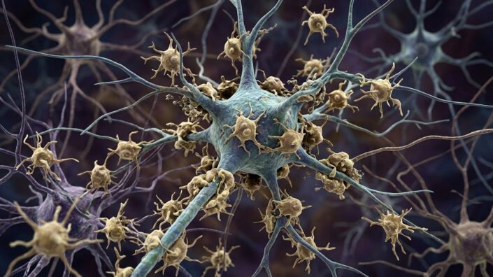
In ALS, Microglia Eat Living Neurons, Mistaking Them for Dead

A new study suggests that in amyotrophic lateral sclerosis – and possibly other neurodegenerative diseases – the brain’s immune cells devour stressed but still living neurons due to altered signaling [1].

The living dead

Amyotrophic lateral sclerosis (ALS), an age-related neurodegenerative disease, is characterized by the progressive loss of motor neurons. Historically, much of ALS research focused on what goes wrong inside the neurons themselves. However, it is becoming clear that neighboring cells, particularly the brain and spinal cord’s resident immune cells (microglia), can strongly influence neurons’ survival.

Microglia are essentially phagocytes: among other jobs, they recognize damaged or dead cells and engulf them. Microglia activation has been linked to several neurodegenerative conditions [2] and is known to be triggered by elevated expression of the tyrosine kinases AXL and MER from the TAM receptor family. TAM machinery recognizes its targets via phosphatidylserine (PtdSer), a lipid normally confined to the inner surface of the cell membrane. During cellular death by apoptosis, it flips to the outer surface and essentially tells phagocytes to attack the cell.

The authors of a new study published in Nature Communications suspected that something in this signaling system goes wrong in ALS, making microglia attack neurons that are not actually dying. “Cells that are dying throw an ‘eat me’ sign out on their surface, and the TAM system recognizes that sign,” explains Greg Lemke, distinguished professor emeritus at Salk Institute and a lead author. “It’s an essential system that clears billions upon billions of dead and dying cells from the body daily. We wondered whether microglia were corrupting this TAM system to kill living neurons in ALS.”

Inside out

The researchers started by investigating whether this machinery is actually engaged in human disease by examining postmortem lumbar spinal cord from six people with sporadic ALS and three age-matched controls. In microglia, MER was about threefold higher in ALS than in healthy controls, while AXL was increased about 16-fold.

They then reproduced this in a mouse model of ALS (SOD1G93A mice). As the disease progressed, Axl and Mer expression rose in the spinal cord. Both receptors were concentrated mostly in microglia rather than in motor neurons or astrocytes.

To see if the ligand part of the pathway was also activated, the authors examined Gas6, one of the proteins that bridges PtdSer on a target cell to TAM receptors on a phagocyte. In human spinal cord tissue and in SOD1 mice, motor-neuron GAS6 expression was significantly higher in ALS than in controls.

Next, the team looked for externalized PtdSer, which usually appears on dying cells and signals to TAM. They detected essentially no externalized PtdSer in normal spinal cord, whereas late-stage SOD1 mice contained many PtdSer-positive cells, including motor neurons.

That could simply mean these neurons were already dying by apoptosis. So, the researchers stained for cleaved caspase-3, a marker of ongoing apoptosis, but most PtdSer-positive neurons tested negative for it. Therefore, the authors concluded that the great majority of neurons displaying the “eat-me” signal were still alive rather than truly apoptotic.

Get ill sooner, die later

When the researchers crossed SOD1 mice with mice lacking both Axl and Mer, this produced an unexpected result. These mice developed their earliest signs of illness about 30 days sooner but reached the terminal clinical endpoint roughly three weeks later than ordinary SOD1 mice.

TAM signaling is important for immune homeostasis, so, TAM-deficient mice develop autoimmunity, inflammation, and several other pathologies sooner. However, as ALS progressed, the knockout produced a large survival advantage. That led to the next question: were motor neurons actually being preserved? Apparently, they were, as SOD1 mice lacking Axl and Mer retained roughly three times as many motor neurons as ordinary SOD1 mice.

“It somehow was not devastating,” says first author Youtong Huang, Ph.D., a former graduate student researcher in Lemke’s lab. “When we looked at how many motor neurons mice without Axl and Mer had, compared to mice with Axl and Mer, we found losing the TAM proteins meant preserving muscle control.”

The team then discovered that it was more than just neuron bodies surviving: in Axl and Mer-deficient SOD1 mice, the neurons’ axons remained connected to muscles, and muscle sizes were preserved compared to regular SOD1 mice.

One design problem needed to be addressed: Axl and Mer having been deleted throughout the body from development onward. The team staged a cleaner experiment by creating a mouse model where deletion of TAM receptors was triggered by tamoxifen – mostly in microglia in adult animals rather than from every cell throughout life.

They turned microglia-confined silencing of Axl and Mer on day 100, when disease was already developing, which substantially reproduced the survival benefit of the germline knockout. On day 160, none of the tamoxifen-treated animals had reached the clinical endpoint, compared with about 60% of vehicle-treated controls. Conditional TAM deletion also preserved significantly more spinal motor neurons and partially rescued neuromuscular connections.

Finally, the authors demonstrated that knocking out the TAM receptors does reduce the “devouring” of motor neurons by microglia. In ordinary SOD1 mice, microglial lysosomes contained abundant neuronal material, but Axl/Mer deletion reduced this material by roughly tenfold.

“The bottom line is, microglia are using the TAM system to eat cells that aren’t dead,” said Lemke. “There is enormous potential for this in clinical translation. Rather than engineering entire cells as immunotherapies – a process that is far more complicated, time-consuming and invasive – we could simply design TAM-based proteins that target any cell you’d like. I’m really excited to see where this discovery goes and how it changes immunotherapy opportunities.”

Very similar TAM activation has been observed in Alzheimer’s and Parkinson’s disease models, including by the same team [3]. The authors therefore speculate that PtdSer-dependent killing of stressed-but-living neurons by microglia may be a general mechanism of neurodegeneration that is not restricted to ALS.

We would like to ask you a small favor. We are a non-profit foundation, and unlike some other organizations, we have no shareholders and no products to sell you. All our news and educational content is free for everyone to read, but it does mean that we rely on the help of people like you. Every contribution, no matter if it’s big or small, supports independent ethical journalism and sustains our future.

Literature

[1] Huang, Y., Mavinkurve, A., Sabikunnahar, B., Stevens, B., & Lemke, G. (2026). Microglia deploy TAM receptors to kill motor neurons in a mouse model of amyotrophic lateral sclerosis. Nature Communications.

[2] Hickman, S., Izzy, S., Sen, P., Morsett, L., & El Khoury, J. (2018). Microglia in neurodegeneration. Nature neuroscience, 21(10), 1359-1369.

[3] Fourgeaud, L., Través, P. G., Tufail, Y., Leal-Bailey, H., Lew, E. D., Burrola, P. G., … & Lemke, G. (2016). TAM receptors regulate multiple features of microglial physiology. Nature, 532(7598), 240-244.

Fat cells

How Old Fat Cells Trigger Inflammation and Raise Risks

Using cohort studies and a mouse model, researchers have found that the circulating factor ANGPTL8, which is produced by senescent fat cells, is related to age-related diseases and mortality in mice and people.

Too much of a regulator leads to dysregulation

This paper introduces itself with a standard explanation of the relationship between senescence and inflammaging, including the contributions of the senescence-associated secretory phenotype (SASP) that senescent cells emit. The authors state the need for a clear understanding of the “molecular mediators” involved, focusing specifically on the contributions of fat (adipose) tissue, which has been identified in previous work as a crucial link [1].

Adipose tissue is not just energy storage; it is an endocrine organ all its own that regulates metabolism [2], and the aging of this tissue pushes it towards dysregulation and inflammation [3]. This includes SASP emissions, which senescent adipose tissue creates in sufficient quantities to stimulate systemic inflammation [4]. This paper makes it clear that aging fatty tissues are a cause, rather than just a consequence, of other aspects of aging [5].

The related molecule on which this paper focuses is ANGPTL8, a metabolic regulator that itself is upregulated when insulin is administered [6]. However, this team has previously pinpointed it as being related to multiple serious metabolic issues, such as diabetes-related kidney disease (nepropathy) [7]. Other teams have corroborated such findings, finding it to be related to diabetic atherosclerosis [8] and a higher risk of future heart attacks in existing cardiovascular disease patients [9]. Additionally, higher circulating ANGPTL8 levels have been found to be associated with all-cause mortality in diabetic patients [10].

A significant predictor

As the first part of this paper, the researchers built their own biomarker-based clock, MBA8-Clock using data from the China Cardiometabolic Disease and Cancer Cohort, which recruited nearly ten thousand people. Comparing five separate algorithms, the team found that a multilayer perceptron-based framework was the best way to utilize this clock, as it gave estimated ages with an error rate of approximately five years.

This clock contained many basic measurements of metabolic health, including ANGPTL8. The researchers found that circulating ANGPTL8 values of approximately 250 nanograms per liter of serum were associated with the lowest biological age estimations. At values higher than that, particularly in people with four times as much or more, biological age was likely to be estimated markedly higher. Removing ANGPTL8 from the clock notably diminished its predictive abilities. The researchers hold that “circulating ANGPTL8 represents a consistent, biologically meaningful contributor to age prediction across individuals.”

The next step was to create another clock, AIMR-Model, this one based on all-cause mortality over a ten-year period. The researchers used five algorithmic analyses, and this time they discovered that an extreme gradient-based framework yielded the most accurate results. Once more, ANGPTL8 was found to be a significant contributor; people who had been found to have higher ANGPTL8 levels were significantly more likely to have died within 10 years of that initial assessment. Further analysis revealed that “ANGPTL8 may amplify age-related clinical vulnerability rather than acting solely as an isolated risk marker,” as it was consistently found to occur alongside high blood pressure and other well-known risk factors for age-related disease.

Mice without ANGPTL8 live longer

The researchers then turned to mice. As in humans, ANGPTL8 naturally increases in mice with age: 4-month-old (young) wild-type mice have significantly less of it than their 22-month-old (old) counterparts. As expected, the source was found to be the adipose tissue rather than the liver or any other organs; while hepatocytes did somewhat increase their production of the protein, that was localized rather than systemic.

In addition, the researchers created a group of mice that do not express the murine gene Angptl8. Compared to their wild-type counterparts, the old Angptl8-less mice performed considerably better on various physical tests at older ages. They had less fat mass, less senescence in their fat, and reductions in circulating SASP factors. They were also able to hang onto a rotarod longer, performed better on a treadmill, engaged in more exploratory behavior, and had more gastrocnemius muscle mass along with more fast-twitch fibers. Finally, and perhaps most importantly, these mice also lived longer.

Survival curve ANGPTL8

The specific pathway involved in the relationship between ANGPTL8 and senescence was identified. This protein was found to bind to AKT2 on the molecular level, affecting the fundamental AKT/mTOR/S6K pathway, and this finding was confirmed in mice; mice with Angptl8 turned off have significantly decreased activity of AKT, mTOR, and S6K. Deriving fat precursor cells (preadipocytes) from older mice confirmed these findings, and experiments using cells with Angptl8 overexpression found that AKT is indeed necessary for ANGPTL8 to contribute to cellular senescence.

These researchers are of the clear opinion that they have hit upon a significant target, stating that “from a translational standpoint, the dual identity of ANGPTL8 as both a circulating biomarker and a functional effector of aging is particularly compelling.” However, this paper did not test any potential method of removing circulating ANGPTL8 from the bloodstreams of wild-type animals. If such a drug can be developed and proven to work in a human clinical trial, it may be possible to grant additional healthspan and lifespan to older people suffering from metabolic issues.

We would like to ask you a small favor. We are a non-profit foundation, and unlike some other organizations, we have no shareholders and no products to sell you. All our news and educational content is free for everyone to read, but it does mean that we rely on the help of people like you. Every contribution, no matter if it’s big or small, supports independent ethical journalism and sustains our future.

Literature

[1] Hotamisligil, G. S. (2017). Inflammation, metaflammation and immunometabolic disorders. Nature, 542(7640), 177-185.

[2] Ou, M. Y., Zhang, H., Tan, P. C., Zhou, S. B., & Li, Q. F. (2022). Adipose tissue aging: mechanisms and therapeutic implications. Cell death & disease, 13(4), 300.

[3] Tchkonia, T., Morbeck, D. E., Von Zglinicki, T., Van Deursen, J., Lustgarten, J., Scrable, H., … & Kirkland, J. L. (2010). Fat tissue, aging, and cellular senescence. Aging cell, 9(5), 667-684.

[4] Dahlquist, K. J., & Camell, C. D. (2022). Aging leukocytes and the inflammatory microenvironment of the adipose tissue. Diabetes, 71(1), 23-30.

[5] Franceschi, C., Garagnani, P., Vitale, G., Capri, M., & Salvioli, S. (2017). Inflammaging and ‘Garb-aging’. Trends in Endocrinology & Metabolism, 28(3), 199-212.

[6] Abu-Farha, M., Ghosh, A., Al-Khairi, I., Madiraju, S. M., Abubaker, J., & Prentki, M. (2020). The multi-faces of Angptl8 in health and disease: Novel functions beyond lipoprotein lipase modulation. Progress in lipid research, 80, 101067.

[7] Pan, L., He, Y., Xiang, Y., Mao, B., Meng, X., Guo, Y., … & Yu, X. (2025). Angiopoietin-like protein 8 mediates inflammation and fibrosis of tubular cells in diabetic kidney disease progression by interacting with Akt2. Metabolism, 156418.

[8] Ye, H., Zhu, Q., Zong, Q., Luo, S., Ji, Z., Zhang, R., & Zou, H. (2026). Elevated ANGPTL8 (Angiopoietin‐Like Protein 8) Levels as a Novel Predictor of Atherosclerosis in Type 2 Diabetes: Beyond Lipid Metabolism. Journal of the American Heart Association, 15(3), e044806.

[9] Morinaga, J., Kashiwabara, K., Torigoe, D., Okadome, Y., Aizawa, K., Uemura, K., … & Oike, Y. (2023). Plasma ANGPTL8 levels and risk for secondary cardiovascular events in Japanese patients with stable coronary artery disease receiving statin therapy. Arteriosclerosis, thrombosis, and vascular biology, 43(8), 1549-1559.

[10] Zou, H., Xu, Y., Chen, X., Yin, P., Li, D., Li, W., … & Yu, X. (2020). Predictive values of ANGPTL8 on risk of all-cause mortality in diabetic patients: results from the REACTION Study. Cardiovascular Diabetology, 19(1), 121.

GenBio Launches a “Virtual Cell” AI Model

This AI startup, which lists Nobel laureate David Baker among its co-founders, has announced a “world model” of a cell that can simulate both its natural state and responses to successive perturbations, potentially transforming biological research.

Living in a simulation

Simulating a living organism on a computer (in silico) has been a dream of both computer scientists and biologists for decades. However, previous attempts have been hampered by the immense complexity of biology, which existing computational tools could not adequately recreate.

Recent advances in AI may have changed the equation considerably, giving scientists a much better shot at the target. Multiple teams are now developing foundation and other large AI models in biology, including Nucleotide Transformer, Evo, ESM, AlphaFold, GeneFormer, and many others. A foundation model is a large model trained on a very broad dataset so that it learns general-purpose representations that can later be reused for many different tasks.

However, even these huge models remain limited in their scope and modalities. Take AlphaFold, an AI model that predicts protein structures from amino acid sequences. Impressive as it is, it covers only a tiny sliver of what we call life. One proposed route toward actually simulating life is therefore to make multiple specialized models work together within a larger system.

The challenge of building a world

GenBio AI, a startup based in Palo Alto, CA, has just announced what may be an early step toward that goal. The company’s list of co-founders includes Nobel laureate David Baker, AI scientist Eric Xing, and other prominent life science and AI researchers. The press release, published today, describes AIDO Cell as a virtual cell “world model” and “the first system capable of simulating a human cell, both in its natural state and in response to drugs and other interventions, across its full biological hierarchy, from DNA and RNA through protein to the whole-cell level.”

“What’s exciting here isn’t that we’ve solved cellular biology – we haven’t, at least not yet,” said Baker, who received the 2024 Nobel Prize in Chemistry for his work in computational protein design. “It’s that, for the first time, we have a system capable of simulating a cell across the full hierarchy of biological scales, from DNA to whole-cell behavior, in one place, which lets you interrogate it computationally.”

An accompanying Perspective in Nature Medicine, published several days ago by three of the co-founders – Eric Xing, Eran Segal, and Le Song – describes how such a system could be built in three stages. Stage 1 involves building strong foundation models for individual biological modalities. At Stage 2, mechanisms are developed to link those models across modalities and biological scales. Finally, at Stage 3, the entire network is jointly aligned and optimized so that it behaves coherently as one system.

Ultimately, the researchers envision AIDO drawing on many forms of information – sequences, structures, pathways, transcriptomics, metabolomics, imaging, and spatial and longitudinal data – and integrating them across biological levels. A crucial feature is the ability to feed information back from higher biological levels to lower ones. AIDO Cell is also stateful, meaning that a sequence of perturbations can build on one another rather than being treated as a series of isolated predictions. A more detailed explanation can be found in the technical paper.

GenBio Stages

In addition to developing a reliable enough “common language” for models operating at very different biological scales without losing important information in translation, another major challenge is preventing small inaccuracies from accumulating as predictions propagate across those scales.

“A key part of AIDO’s design is to avoid treating biology as a one-way chain of predictions, where a small error at the molecular level could become a much larger error by the time you reach the cell or patient level,” said Le Song, co-founder and CTO. “Instead, AIDO uses feedback across biological scales: predictions made at the molecular, cellular, and higher levels can be checked against real measurements and biological outcomes, and that feedback can be used to improve the system as a whole. You can think of it somewhat like the feedback and alignment process used to improve large language models.”

Experimental and accessible

What can AIDO Cell actually do? In one early demonstration, GenBio used the system to model the effects of imatinib on leukemia cells and found that it could reproduce the drug’s known mechanism of action across several levels of cellular biology.

AIDO Cell currently supports K562 and HepG2, two of the most widely used immortalized human cell lines in biomedical research, derived, respectively, from a patient with chronic myeloid leukemia and from a liver tumor. They are commonly used as laboratory models of blood cancer and liver biology. While additional cell types are in development, the current version remains an early-stage system. GenBio itself describes it as a preview and an early functional demonstration.

GenBio is also preparing to launch an early-access academic collaborator program for scientists across academia, biotech, and pharma, and it plans to release more advanced versions of the system later this year and over the next year.

“Unlike our competitors, we do not believe that virtual cell models belong behind closed doors,” said another co-founder, Emma Lundberg, professor at Stanford University and Co-Director of the Human Protein Atlas. “We’ve not only built a virtual cell model, we’ve also built an AI that can rapidly build a tailored virtual cell for your question, on your data. Bring your data, and we’ll build it for you.”

In the near term, AIDO Cell could serve as a virtual testing ground for drugs, allowing researchers to explore cellular effects in silico before committing to physical experiments. If sufficiently accurate, that could help eliminate weaker candidates earlier and focus resources on the most promising ones.

“Having led AI for R&D at a major pharmaceutical company, the major challenge we were facing was the inability to accurately determine how a drug behaves in a real cellular environment,” said Ziv Bar-Joseph, co-founder and CSO. “This often led to many clinical trial failures. A system like AIDO Cell enables companies to quickly and accurately evaluate several drug candidates leading to better, safer and more efficient treatments years earlier than is possible today.”

There is a broader scientific promise as well. AIDO Cell and similar systems could eventually become powerful tools for basic biological research, allowing scientists to perturb genes, proteins, or pathways, follow the predicted consequences across biological levels, generate hypotheses, and identify relationships that might take much longer to uncover in the lab.

We would like to ask you a small favor. We are a non-profit foundation, and unlike some other organizations, we have no shareholders and no products to sell you. All our news and educational content is free for everyone to read, but it does mean that we rely on the help of people like you. Every contribution, no matter if it’s big or small, supports independent ethical journalism and sustains our future.
Brain cell types

How Senescence Spreads Between Brain Cells

Researchers have gone into deep detail regarding how each of five senescent brain cell types expresses and receives factors that encourage other cells to become senescent.

The contagion of age

This paper revolves around paracrine senescence: SASP factors being emitted by primary senescent cells to drive secondary senescence in other cells [1]. Senescent cells differ by both tissue type and senescence source, and primary senescent cells, which were driven senescent through other stressors, do not necessarily exhibit the same features or function in the same ways as secondary senescent cells [2]. Recent work has explored many of those differences in detail [3, 4].

The molecular causes of this spreading senescence are fairly well-established. The researchers specifically name the inflammatory factors TGFβ, IGF1, IL-6, IL-8, and CCL2 as being part of the SASP [5], and they mention the role of reactive oxygen species (ROS) and the inflammatory NFκ-B pathway as mediating the process within cells [6].

However, only limited amounts of this research have been done on brain cells. In this study, the researchers sought to fill that gap by taking a closer look at how SASP factors affect various types of these cells and what might be done about them.

Five brain cell types express the SASP very differently

This study used the expression of the well-known biomarker SA-β-gal to determine if a cell had become senescent. It used five cell types commonly found in the brain: astrocytes, endothelial cells, microglia, oligodendrocytes, and neurons. To create primary senescent cells, the researchers treated each of these types with 5-bromodeoxyuridine (BrdU) for a week, a technique that they had used in a previous study [3].

Each of these cell types developed a specific SASP profile after senescence was induced. A panel of 286 cytokines revealed that, while a small handful of cytokines was common between cell types, each type was expressing its own combination of factors, particularly microglia, oligodendrocytes, and neurons, which expressed more factors compared to astrocytes; endothelial cells were found to express very few of these factors at all. Oligodendrocytes and microglia expressed a considerable number of factors that were only common between these two groups.

The cells also responded much differently when treated with conditioned media derived from these groups (BrdU CM), a technique that would presumably induce secondary senescence. Interestingly, this did not cause neurons and oligodendrocytes to become secondarily senescent, regardless of the source. Despite their few SASP factors under direct BrdU exposure, astrocytes and endothelial cells would still become senescent when treated with BrdU CM derived from microglia or astrocytes. Microglia were found to be particularly susceptible to secondary senescence; any BrdU CM source other than endothelial cells would drive them senescent in this way.

There was a very interesting result: When normal astrocytes were exposed to BrdU CM from other astrocytes, their expression of CDNK1A, which encodes for the senescence-associated factor p21, would significantly increase. However, when microglia were exposed to the same astrocytic SASP, their expression of CDNK1A significantly decreased instead. Unsurprisingly, however, both astrocyte and microglia SASP increased the expression of the DNA damage marker γH2AX in both types of cells.

Neurons and oligodendrocytes were also found to not be completely immune to the SASP. Despite not becoming senescent, neurons were affected by BrdU CM from astrocytes and microglia, expressing extra CDNK1A along with CDNK2A, which increases p16. Oligodendrocytes’ related biomarkers were affected by the SASP of astrocytes, microglia, and, interestingly, endothelial cells.

Potential targets

This paper goes into the relationship of various SASP factors in exacting detail. The researchers frequently noted that the CM of these senescent cells did not always induce senescence; on some occasions, it was found to downregulate inflammatory factors instead, and there were also effects on MIF, a double-edged factor whose age-related effects are dependent on context [7].

The researchers also performed an extensive series of experiments using various drugs that inhibit the spreading of the SASP. Most of these drugs found partial successes; most notable was the drug Bindarit, which had significant, if incomplete, effects on senescence transmission between astrocytes. The factors CCL2, MIF, CXCR7, and DPP4 were identified as potential targets for future therapeutic work.

This study provides an illuminating look into the complexity inherent in dealing with the wide panoply of factors that make up the overall circulating SASP. However, even this level of detail only dealt with SASP factors taken from five cell types driven primarily senescent by chemical exposure; CM derived from the secondary senescent cells themselves was not studied here.

Additionally, this was only a cellular study; these findings were not confirmed in an animal model, and the researchers noted that conditioned media do not reflect the complete variety of factors found in the human brain. Animal experiments and human trials will have to be done in order to determine which, if any, of these factors might be a valuable target for reducing unwanted senescence in the brain.

We would like to ask you a small favor. We are a non-profit foundation, and unlike some other organizations, we have no shareholders and no products to sell you. All our news and educational content is free for everyone to read, but it does mean that we rely on the help of people like you. Every contribution, no matter if it’s big or small, supports independent ethical journalism and sustains our future.

Literature

[1] Martin, L., Schumacher, L., & Chandra, T. (2023). Modelling the dynamics of senescence spread. Aging Cell, 22(8), e13892.

[2] Teo, Y. V., Rattanavirotkul, N., Olova, N., Salzano, A., Quintanilla, A., Tarrats, N., … & Chandra, T. (2019). Notch signaling mediates secondary senescence. Cell reports, 27(4), 997-1007.

[3] Russo, T., Plessis-Belair, J., Sher, R., & Riessland, M. (2025). Systematic profiling reveals distinct senescence signatures and regulators across human brain cell types. Nature Communications, 16(1), 11059.

[4] Sweeney, E. M., Abate, G., Bakker, B. R., Mastinu, A., Lai, Y., Uberti, D., … & Tambaro, S. (2026). Cell Type‐Specific Expression of p16, p21, and p53 Reveals Age‐Dependent Glial Senescence in the AppNL‐G‐F Mouse Model of Alzheimer’s Disease. Aging Cell, 25(4), e70478.

[5] Admasu, T. D., Rae, M. J., & Stolzing, A. (2021). Dissecting primary and secondary senescence to enable new senotherapeutic strategies. Ageing research reviews, 70, 101412.

[6] da Silva, P. F., Ogrodnik, M., Kucheryavenko, O., Glibert, J., Miwa, S., Cameron, K., … & von Zglinicki, T. (2019). The bystander effect contributes to the accumulation of senescent cells in vivo. Aging cell, 18(1), e12848.

[7] Altulea, A., Nehme, J., & Demaria, M. (2026). Dual role of MIF in aging and cellular senescence. Cytokine & Growth Factor Reviews.

No getting fat

A Million-Person Study Finds a Protective Metabolic Gene

A massive human genetics study identified rare folliculin-interacting protein 1 (FNIP1) mutations associated with favorable metabolism and much lower cardiometabolic disease risk. Experiments in human liver cells and mice suggest a therapeutic route [1].

Longevity hidden in the genome

For decades, aging researchers have hoped that human genetics might reveal genuine “longevity genes” whose altered activity produces substantially healthier or longer lives and therefore provides an obvious therapeutic target. However, unlike in some simpler organisms, human aging seems to be highly influenced by numerous genes (polygenic), most of which have relatively small effects. The APOE gene, strongly associated with dementia, might count as an exception, as well as FOXO3, although the latter’s effect is not nearly as strong.

Importantly, that does not necessarily mean that genetics contributes little to lifespan. Some recent research argues that intrinsic human lifespan may be highly heritable while still being genetically complex [2]. Although these results are still a matter of active debate, there is no denying that our genes do affect our lifespan.

In a new study coming from the biotech company Regeneron and published in Nature, the researchers leveraged 11 large cohorts to analyze the protein-coding parts of the genome (the exomes) of more than one million people with extensive linked health and phenotype data.

60% risk reduction

The team focused on energy metabolism because metabolic dysfunction contributes to obesity, type 2 diabetes, cardiovascular disease, and metabolic-dysfunction-associated steatotic liver disease (MASLD). Rather than starting with one candidate gene, they used the ratio of triglyceride to high-density-lipoprotein cholesterol (TG:HDL) as a marker of metabolic state and searched for genetic variants associated with unusually high or low values.

The authors’ bet was that TG:HDL genuinely captures something meaningful about whole-body energy metabolism, and their analysis using a remarkably broad set of metabolic measurements confirmed that: a higher TG:HDL consistently tracked a worse metabolic phenotype. Importantly, this was not just cross-sectional but also longitudinal. People with higher baseline TG:HDL subsequently had higher rates of type 2 diabetes, myocardial infarction, MASLD, and cirrhosis. The relationships appeared across several ancestry groups.

Analyzing their one-million-strong dataset, the researchers found 59 genes in which rare protein-altering variants had independent, statistically robust effects on TG:HDL. Only 15 had been identified in the previous largest rare-variant TG:HDL analysis, meaning that 44 were new associations.

The 59 genes were strongly enriched for genes expressed in the liver and adipose tissue, the organs that govern lipid storage and energy metabolism. Many belonged to familiar pathways. 31 of the 59 encode known drug targets, and 23 are already targeted by approved drugs or agents in human clinical development – an encouraging sign that their genetic screen was picking up biologically and therapeutically relevant pathways.

People carrying ultra-rare loss-of-function mutations in one copy of the gene FNIP1 had much lower TG:HDL and one of the strongest favorable metabolic signatures in the entire study: the carriers had lower triglycerides and ApoB, less liver fat, better glycemic control, and a more favorable distribution of body fat.

Most importantly, heterozygous FNIP1 loss-of-function carriers had about 60% lower odds of a composite cardiometabolic disease outcome, which consisted of coronary artery disease (CAD), type 2 diabetes, MASLD, and cirrhosis. However, the estimate was based on relatively few mutation carriers, and the reductions in CAD and cirrhosis individually did not reach statistical significance.

“This study implicates the FNIP1 pathway in human energy metabolism and in the risk of common cardiometabolic diseases in the general population,” said Dr. Luca Andrea Lotta, Head of Cardiometabolic and Musculoskeletal Disease Genetics at the Regeneron Genetics Center and the study’s corresponding author, to Lifespan News. “It illustrates the power of large-scale human genetics to reveal new and important biology and identifies a pathway that may be modified for therapeutic benefit in these common diseases.”

A metabolic break

The next question was about why having one defective copy of FNIP1 would produce such a phenotype. FNIP1 partners with folliculin (FLCN) in a nutrient-sensing pathway downstream of AMPK, one of the cell’s central sensors of energy availability. Previous work suggested that reducing FNIP1/FLCN activity might increase mitochondrial activity, fuel oxidation, and cellular energy expenditure [3].

“The FNIP1 pathway can be considered a ‘metabolic brake,’ curbing the consumption of energy so that calories can be saved for a ‘rainy day’ when we may undergo prolonged fasting/starvation,” said Lotta. “This happened all the time over millennia of human evolution but almost never happens in the modern calorie-rich environment, so this pathway now leads to cardiometabolic diseases, and mutations that switch it off are protective.”

Knocking down FNIP1 in primary human hepatocytes activated a transcriptional program associated with lysosomal activity and lipid breakdown. Disrupting the FNIP1-FLCN pathway specifically in the livers of mice fed a high-fat, high-fructose diet protected against obesity, fatty liver, and insulin resistance. Importantly, however, in mice, the effect hinged on also inhibiting the closely related protein FNIP2. This was not the case in human liver cells. The researchers attribute this discrepancy to inter-species differences, but this might have consequences for clinical translation.

“Developing new medicines is always hard and this makes it more difficult to study this pathway in model organisms and preclinical models, so it adds a layer of complexity,” Lotta said. “However, our human cell experiments suggest that, in humans, silencing of FNIP1 alone should be enough. We are pursuing more testing to better understand the biology of this pathway.”

We would like to ask you a small favor. We are a non-profit foundation, and unlike some other organizations, we have no shareholders and no products to sell you. All our news and educational content is free for everyone to read, but it does mean that we rely on the help of people like you. Every contribution, no matter if it’s big or small, supports independent ethical journalism and sustains our future.

Literature

[1] Hindy, G., Adam, R. C., Sosina, O., Pryce, D., Blair, D., Herman, J., … & Lotta, L. A. (2026). FNIP1 variants are associated with favourable metabolism in 1 million humans. Nature, 1-10.

[2] Shenhar, B., Pridham, G., De Oliveira, T. L., Raz, N., Yang, Y., Deelen, J., … & Alon, U. (2026). Heritability of intrinsic human life span is about 50% when confounding factors are addressed. Science, 391(6784), 504-510.

[3] Malik, N., Ferreira, B. I., Hollstein, P. E., Curtis, S. D., Trefts, E., Weiser Novak, S., … & Shaw, R. J. (2023). Induction of lysosomal and mitochondrial biogenesis by AMPK phosphorylation of FNIP1. Science, 380(6642), eabj5559.

Three Against Cancer

A Drug Combination Fights Both Senescence and Cancer

In Aging, the Conboys and their team have described how a combination treatment kills both senescent and cancer cells and lengthens the lives of old mice.

Of senescence and cancer

Just as it does in mice, the incidence of cancer rises with age in people [1]. Among other things, senescence is an evolved defense against cancer, and the activation of cancer-related genes (oncogenes) can cause cells to stop dividing instead. On the other hand, SASP factors can encourage tumors to form [2] and remodel the extracellular matrix in a cancer-promoting way [3]. This can make chemotherapies and radiotherapies counterproductive over the long term; many such therapies work by inducing senescence, preventing cancer cells from proliferating, but the senescent cells then contribute to cancer [4].

In experiments, senolytics, which are usually meant to exclusively kill senescent cells, are the go-to approach. Some such drugs, including ABT-263 (navitoclax), also kill cancer cells. However, this is a BCL-2 family inhibitor that depletes the BCL-XL needed for platelets to function; at its normally effective doses, this leads to thrombocytopenia, a condition that leads to uncontrolled bleeding. Despite some progress in clinical trials, navitoclax currently remains unapproved by the FDA for human use. Additionally, depleting BCL-2 does not work against all cancers and can stabilize some cancer cells instead [5].

Interestingly, both senescent cells and cancer cells have altered mitochondrial energy generation. They do not produce as much ATP as healthy cells [6], have problems with ion leakage [7], and are more likely to engage in glycolysis for energy [8]. Targeting the mitochondria, then, is a logical approach and has been attempted in previous work [9]; however, no one has yet managed to refine this method into something appropriate for the clinic.

These researchers’ attempt to do so involves dichloroacetate and metformin, two drugs that have been previously reported to have antisenescence and anticancer properties [10, 11]. Combining the two has already been investigated as a method of fighting cancer [12]. However, this paper’s combination also includes navitoclax, albeit at a much lower dose than normal; the researchers termed this combination DMA.

More than the sum of its parts

To create their target, the researchers drove cells senescent using the toxin etoposide, creating damage-induced senescent (DIS) cells. These cells were used to determine the effective doses used in the final DMA cocktail: 5 mM of DCA, 5 mM of metformin, and 1μM of navitoclax, which comes out to roughly one-tenth of the 50 mg/kg normally used in mouse studies. Using these drugs at these doses separately was found to have little effect; the combination is only effective when used as a whole, killing roughly 60% of DIS cells while leaving control, non-senescent cells unscathed. Using this combination on cells kept at a much lower oxygen concentration, which mimics natural physiology, slightly decreased normal cell viability but killed 90% of the DIS cells.

Further cellular work confirmed the synergistic effects. A combination of DCA, metformin, and the BCL-2 inhibitor ABT-737 was more effective against senescent cells than ABT-737 alone. Additionally, DMA was found to significantly, but not completely, reduce the amount of SASP secreted by surviving senescent cells, even at very low doses.

While there appeared to be a slight downward trend, DMA did not have a significant effect on platelet count; meanwhile, navitoclax at its usual dose was confirmed to have a stark and significant effect in this area.

Effectiveness against cancer

The researchers first confirmed previous work on navitoclax’s relationship to cancer cells, using three distinct lines. Navitoclax killed the vast majority of SW480 cancer cells and only a quarter of HeLa cancer cells, and it did nothing against MCF-7, a line of cancer that is unaffected by this BCL-2 inhibitor as it uses a different version, MCL-1, to survive.

DMA, on the other hand, was significantly effective against all three. After a week of exposure and using DMSO for the control group, the researchers had found that it had killed roughly three-fourths of HeLa cells, four-fifths of SW480 cells, and practically all the MCL-7 cells: 0% detected viability. Similarly to the senescence experiments, individual components of this cocktail were ineffective against cancer. In the MCF-7 cells, proliferation was substantially diminished, and death by apoptosis was substantially increased.

DMA against cancer

This effectiveness against MCF-7 cells had nothing to do with MCL-1. Instead, it was due to DMA’s effects against ATP within the cells, in senescence and cancer. Both DCA and metformin are known to deplete ATP, particularly in already damaged cells, and navitoclax adds to this effect. The net effect of DMA was to starve DIS cells, replicatively senescent cells, and MCF-7 cells of energy, while normal cells were robust enough to survive. This was particularly evident under stress induced by FCCP, which causes cells to undergo maximal respiration. Normal cells treated with DMA were still able to respond to FCCP, but senescent and cancer cells were not, demonstrating their failure to produce sufficient ATP.

Increases in mouse lifespan

The researchers then applied DMA for two weeks, five days a week, to 18- to 24-month-old male and female mice. The injected mice performed better on treadmill tests than their uninjected counterparts, although hanging tests and overall frailty were unaffected. SASP molecules were broadly reduced, although not every molecule’s reduction reached the threshold of statistical significance.

A different experiment involved injecting 18-month-old mice with DMA for two weeks, five days a week, with eight-week breaks between administrations, until the mice died of age-related diseases. The effects on lifespan were substantial:

Interestingly, despite being treated with a combination of chemotherapeutic drugs, the average lifespan was not shortened but was instead extended by an average of 102.6 days, or a 41.7% increase post-injection.

The researchers describe their treatment as exploiting the vulnerability of senescent and cancer cells’ poor metabolic health, creating an environment that healthy cells tolerate but which such diseased cells cannot survive. They hint that this treatment may improve the long-term effectiveness of chemotherapy. They note that both DCA and metformin are already approved by the FDA, and clinical trials have already been conducted with navitoclax — which, in DMA, is not being used at a dose that should cause significant bleeding problems. However, only a proper clinical trial of DMA could determine if it is effective against senescence or any kind of cancer in human beings.

We would like to ask you a small favor. We are a non-profit foundation, and unlike some other organizations, we have no shareholders and no products to sell you. All our news and educational content is free for everyone to read, but it does mean that we rely on the help of people like you. Every contribution, no matter if it’s big or small, supports independent ethical journalism and sustains our future.

Literature

[1] Li, L., Shan, T., Zhang, D., & Ma, F. (2024). Nowcasting and forecasting global aging and cancer burden: analysis of data from the GLOBOCAN and Global Burden of Disease Study. Journal of the National Cancer Center, 4(3), 223-232.

[2] Kumari, N., Dwarakanath, B. S., Das, A., & Bhatt, A. N. (2016). Role of interleukin-6 in cancer progression and therapeutic resistance. Tumor Biology, 37(9), 11553-11572.

[3] Chidambaram, D., Subashini, V., Nanthanalaxmi, M., Saranya, I., & Selvamurugan, N. (2025). Regulation of matrix metalloproteinase-13 in cancer: Signaling pathways and non-coding RNAs in tumor progression and therapeutic targeting. World Journal of Clinical Oncology, 16(6), 105996.

[4] Wyld, L., Bellantuono, I., Tchkonia, T., Morgan, J., Turner, O., Foss, F., … & Kirkland, J. L. (2020). Senescence and cancer: a review of clinical implications of senescence and senotherapies. Cancers, 12(8), 2134.

[5] Wang, B., Ni, Z., Dai, X., Qin, L., Li, X., Xu, L., … & He, F. (2014). The Bcl-2/xL inhibitor ABT-263 increases the stability of Mcl-1 mRNA and protein in hepatocellular carcinoma cells. Molecular cancer, 13(1), 98.

[6] Miwa, S., Kashyap, S., Chini, E., & von Zglinicki, T. (2022). Mitochondrial dysfunction in cell senescence and aging. The Journal of clinical investigation, 132(13).

[7] Hutter, E., Renner, K., Pfister, G., Stöckl, P., Jansen-Duerr, P., & Gnaiger, E. (2004). Senescence-associated changes in respiration and oxidative phosphorylation in primary human fibroblasts. Biochemical Journal, 380(3), 919-928.

[8] Korolchuk, V. I., Miwa, S., Carroll, B., & Von Zglinicki, T. (2017). Mitochondria in cell senescence: is mitophagy the weakest link?. EBioMedicine, 21, 7-13.

[9] Hubackova, S., Davidova, E., Rohlenova, K., Stursa, J., Werner, L., Andera, L., … & Neuzil, J. (2019). Selective elimination of senescent cells by mitochondrial targeting is regulated by ANT2. Cell Death & Differentiation, 26(2), 276-290.

[10] Michelakis, E. D., Webster, L., & Mackey, J. (2008). Dichloroacetate (DCA) as a potential metabolic-targeting therapy for cancer. British journal of cancer, 99(7), 989-994.

[11] Blandino, G., Valerio, M., Cioce, M., Mori, F., Casadei, L., Pulito, C., … & Strano, S. (2012). Metformin elicits anticancer effects through the sequential modulation of DICER and c-MYC. Nature communications, 3(1), 865.

[12] Voltan, R., Rimondi, E., Melloni, E., Gilli, P., Bertolasi, V., Casciano, F., … & Secchiero, P. (2016). Metformin combined with sodium dichloroacetate promotes B leukemic cell death by suppressing anti-apoptotic protein Mcl-1. Oncotarget, 7(14), 18965.

Forever Healthy Foundation

Forever Healthy Launches the Evipedia Browser Extension

Staying informed about health and longevity interventions can be tedious and time-consuming. Often it involves manually decoding a supplement label with numerous compounds, reading a blog post about some therapy, or an X-post that name-drops a peptide — and having to look up every item manually.

The Evipedia browser extension takes this challenge head-on.

It scans whatever page the user is on, recognizes any intervention Evipedia covers, and underlines it right there in the text. Hover for an instant evidence summary, or click through to the full review — no more switching tabs to look things up by hand.

The extension currently recognizes 3,700+ terms across 630+ evidence reviews.

It is free to use and available for Chrome, Firefox & Safari. Installing the extension is a simple one-click process from Evipedia’s extension page.

Evipedia Browser Extension: https://evipedia.ai/extension

What’s in Evipedia

  • 630+ Evidence Reviews Evipedia covers a wide array of subjects, including first-generation rejuvenation therapies, peptides, psychedelics, supplements, botanicals, lifestyle protocols, and many more. The encyclopedia is constantly expanding its library and actively invites users to suggest new reviews.
  • A dual structure for every entry Each intervention has a one-page Quick Reference Sheet for at-a-glance protocol, benefits, risks, contraindications, and monitoring, plus a Full Evidence Review for in-depth analysis.
  • Continuous updates Entries are refreshed every 4-6 weeks to reflect new research, keeping reviews current rather than freezing them in time.
  • Stable, shareable permalinks Every intervention has a fixed, short URL and a purpose-designed social sharing card — ideal for citing a compound in a supplement stack or anchoring a claim in an online discussion.
  • Full Audit & Quality Transparency Every “Quick Reference Sheet” and “Evidence Review” on Evipedia is accompanied by its audit report, which outlines the detailed audit criteria and the history of audits and fixes applied to the documents.
  • AI & Agent-friendly, Extensive Integration Support Built as a backbone service for the longevity and rejuvenation community, Evipedia features a variety of integration tools and an easily accessible API. All free to use, under an AI and agent-friendly site policy and a Creative Commons 4.0 license.
  • Based on AI4L Evipedia is built on top of Forever Healthy’s open-source AI4L framework, which enables anyone to create high-quality, evidence-based reviews of health and longevity interventions. At the core of AI4L is its novel “Audit-Driven Prompting” approach, which generates hallucination-free, accurate, and well-structured reviews using frontier AI models.

About the Forever Healthy Foundation

The Forever Healthy Foundation gGmbH is a German nonprofit with a single mission: to enable people to extend their healthy lifespan and benefit from the rapidly approaching breakthroughs in human rejuvenation. More at forever-healthy.org.

Resources

Press contact

hello@forever-healthy.org

We would like to ask you a small favor. We are a non-profit foundation, and unlike some other organizations, we have no shareholders and no products to sell you. All our news and educational content is free for everyone to read, but it does mean that we rely on the help of people like you. Every contribution, no matter if it’s big or small, supports independent ethical journalism and sustains our future.

Thymus in body

The Thymus Hormone Thymulin Reduces Inflammaging in Mice

A recent study identified the thymus hormone thymulin as a molecule with the potential to reduce inflammation in an age-dependent manner. Treating cancer-bearing mice with this molecule made other cancer treatments more effective [1].

The aging immune system

Aging has a profound impact on the immune system. It leads to a progressive decline in immune function [2] and to a chronic, systemic inflammatory state known as inflammaging. Chronic inflammation is a contributor to cancer progression and its resistance to therapies, but the connection between those two processes is still not well understood. The researchers of this study focused on identifying circulating factors with the potential to reduce inflammaging and cancer progression.

They started by identifying cell populations that produce pro-inflammatory factors. They observed increased levels of those cells, along with elevated levels of pro-inflammatory factors, in aged mice and humans as well as in tumors from aged patients. A similar pattern was observed in breast cancer and melanoma mouse models. In those mouse models, they also observed faster tumor progression and reduced survival in aged mice. This was accompanied by slightly elevated levels of pro-inflammatory cells in mice with tumors (both young and old) compared to healthy mice. While tumor presence contributed to inflammation, its impact was much lower than that of age, suggesting that aging is the main driver of systemic inflammation.

Exchanging blood

Some of the identified pro-inflammatory molecules were previously described as characteristic of inflammaging and were elevated in some cancers. Moreover, they have been associated with tumor progression, metastasis, and resistance to therapies, while inhibiting them was shown to improve the efficacy of antitumor therapies [3-5].

This suggests that a dysregulated immune system and inflammation prevent effective cancer immunotherapy. Restoring proper immune function can enhance the effectiveness of cancer therapy in older people. However, full rejuvenation of the immune system is currently out of reach; therefore, this study’s authors turned to heterochronic parabiosis, a process in which the circulatory systems of young and aged animals are surgically connected. Previous experiments that used heterochronic parabiosis showed a reduction in inflammatory markers in aged animals who underwent such a procedure [6].

When the circulatory systems of aged and young mice were connected, the levels of circulating cytokine-producing cells were reduced to levels similar to those in the young control animals. Connecting aged and young tumor-bearing mice resulted in benefits for the aged mice, including reduced circulating pro-inflammatory cells, delayed tumor progression, and improved survival, but young mice in this pair suffered from increased tumor progression and worse survival than their age-matched controls.

Narrowing down the search

In the next step, the authors aimed to identify circulating factors from young mice that reduce the activation of cells that produce pro-inflammatory factors. Their initial experiments using mice with transplanted bone marrow showed that non-bone marrow-derived circulating factors are important for pro-inflammatory cytokine production levels and the speed of tumor progression.

What were these factors? Using their experimental data combined with bioinformatics analysis, the researchers narrowed their search to three candidates. There was a common theme among those candidates: inactivating each one leads to thymic atrophy [7-9], suggesting that the thymus might play a role in regulating age-associated inflammation. Among the thymus-related candidates, thymulin, a thymus-produced hormone, showed the highest potential since previous studies reported thymulin’s role in suppressing pro-inflammatory cytokine production in vitro [10]. Additionally, thymulin activity decreases with age [11] and in cancer patients [12]. The researchers, therefore, performed more testing on thymulin as a regulator of inflammatory cytokines.

In their experiments, thymulin reduced the expression of pro-inflammatory cytokines in human peripheral blood cells grown in the lab and in mice. However, this reduction only occurred in older cells and older animals, not in young ones.

“The thymus is best known for producing T-cells that allow the immune system to fight infections and cancer, but our findings show it also helps keep age-related inflammation in check,” said Fumito Ito, MD, PhD, professor of surgery and immunology and immune therapeutics at the Keck School of Medicine and lead author of the study.

“This is the first evidence of a substance that is naturally produced in the thymus, declines with age, and has the power to reverse age-related inflammation,” Ito added.

Beyond suppressing pro-inflammatory cytokines, the researchers showed that thymulin delayed the growth of various tumors, improved survival in aged mice with tumors, and lowered the number of pro-inflammatory cytokines in aged mice, but there was almost no effect in young mice. What’s more, while aged mice do not respond well to cancer immunotherapy, thymulin treatment made tumors in aged mice responsive to this type of therapy, leading to increased survival and better tumor control.

These age-dependent effects suggest that thymulin treatment does not enhance the immune system but restores age-associated immune dysfunction. If these results could be replicated in humans, they could have a clinical application for older people undergoing cancer immunotherapy.

Linking the thymus and systemic inflammation

“Together, these findings uncover a pathway linking aging, inflammation and cancer immunity, and suggest thymulin as a potential strategy to improve cancer immunotherapy in older individuals,” said Ito.

The link between the thymus, cancer, and systemic inflammation was also reported in previous studies. For example, removing the thymus in adults leads to an increase in pro-inflammatory cytokines, increased cancer risk, and higher mortality [13]. All in all, these results “support a model in which age-related thymic decline contributes to inflammaging and shapes cancer susceptibility and therapeutic response.”

The authors also point out one important consideration that their results suggest: since an animal’s immune system undergoes changes with age, using young mice to study cancer might not fully reflect the impact of inflammation on potential treatments, and older mice may be a better choice. “When using young mice, we may be underestimating the impact of age-related chronic inflammation,” Ito said. “Studying older animals may be critical for understanding diseases of aging.”

We would like to ask you a small favor. We are a non-profit foundation, and unlike some other organizations, we have no shareholders and no products to sell you. All our news and educational content is free for everyone to read, but it does mean that we rely on the help of people like you. Every contribution, no matter if it’s big or small, supports independent ethical journalism and sustains our future.

Literature

[1] Kanemaru, H., Luong, S., Yamamoto, Y., Mizukami, Y., & Ito, F. (2026). Thymulin restrains age-associated myeloid inflammation and enhances cancer immunotherapy. Nature communications, 17(1), 6534.

[2] Dolan, M., Libby, K. A., Ringel, A. E., van Galen, P., & McAllister, S. S. (2025). Ageing, immune fitness and cancer. Nature reviews. Cancer, 25(11), 848–872.

[3] Garner, H., Martinovic, M., Liu, N. Q., Bakker, N. A. M., Velilla, I. Q., Hau, C. S., Vrijland, K., Kaldenbach, D., Kok, M., de Wit, E., & de Visser, K. E. (2025). Understanding and reversing mammary tumor-driven reprogramming of myelopoiesis to reduce metastatic spread. Cancer cell, 43(7), 1279–1295.e9.

[4] Harris, M. A., Savas, P., Virassamy, B., O’Malley, M. M. R., Kay, J., Mueller, S. N., Mackay, L. K., Salgado, R., & Loi, S. (2024). Towards targeting the breast cancer immune microenvironment. Nature reviews. Cancer, 24(8), 554–577.

[5] Hailemichael, Y., Johnson, D. H., Abdel-Wahab, N., Foo, W. C., Bentebibel, S. E., Daher, M., Haymaker, C., Wani, K., Saberian, C., Ogata, D., Kim, S. T., Nurieva, R., Lazar, A. J., Abu-Sbeih, H., Fa’ak, F., Mathew, A., Wang, Y., Falohun, A., Trinh, V., Zobniw, C., … Diab, A. (2022). Interleukin-6 blockade abrogates immunotherapy toxicity and promotes tumor immunity. Cancer cell, 40(5), 509–523.e6.

[6] Lagunas-Rangel F. A. (2024). Aging insights from heterochronic parabiosis models. npj aging, 10(1), 38.

[7] Ribeiro, C., Ferreirinha, P., Landry, J. J. M., Macedo, F., Sousa, L. G., Pinto, R., Benes, V., & Alves, N. L. (2024). Foxo3 regulates cortical and medullary thymic epithelial cell homeostasis with implications in T cell development. Cell death & disease, 15(5), 352.

[8] Hale, J. S., Frock, R. L., Mamman, S. A., Fink, P. J., & Kennedy, B. K. (2010). Cell-extrinsic defective lymphocyte development in Lmna(-/-) mice. PloS one, 5(4), e10127.

[9] Zhang, Q., Liang, Z., Zhang, J., Lei, T., Dong, X., Su, H., Chen, Y., Zhang, Z., Tan, L., & Zhao, Y. (2021). Sirt6 Regulates the Development of Medullary Thymic Epithelial Cells and Contributes to the Establishment of Central Immune Tolerance. Frontiers in cell and developmental biology, 9, 655552.

[10] Safieh-Garabedian, B., Ahmed, K., Khamashta, M. A., Taub, N. A., & Hughes, G. R. (1993). Thymulin modulates cytokine release by peripheral blood mononuclear cells: a comparison between healthy volunteers and patients with systemic lupus erythematosus. International archives of allergy and immunology, 101(2), 126–131.

[11] Bach, J. F., Dardenne, M., Pleau, J. M., & Bach, M. A. (1975). Isolation, biochemical characteristics, and biological activity of a circulating thymic hormone in the mouse and in the human. Annals of the New York Academy of Sciences, 249, 186–210.

[12] Consolini, R., Cei, B., Cini, P., Bottone, E., & Casarosa, L. (1986). Circulating thymic hormone activity in young cancer patients. Clinical and experimental immunology, 66(1), 173–180.

[13] Kooshesh, K. A., Foy, B. H., Sykes, D. B., Gustafsson, K., & Scadden, D. T. (2023). Health Consequences of Thymus Removal in Adults. The New England journal of medicine, 389(5), 406–417.