The Blog

Building a Future Free of Age-Related Disease

Public Longevity Group

Lifespan Research Institute Launches Public Longevity Group

[Mountain View, September 17, 2025] — Lifespan Research Institute (LRI) today announced the launch of the Public Longevity Group (PLG), a new initiative focused on bridging the cultural gap between scientific breakthroughs in aging and their public acceptance. To kickstart its work, PLG has opened a crowdfunding campaign to develop tools that measure and strengthen public trust in longevity science.

While the science of longevity biotechnology continues to advance, skepticism and cultural resistance limit progress, with some studies showing that more than half of Americans would reject a safe, proven therapy to extend life. This hesitation poses risks of raising costs, delaying health-promoting regulation, and slowing the delivery of treatments that could combat age-related diseases and extend healthy lifespan.

“The breakthrough that unlocks all other breakthroughs is public trust,” said Sho Joseph Ozaki Tan, Founder of PLG. “Without it, even the most promising therapies may never reach the people they’re meant to help. PLG exists to change that.”

“Persuasion is a science too,” said Keith Comito, CEO of Lifespan Research Institute. “To bring health-extending technologies to the public as quickly as possible, we must approach advocacy with the same rigor as our research. With PLG, we’ll be able to systematically measure and increase social receptivity, making the public’s appetite for credible longevity therapies unmistakable to policymakers, investors, and the public itself.”

PLG is developing the first data-driven cultural intelligence system for longevity—a platform designed to track real-time sentiment, test narratives, and identify which messages resonate and which backfire. Early tools include:

  • The Longevity Cultural Clock: a cultural barometer mapping readiness and resistance across demographics and regions.
  • Sentiment Dashboards: real-time monitoring of public, investor, and policymaker perceptions.
  • Narrative Testing Tools: data-driven analysis that will enable robust pathways to public support.

The crowdfunding campaign will provide the initial $100,000 needed to launch these tools, creating the cultural foundation required for healthier, longer lives.

With a lean, data-driven team, the group aims to provide open-access cultural insights for advocates and policymakers while offering advanced analytics to mission-aligned partners.

Campaign Timeline:

  • Campaign completion: November 2, 2025
  • Dashboard development: Dec 2025 – Feb 2026
  • First survey deployment: Feb – Apr 2026
  • Beta dashboard launch: May 2026
  • First public insight report: June 2026

Supporters can contribute directly at: https://lifespan.io/campaigns/public-longevity-group/

The PLG campaign is sponsored by the members of LRI’s Lifespan Alliance, a consortium of mission-aligned organizations that believe in the promise of extending healthy human lifespan. Newly-joined members include OpenCures, AgelessRx, and Lento Bio.

About Lifespan Research Institute

Lifespan Research Institute accelerates the science and systems needed for longer, healthier lives by uniting researchers, investors, and the public to drive lasting impact. LRI advances breakthrough science, builds high-impact ecosystems, and connects the global longevity community.

Media Contact:

Christie Sacco

Marketing Director

Lifespan Research Institute

christie.sacco@lifespan.io

(650) 336-1780

We would like to ask you a small favor. We are a non-profit foundation, and unlike some other organizations, we have no shareholders and no products to sell you. All our news and educational content is free for everyone to read, but it does mean that we rely on the help of people like you. Every contribution, no matter if it’s big or small, supports independent ethical journalism and sustains our future.
Proteins in the brain

Menopausal Serum Protein Changes May Predict Brain Health

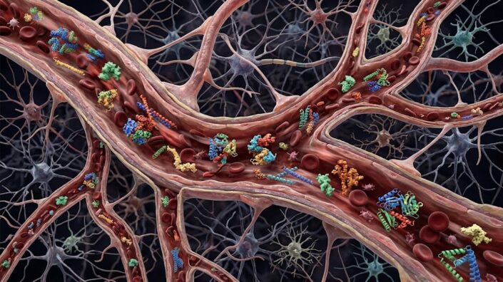
A recent analysis of serum protein changes identified 16 proteins related to inflammatory, synaptic, metabolic, and Alzheimer’s disease processes whose levels change during the menopausal transition [1]. These changes were linked to cognitive health later in life.

The time for interventions

Menopause is mostly known as a stage that marks the end of female reproduction, but the effects of menopause go beyond the reproductive system and affect many organs and systems, with the impact on the brain being widely discussed and researched. For example, multiple studies have reported that women experience various cognitive difficulties during menopause, such as word-finding or attentional difficulties. A recent review estimated that such problems affect between 26 and 95% of menopausal women [2]. Additionally, different studies suggested that the aging of the hypothalamus plays a causal role in ovarian senescence and menopause induction [3]. These data suggest a strong connection between the brain and menopause, creating a window of opportunity during the menopausal transition for improving brain aging and affecting dementia risk.

“Menopause is a normal physiological process that every person who has ovaries who lives long enough will go through,” said Madeline Wood Alexander, a graduate student at the University of Toronto. “We don’t want it to be viewed as a bad thing, but as an opportunity to better understand and maybe modify the biology of brain aging in women.”

Kaitlin Casaletto, Ph.D., an associate professor at the UCSF Fein Memory and Aging Center and co-senior author of the study, added, “Midlife is a critical window for both men and women where we can really set up the brain to be more resilient or more vulnerable to dementia in later life.” “For women, menopause may be this especially powerful time to intervene.”

Menopause proteomic score

Although many studies suggest a connection between menopause and brain aging, human data remain scarce, limiting the ability to understand the underlying molecular pathways driving these processes.

The researchers of this study began by investigating blood biomarkers associated with spontaneous menopause in healthy midlife women (43-58 years old): 30 premenopausal, 26 perimenopausal, and 24 postmenopausal women, along with 36 age-matched men for comparison.

Comparing serum protein levels (proteomics) showed an increase in 16 proteins in postmenopausal women compared to premenopausal women after adjusting for age. These proteins were related to inflammatory, synaptic, metabolic, and Alzheimer’s disease processes, with inflammatory molecules predominant, consistent with previous studies showing increased systemic inflammation after menopause [4].

Because the cohort used to identify these protein changes was relatively small, they also analyzed data from almost 3,000 women in the UK Biobank. 13 of the 16 proteins identified as showing menopause-stage differences were measured in both cohorts; of those, 9 replicated across cohorts.

The observed changes in synaptic and Alzheimer’s disease blood markers align with previous animal studies showing synaptic plasticity and spine density disruptions [5, 6], along with increases in Aβ and tau positron emission tomography (PET) across menopause [7, 8].

Using those 16 proteins, the researchers created a proteomic score that holistically reflects major menopause-related changes with less data noise. Using this score, they showed that compared to premenopausal women, perimenopausal status was associated with an increase of 1.36 units in menopause scores, and postmenopausal status with a 3.25-unit increase. Importantly, the menopausal stage, rather than chronological age, drove the observed changes.

Hormonal changes and menopausal symptoms

One of the most important changes during menopause is hormonal changes, such as reductions in the ovarian hormones 17β-estradiol and progesterone along with increases in follicle-stimulating hormone (FSH).

In this study, the authors found a positive association between their menopause proteomic score and FSH levels, an inverse association between this score and estradiol levels, and no association with progesterone levels. At a more granular level, they reported that some inflammation-related protein levels were inversely associated with 17β-estradiol, which aligns with the role of estrogens in modulating immune responses [9], while other proteins related to inflammatory, synaptic, metabolic, and Alzheimer’s disease processes were positively associated with FSH.

The authors note that this aligns with previous studies showing that “FSH receptors are expressed abundantly in AD-vulnerable brain regions” and that data from animal models suggest FSH’s role in the development of Alzheimer’s disease, neuroinflammation, and cognitive deficits [10]. In their analysis, the identified proteins were clustered into two separate groups, which suggests that each hormone might be linked to different biological pathways.

Menopause is also widely known as a stage in life when women experience various vasomotor symptoms, such as night sweats or hot flashes, and some of those vasomotor symptoms have been associated with vascular inflammation [11, 12], cardiovascular risk, and Alzheimer’s disease-related brain changes [13]. In this study, the authors analyzed different cohorts for the association between menopausal score and vasomotor symptoms. The results were inconsistent across the cohorts, but some suggested a link between some vasomotor symptoms and menopausal score.

Predicting future cognitive health

Beyond many previously reported links between menopausal changes and brain health, these researchers tested how relevant changes in serum proteins are for later-life cognitive health. They analyzed data from four independent cohorts of older women and found a consistent association between higher (more postmenopausal) baseline menopause scores and worse cognitive health later in life and a higher risk for Alzheimer’s disease dementia, suggesting that changes in certain protein levels during menopause may remain relevant in later decades of life.

“We don’t think that menopause is directly causing dementia,” said Casaletto. “But it’s possible that we may be able to predict a woman’s risk for dementia decades later by the levels of these molecules around menopause.”

While all women will undergo menopause, only a fraction will develop dementia. Molecular signatures during the menopausal transition can provide the earliest signals of future risk of developing the disease.

“We’re identifying molecular shifts in blood, in midlife post-menopausal women, that are also predicting cognition later in life,” said co-senior author Rowan Saloner, Ph.D., assistant professor at the UCSF Fein Memory and Aging Center. “So, we think these molecules are really important markers of both menopause and cognitive aging.”

We would like to ask you a small favor. We are a non-profit foundation, and unlike some other organizations, we have no shareholders and no products to sell you. All our news and educational content is free for everyone to read, but it does mean that we rely on the help of people like you. Every contribution, no matter if it’s big or small, supports independent ethical journalism and sustains our future.

Literature

[1] Wood Alexander, M., Rabin, J. S., Caunca, M., Iadipaolo, A., Cornelis, L., Warrier, R., Walker, K. A., Miolane, N., Bot, V. A., Wood, B., Oh, H. S., Wyss-Coray, T., Pham, A., Borger, J., Diaz, V., Paolillo, E. W., Kramer, J., Pritschet, L., Taylor, C., Panizzon, M. S., … Casaletto, K. B. (2026). Blood proteomics of menopause map to brain aging and dementia risk. Nature medicine, 10.1038/s41591-026-04648-4. Advance online publication.

[2] Gurvich, C., Spector, A., & Hickey, M. (2026). Advances in understanding of cognitive symptoms during menopause. The Lancet Obstetrics, Gynaecology, & Women’s Health, 2(4), e335–e345.

[3] Bacon, E. R., Mishra, A., Wang, Y., Desai, M. K., Yin, F., & Brinton, R. D. (2019). Neuroendocrine aging precedes perimenopause and is regulated by DNA methylation. Neurobiology of aging, 74, 213–224.

[4] McCarthy, M., & Raval, A. P. (2020). The peri-menopause in a woman’s life: a systemic inflammatory phase that enables later neurodegenerative disease. Journal of neuroinflammation, 17(1), 317.

[5] Hara, Y., Waters, E. M., McEwen, B. S., & Morrison, J. H. (2015). Estrogen Effects on Cognitive and Synaptic Health Over the Lifecourse. Physiological reviews, 95(3), 785–807.

[6] Russell, J. K., Jones, C. K., & Newhouse, P. A. (2019). The Role of Estrogen in Brain and Cognitive Aging. Neurotherapeutics : the journal of the American Society for Experimental NeuroTherapeutics, 16(3), 649–665.

[7] Buckley, R. F., O’Donnell, A., McGrath, E. R., Jacobs, H. I. L., Lois, C., Satizabal, C. L., Ghosh, S., Rubinstein, Z. B., Murabito, J. M., Sperling, R. A., Johnson, K. A., Seshadri, S., & Beiser, A. S. (2022). Menopause Status Moderates Sex Differences in Tau Burden: A Framingham PET Study. Annals of neurology, 92(1), 11–22.

[8] Rahman, A., Schelbaum, E., Hoffman, K., Diaz, I., Hristov, H., Andrews, R., Jett, S., Jackson, H., Lee, A., Sarva, H., Pahlajani, S., Matthews, D., Dyke, J., de Leon, M. J., Isaacson, R. S., Brinton, R. D., & Mosconi, L. (2020). Sex-driven modifiers of Alzheimer risk: A multimodality brain imaging study. Neurology, 95(2), e166–e178.

[9] Straub R. H. (2007). The complex role of estrogens in inflammation. Endocrine reviews, 28(5), 521–574.

[10] Xiong, J., Kang, S. S., Wang, Z., Liu, X., Kuo, T. C., Korkmaz, F., Padilla, A., Miyashita, S., Chan, P., Zhang, Z., Katsel, P., Burgess, J., Gumerova, A., Ievleva, K., Sant, D., Yu, S. P., Muradova, V., Frolinger, T., Lizneva, D., Iqbal, J., … Ye, K. (2022). FSH blockade improves cognition in mice with Alzheimer’s disease. Nature, 603(7901), 470–476.

[11] Thurston, R. C., El Khoudary, S. R., Sutton-Tyrrell, K., Crandall, C. J., Gold, E., Sternfeld, B., Selzer, F., & Matthews, K. A. (2011). Are vasomotor symptoms associated with alterations in hemostatic and inflammatory markers? Findings from the Study of Women’s Health Across the Nation. Menopause (New York, N.Y.), 18(10), 1044–1051.

[12] Bechlioulis, A., Naka, K. K., Kalantaridou, S. N., Kaponis, A., Papanikolaou, O., Vezyraki, P., Kolettis, T. M., Vlahos, A. P., Gartzonika, K., Mavridis, A., & Michalis, L. K. (2012). Increased vascular inflammation in early menopausal women is associated with hot flush severity. The Journal of clinical endocrinology and metabolism, 97(5), E760–E764.

[13] Thurston, R. C., Maki, P., Chang, Y., Wu, M., Aizenstein, H. J., Derby, C. A., & Karikari, T. K. (2024). Menopausal vasomotor symptoms and plasma Alzheimer disease biomarkers. American journal of obstetrics and gynecology, 230(3), 342.e1–342.e8.


Vegan buffet

Low-Fat Vegan Diet Shows Benefits in Type 2 Diabetes

A human trial found that even without caloric restriction, a low-fat, plant-based diet improved blood sugar control, reduced liver fat, and lowered the quantity of advanced glycation end products (AGEs) in the skin [1].

An all-you-can-eat vegan buffet

Type 2 diabetes is an age-related metabolic disorder in which the body becomes less able to regulate blood glucose. Two major problems usually underlie disease onset: tissues respond less strongly to insulin signaling, meaning that they take up less glucose from the bloodstream, and pancreatic beta cells, which produce insulin, become less able to meet the body’s needs.

As with many biological processes, a self-reinforcing loop can develop: insulin resistance increases the demand for insulin, while insufficient secretion allows glucose levels to rise, further impairing beta-cell function. The accumulation of fat surrounding the internal organs (visceral fat) and fat deposited outside its usual storage sites (ectopic fat), such as inside liver and muscle cells, is thought to be a major contributor to this metabolic dysfunction [2].

Dietary interventions for type 2 diabetes usually require reducing caloric intake and can be hard to adhere to. A new clinical trial, led by researchers at the Physicians Committee for Responsible Medicine, in collaboration with the Yale School of Medicine, and published in BMJ Open Diabetes Research & Care, tried a different approach: an all-you-can-eat low-fat plant-based diet. While previous studies have investigated plant-based diets in obesity and diabetes, this is “the first to simultaneously measure liver fat, muscle fat, visceral fat, and tissue advanced glycation end-products (AGEs) in a single plant-based dietary intervention in people with type 2 diabetes,” according to the accompanying press release.

Fewer calories without counting

25 adults with type 2 diabetes began a 16-week low-fat vegan diet, and all but one completed it. They averaged 59 years of age, had an average BMI of about 31, and had lived with diabetes for about 6.6 years. People using insulin, sulfonylureas, or weight-loss medications were excluded, while diabetes medications such as metformin were allowed.

The prescribed diet was based on vegetables, grains, legumes, and fruit, with animal products and added fats completely excluded. Its target composition was approximately 75% carbohydrate, 15% protein, and 10% fat. Participants were told to eat to satiety, without counting calories, and to maintain their exercise habits.

Despite that, participants reported eating about 346 fewer calories per day. Average carbohydrate consumption increased only modestly, from 198 to 259 grams a day; fiber consumption increased almost twofold; total fat consumption dropped by more than three times, and saturated fat consumption by more than five times; and protein consumption decreased only slightly, from 70 to 59 grams a day.

Participants lost 6 kilograms on average, or 6.6% of their starting weight. Fat mass fell by 4.2 kilograms, and visceral fat volume decreased by approximately 290 cm³, from 2,168 to 1,878 cm³. Liver fat decreased substantially, by 33% on average. Some lean mass was lost as well, which is something that future trials may address.

Importantly, these changes did not come from more exercise, as reported physical activity actually decreased. Neither fasting nor post-meal energy expenditure changed significantly, as well as the increase in energy expenditure after eating (the thermic effect of food). Among the 16 metformin users who completed the study, five reduced their dose and two discontinued it. No participant started or increased a glucose-lowering medication.

Better glucose control and less AGEs

The “under the hood” results were impressive as well, with blood glucose control improving substantially. Average fasting glucose fell from 155 mg/dL to 116 mg/dL, fasting insulin from 13.5 µU/mL to 8.8 µU/mL, and harmful LDL cholesterol from 108 mg/dL to 86 mg/dL. HbA1c, a marker of long-term glucose trends, fell from 7.6% to 6.6%, dipping below the usual treatment target of 7% for many adults with diabetes. On the other hand, beneficial HDL cholesterol also fell, from approximately 55 to 47 mg/dL, and triglycerides did not change significantly.

Several metrics suggested that beta cells became more responsive to glucose. Estimated insulin secretion increased at higher glucose concentrations, and rate sensitivity improved: beta cells responded more strongly to the speed at which glucose was rising.

AGEs are compounds formed when sugars react with proteins or fats. They accumulate with age and can impair tissue function by stiffening structural proteins and promoting inflammation. In this study, one of the first to measure AGEs in this context, skin AGE score fell from 3.1 to 2.7.

“For years, people with diabetes have been told to count calories and measure portions,” said Hana Kahleova, MD, PhD, the lead author of the study and director of clinical research at the Physicians Committee for Responsible Medicine. “This study shows that a simple shift in what you eat – toward vegetables, whole grains, legumes, and fruit – can reduce the fat clogging the liver by a third and improve how the pancreas makes insulin, without anyone having to count a single calorie.”

We would like to ask you a small favor. We are a non-profit foundation, and unlike some other organizations, we have no shareholders and no products to sell you. All our news and educational content is free for everyone to read, but it does mean that we rely on the help of people like you. Every contribution, no matter if it’s big or small, supports independent ethical journalism and sustains our future.

Literature

[1] Kahleova, H., Petersen, K., Shulman, G. I., Znayenko-Miller, T., Dufour, S., Brennan, H., … & Barnard, N. D. (2026). Changes in ectopic lipids, insulin sensitivity, and beta cell function after a low-fat vegan diet in adults with type 2 diabetes: a 16-week single-arm clinical trial. BMJ Open Diabetes Research & Care, 14(5).

[2] Chang-Chen, K. J., Mullur, R., & Bernal-Mizrachi, E. (2008). β-cell failure as a complication of diabetes. Reviews in Endocrine and Metabolic Disorders, 9(4), 329-343.

[3] Semba, R. D., Nicklett, E. J., & Ferrucci, L. (2010). Does accumulation of advanced glycation end products contribute to the aging phenotype?. Journals of Gerontology Series A: Biomedical Sciences and Medical Sciences, 65(9), 963-975.

Inflamed lower back discs

An Anti-Senescence Gene May Fight Spinal Disc Deterioration

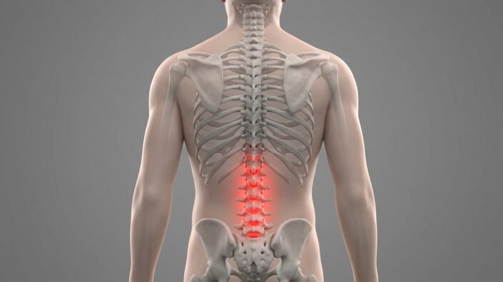
In Aging Cell, a team of researchers has described how a little-known gene, ZNF865, has a significant influence on back pain through the regulation of cellular senescence.

Lower back pain and senescence are related

The link between cellular senescence and back pain has been thoroughly documented, particularly in the context of intervertebral disc degeneration (IVDD) [1], and in both rats and people, senescence accumulates as discs deteriorate [2]. These cells and their SASP contribute to the pain both directly and indirectly, as they both inflame the area and cause the extracellular matrix to deteriorate [3]. Targeting these cells through senolytics has shown some results in animal models; giving 14- to 23-month-old mice the senolytic combination of dasatinib and quercetin was found to significantly reduce their IVDD [4].

Using CRISPR technology, this team has previously investigated ZNF865 in the context of bioengineered intervertebral disc cells, finding that it increases cartilage, maturation, and overall performance [5]. Little other work has been done on ZNF865, although one previous paper has noted that people with an ineffective version of this gene have unique problems with neurological development and other organs [6]. As this team’s previous RNA sequencing work has also linked ZNF865 to senescence [5], the researchers took another look, focusing on its effects in that context.

Little known but highly necessary

These researchers first performed another RNA sequencing analysis, using degenerative human nucleus pulposus cells (dhNPCs) along with cells derived from fat tissue and the well-known human embryonic kidney cell line HEK293. In all three cell types, ZNF865 was found to be related to cellular replication, DNA repair, mTOR, and senescence.

In dhNPCs, ZNF865 is substantially lower than in healthy human nucleus pulposus cells (hNPCs). However, in hNPCs, ZNF865 decreases with age. This was measured with samples taken from lower back pain patients and trauma patients. In mice, this was confirmed with analyses at 4, 12, and 24 months of naturally aged wild-type animals. Unsurprisingly, dhNPCs were found to have significantly more markers of DNA damage than hNPCs.

The researchers then used CRISPR technology to silence the ZNF865 gene in hNPCs. Compared to an unsilenced control group, the affected cells had roughly five times the markers of cellular senescence along with a significant increase in markers of DNA damage.

The opposite experiment was then performed: using CRISPR to boost ZNF865 in dhNPCs. The treated cells had more rapid cellular proliferation and had their markers of cellular senescence reduced nearly to those of hNPCs, including those of SASP expression and inflammation. DNA damage was also significantly reduced, and the treatment group expressed more collagen as well, suggesting benefits for the extracellular matrix.

Promising cellular and rat data

A chromatin accessibility analysis confirmed these benefits. Administering ZNF865 to dhNPCs restored their chromatin accessibility to be much more like that of hNPCs, reducing the number of differences in this area by 94% and giving them no upregulated pathways compared to hNPCs. RNA expression analysis bolstered these findings, showing that ZNF865 upregulation had given these cells a healthier overall gene expression phenotype.

The researchers then confirmed the effects of ZNF865 in living animals. Silencing its ortholog in rat NP cells using a CRISPR vector and injecting those cells directly into the rats’ vertebral discs, the team found that the animals developed signs of degenerative disc disease in less than two months, including increased pain sensitivity, along with increases in cellular senescence, SASP proteins, and genetic damage.

While these results are informative, they are limited to cells derived from a limited number of people and an experiment that demonstrated the results of depriving it from rats. The researchers note that the molecular mechanisms of ZNF865’s effectiveness are undetermined, and they lament that there are no treatments other than CRISPR that are known to directly affect ZNF865 in cells; there are no antibodies, inhibitors, or activators yet developed for it. For this research to be made into a therapy suitable for human use, a workable approach will first have to be developed and given to an animal model to demonstrate that ZNF865 can mitigate IVDD or another senescence-related disease in vivo.

We would like to ask you a small favor. We are a non-profit foundation, and unlike some other organizations, we have no shareholders and no products to sell you. All our news and educational content is free for everyone to read, but it does mean that we rely on the help of people like you. Every contribution, no matter if it’s big or small, supports independent ethical journalism and sustains our future.

Literature

[1] Wang, F., Cai, F., Shi, R., Wang, X. H., & Wu, X. T. (2016). Aging and age related stresses: a senescence mechanism of intervertebral disc degeneration. Osteoarthritis and cartilage, 24(3), 398-408.

[2] Gruber, H. E., Ingram, J. A., Norton, H. J., & Hanley Jr, E. N. (2007). Senescence in cells of the aging and degenerating intervertebral disc: Immunolocalization of senescence-associated β-galactosidase in human and sand rat discs. Spine, 32(3), 321-327.

[3] Zhang, G. Z., Liu, M. Q., Chen, H. W., Wu, Z. L., Gao, Y. C., Ma, Z. J., … & Kang, X. W. (2021). NF‐κB signalling pathways in nucleus pulposus cell function and intervertebral disc degeneration. Cell proliferation, 54(7), e13057.

[4] Novais, E. J., Tran, V. A., Johnston, S. N., Darris, K. R., Roupas, A. J., Sessions, G. A., … & Risbud, M. V. (2021). Long-term treatment with senolytic drugs Dasatinib and Quercetin ameliorates age-dependent intervertebral disc degeneration in mice. Nature communications, 12(1), 5213.

[5] Levis, H., Lewis, C., Fainor, M., Lawal, A., Stockham, E., Weston, J., … & Bowles, R. D. (2025). Targeted CRISPR regulation of ZNF865 enhances stem cell cartilage deposition, tissue maturation rates, and mechanical properties in engineered intervertebral discs. Acta biomaterialia, 191, 276-291.

[6] Bradbrook, S. M., Graham, G., Carter, M. T., Kibaek, M., Fagerberg, C., Larsen, M. J., … & Marcadier, J. (2026). De novo truncating variants in ZNF865 cause a novel neurodevelopmental disorder. American Journal of Medical Genetics Part A, 200(1), 244-252.

Gut bacteria

Gut Bacteria Molecule Slows Atherosclerosis in Mice

A new study highlights a fatty acid produced by a particular genus of bacteria as a “natural statin,” suggesting a new way of treating atherosclerosis [1].

Bacteria and cholesterol

Atherosclerosis develops when cholesterol-rich particles accumulate in arterial walls, driving inflammation and plaque buildup. The liver helps mitigate this process by removing low-density lipoprotein (LDL), which is known for its negative effects, from the bloodstream through LDL receptors that capture those particles and deposit them in cells.

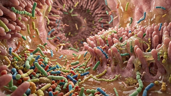
Gut bacteria are known to be able to influence cholesterol metabolism through the molecules they produce [2]. Previous research has found that several species of the genus Bacteroides are less abundant in people with atherosclerotic cardiovascular disease. A group of Chinese researchers decided to dig deeper into this phenomenon and published their results in Nature.

First, the researchers analyzed gut microbial sequencing data from people with atherosclerotic cardiovascular disease as well as healthy controls. B. uniformis stood out as the most abundant bacterial species associated with the healthy group.

They then administered the bacterium to male mice lacking Apoe, a gene involved in clearing cholesterol-rich particles. These mice quickly develop atherosclerosis when on a high-fat diet. Atorvastatin, an established cholesterol-lowering drug, was used as positive control.

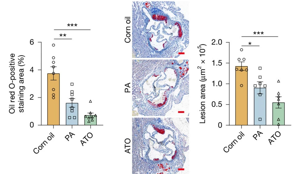
Twelve weeks of B. uniformis treatment reduced plaque area in the aorta and aortic root. It also lowered the overall plaque burden, circulating LDL cholesterol, total cholesterol, and triglycerides. HDL cholesterol did not change significantly.

Importantly, this is compared to vehicle-fed controls by the end of the experiment, rather than to baseline, which was not measured. Hence, the experiment demonstrated slowing, but not necessarily reversing, atherosclerosis. Atorvastatin produced comparable or slightly better results.

The treatment also reduced the number of plaque-associated macrophages, the immune cells that take up lipids and contribute to plaque development, and shifted the local immune environment toward a less inflammatory profile.

Dampening cholesterol production

The researchers also examined gene and protein activity in the liver. B. uniformis increased expression of LDL receptors and activated SREBP2, a regulatory protein that responds to low cellular cholesterol.

To test whether increased LDL uptake actually mattered, the researchers disabled the LDL receptor gene selectively in the liver. Without these receptors, the bacterium lost its ability to improve blood lipids, plaque burden, or inflammation, showing that working liver LDL receptors are necessary for the protective effect.

But why was this low-cholesterol response activated? The researchers found that B. uniformis actually reduced cholesterol synthesis in the liver. They hypothesized that decreased cholesterol production in liver cells prompts them to upregulate LDL receptors and draw more cholesterol out of the bloodstream, causing blood LDL levels to drop.

Next, the researchers asked whether the effect required living bacteria or could be reproduced by something they released. Dead bacteria did not do the trick, but an extract of the bacteria’s culture fluid did. Together, these results implicated a molecule produced by metabolically active bacteria.

The molecule in question was identified as pentadecanoic acid – a saturated fatty acid with 15 carbon atoms (“odd-chain”), also known as C15:0. It activated the same SREBP2-LDL receptor pathway.

Same pathway, smaller effect than atorvastatin

The researchers then gave purified C15:0 to atherosclerosis-prone mice for eight weeks, again with atorvastatin as a positive control, both at 25 mg/kg three times weekly. C15:0 reduced plaque burden by about 50% and lowered total and LDL cholesterol. It also reduced macrophage accumulation in plaques and systemic inflammatory markers. Atorvastatin, however, performed noticeably better.

Atorvastatin comparison

Like the bacterium, C15:0 reduced cellular and liver cholesterol. The researchers then investigated HMG-CoA reductase, an enzyme controlling a key step in cholesterol synthesis and the target of statin drugs. The experiments showed that C15:0 directly bound the enzyme and inhibited its activity.

However, C15:0 is a weaker inhibitor than atorvastatin, consistent with its more modest effect on atherosclerosis. The authors suggest that its greater persistence and exposure in mice might partly compensate for this.

Finally, the researchers measured C15:0 in people with abnormally high blood lipid levels (dyslipidemia) and healthy controls. The former had significantly lower C15:0 concentrations. Reanalysis of existing microbial sequencing datasets also showed lower levels of C15:0-production genes in people with atherosclerotic cardiovascular disease.

The findings underline new ways in which the microbiome might be harnessed to protect the heart, said study co-author Wenjing Zhao, a microbiologist at Sun Yat-sen University in Shenzhen, to Nature. However, more studies – especially in humans – are needed. Currently, it is not clear that C15:0, which can be purchased as a supplement, is superior to statins in any way. However, this still might prove to be the case; for instance, if further research shows a better safety profile or a larger tolerable dose.

We would like to ask you a small favor. We are a non-profit foundation, and unlike some other organizations, we have no shareholders and no products to sell you. All our news and educational content is free for everyone to read, but it does mean that we rely on the help of people like you. Every contribution, no matter if it’s big or small, supports independent ethical journalism and sustains our future.

Literature

[1] Yin, C., Chen, Y., Lin, G. et al. (2026). A gut microbial odd-chain fatty acid alleviates atherosclerosis in mice. Nature.

[2] Haghikia, A., Zimmermann, F., Schumann, P., Jasina, A., Roessler, J., Schmidt, D., … & Landmesser, U. (2022). Propionate attenuates atherosclerosis by immune-dependent regulation of intestinal cholesterol metabolism. European heart journal, 43(6), 518-533.

[3] Jie, Z., Xia, H., Zhong, S. L., Feng, Q., Li, S., Liang, S., … & Kristiansen, K. (2017). The gut microbiome in atherosclerotic cardiovascular disease. Nature communications, 8(1), 845.

Proteins or extracellular vesicles

Untangling Differences in Senescent Cellular Signaling

In Aging, researchers have described the differences between the secretions of cells driven senescent through a cancer gene and cells that stopped proliferating due to p16 expression.

A difference of kind

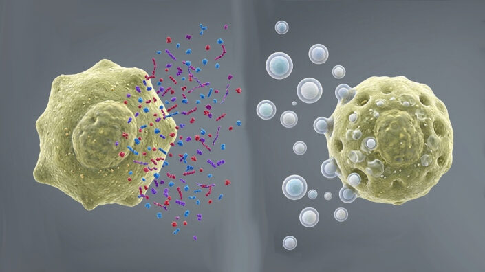
The SASP is a mix of many components that have various effects on the cells exposed to it. While inflammatory factors, such as cytokines and matrix metalloproteinases, are the most well-known of these, it also consists of ions, various metabolites, and exosomes: encapsulated cellular contents that cells use to signal one another. While exosomes have been researched for their therapeutic value [1], and we have reported on beneficial exosomes’ life-extending effects, exosomes secreted by senescent cells do the opposite [2].

This paper focuses on cells exhibiting the biomarker p16, which is encoded by the gene CDKN2A. This particular biomarker has been heavily linked to senescence, and purging p16-expressing cells from mice lengthens their lives [3]. However, Judith Campisi’s lab had noted that cells that were induced into senescence through p16 express different SASP components [4].

A SASP without inflammation

These researchers took a closer look at such differences, using IMR-90 fibroblasts to directly compare the secretions of ordinary senescent cells to those of p16-expressing cells and non-senescent cells. The first group was driven senescent through the oncogene H-RASG12V (RAS), while the second was made to express p16 through a specific viral vector.

As expected, the secretions of all three groups was substantially different. The RAS group secreted the inflammatory SASP component IL-1β, while the other two did not secrete it at all. While mRNA analysis found that the p16 group was producing IL-6, it secreted even less of it into its environment than the control group did. However, while the control group did not secrete any small extracellular vesicles (sEVs), the RAS group secreted many, and the p16 group secreted slightly more.

Cells spread their own type of senescence

These sEVs caused recipient non-senescent cells to behave much like the cells that secreted them. sEVs from p16 cells induced p16 expression in their recipients, yet they did not encourage the recipients to secrete inflammatory factors such as IL-6 and IL-8. sEVs from RAS cells were inconsistent in encouraging p16 expression, but they did encourage the production of these inflammatory cytokines. sEVs from both types stimulated DNA damage as measured by the biomarker γH2AX.

The researchers confirmed their findings by exposing babies’ fibroblasts in vitro to sEVs derived from the fibroblasts of older people. Even without the rest of the SASP, these older EVs were fully capable of driving the young cells into senescence, as measured by the biomarker SA-β-gal.

This paper holds that the SASP is fundamentally divided into two different portions: a soluble fraction (the sSASP), which consists of proteins and other relatively simple components, and a fraction that consists of extracellular vesicles (the evSASP). While the p16-expressing cells used in this experiment did not secrete the sSASP, they secreted the evSASP, which was found to have negative effects even without classical inflammatory factors.

This is exploratory cellular research, and the specific contents of the sEVs were not discovered, nor was any RNA transcription done in the recipient cells. The researchers suggest that their cargo may be genotoxic in nature and activates stress responses in the recipient cells. Whether or not it is feasible to target sEVs or their originating machinery is not yet clear.

We would like to ask you a small favor. We are a non-profit foundation, and unlike some other organizations, we have no shareholders and no products to sell you. All our news and educational content is free for everyone to read, but it does mean that we rely on the help of people like you. Every contribution, no matter if it’s big or small, supports independent ethical journalism and sustains our future.

Literature

[1] Ripoll, L., Zickler, A. M., Vader, P., El Andaloussi, S., Verweij, F. J., & Van Niel, G. (2026). Biology and therapeutic potential of extracellular vesicle targeting and uptake. Nature Reviews Molecular Cell Biology, 27(5), 358-376.

[2] Takasugi, M., Okada, R., Takahashi, A., Virya Chen, D., Watanabe, S., & Hara, E. (2017). Small extracellular vesicles secreted from senescent cells promote cancer cell proliferation through EphA2. Nature communications, 8(1), 15729.

[3] Baker, D. J., Childs, B. G., Durik, M., Wijers, M. E., Sieben, C. J., Zhong, J., … & Van Deursen, J. M. (2016). Naturally occurring p16Ink4a-positive cells shorten healthy lifespan. Nature, 530(7589), 184-189.

[4] Coppé, J. P., Rodier, F., Patil, C. K., Freund, A., Desprez, P. Y., & Campisi, J. (2011). Tumor suppressor and aging biomarker p16INK4a induces cellular senescence without the associated inflammatory secretory phenotype. Journal of Biological Chemistry, 286(42), 36396-36403.

Forever Healthy Foundation

Evipedia Goes Hugging Face

With more than 860 evidence reviews on health and longevity interventions, Evipedia is now probably one of the deepest online sources in this area.

We have always been extremely open to sharing our data and making it accessible in any form that supports projects and tools that enable people to make better decisions about their health and longevity.

Now, we are taking it to the next level by making all our reviews available on HuggingFace. Our whole dataset, including all updates, published and refreshed daily.  One row per review with all metadata and full Markdown, ready to drop into any ML or RAG pipeline.

What’s in Evipedia

Evipedia is a comprehensive, continuously updated online encyclopedia that provides much-needed, accurate, and up-to-date information on a wide range of health and longevity-related interventions.

  • Free to Use Evipedia is completely free to use and open to everyone. We built Evipedia as a backbone tool for the whole longevity and rejuvenation community. Our goal is to empower individuals with knowledge to make informed decisions about their health and longevity.
  • 870+ Evidence Reviews Including whole-body therapies, brain health, skin rejuvenation, hormone optimization, peptides, psychedelics, medications, blood and plasma therapies, complementary cancer approaches, foundational habits, diets, foods, probiotics, botanicals, isolates, and many more.
  • A Dual Structure for Every Entry Each intervention has a one-page Quick Reference Sheet for an at-a-glance protocol, benefits, risks, contraindications, and monitoring, plus a Full Evidence Review for in-depth analysis.
  • Continuous Updates Entries are continuously updated to reflect the latest developments in health and longevity. On average, reviews are refreshed every 4-6 weeks, ensuring access to the most current information available.
  • Full Audit and Quality Transparency Every “Quick Reference Sheet” and “Evidence Review” on Evipedia includes an Audit Report that outlines the detailed audit criteria and the history of audits and fixes applied to the documents.
  • Stable Permalinks – Beautiful Sharing Cards Every intervention has a fixed, short URL and a purpose-designed social sharing card — ideal for citing a compound in a supplement stack or anchoring a claim in an online discussion.
  • AI & Agent-friendly, Extensive Integration Support Built as a backbone service for the longevity and rejuvenation community, Evipedia features a wide variety of AI integration tools and an easily accessible API. All are free to use, under an AI- and agent-friendly site policy and the Creative Commons 4.0 license.
  • Built on AI4L Evipedia is built on top of our open-source AI4L framework, which enables anyone to create high-quality, evidence-based reviews of health and longevity interventions. At the core of AI4L is our novel “Audit-Driven Prompting” approach to generate hallucination-free, accurate, and well-structured reviews using frontier AI models.

About Forever Healthy

Forever Healthy Foundation is a private, humanitarian initiative with the mission of enabling people to vastly extend their healthy lifespan. More at forever-healthy.org

We would like to ask you a small favor. We are a non-profit foundation, and unlike some other organizations, we have no shareholders and no products to sell you. All our news and educational content is free for everyone to read, but it does mean that we rely on the help of people like you. Every contribution, no matter if it’s big or small, supports independent ethical journalism and sustains our future.
Cell and organelles

Declining Autophagy Impairs Senescent Cell Clearance

A new study links an age-related decline in cellular maintenance to impaired immune clearance. Activating the pathway reduced signs of senescence in aged mice [1].

Senescence and autophagy

Cellular senescence, which occurs when cells stop dividing and start emitting largely pro-inflammatory stress signals, the senescence-associated secretory phenotype (SASP), is a double-edged sword. On the one hand, it plays an important role in processes like development and wound healing as well as in regeneration in some animals. On the other hand, with age, senescent cells accumulate in tissues, driving inflammation, fibrosis, and cancer, which earned cellular senescence a place on the Hallmarks of Aging list.

One of the reasons for the age-related accumulation of senescent cells might be the declining ability of immune cells to clear them away. A new study from the Albert Einstein College of Medicine, published in Nature Aging, investigates a mechanism that might be responsible for that.

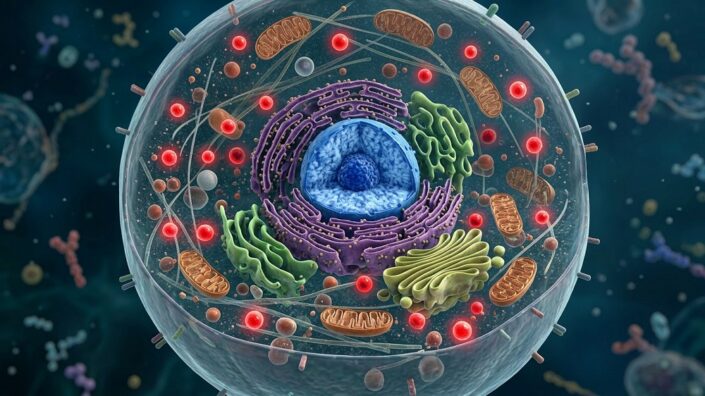
The researchers focused on chaperone-mediated autophagy (CMA). Autophagy is the process of degrading and removing intracellular junk, such as misfolded proteins, and CMA is a distinct pathway mediated by the chaperone protein HSC70, which recognizes suitable proteins and delivers them to a receptor called LAMP2A. The proteins then unfold and pass into the cell’s lysosomes for degradation. This process differs from macroautophagy, the pathway that delivers cellular material to lysosomes in tiny vesicles.

Previous research showed that CMA generally declines with age [2] and rises when cells enter senescence [3], suggesting that it mediates this transition. The researchers asked whether this failure to increase CMA alters the course of senescence onset and whether CMA decline also compromises immune clearance.

CMA mediates senescence and macrophage function

The researchers isolated ear fibroblasts from 4-month-old (young) and 23-month-old (old) mice. The old fibroblasts were not initially more senescent according to the markers used. Both young and old cells could be induced into senescence with the drug palbociclib, but young fibroblasts increased CMA after senescence induction, while old fibroblasts started with lower activity levels and failed to increase them.

The team then reduced LAMP2A in mouse fibroblasts to ask whether CMA loss itself causes senescence. CMA-deficient cells acquired some senescence-like features but remained proliferative until given a senescence-inducing treatment. After palbociclib treatment, both normal and CMA-deficient cells stopped proliferating, but their overall marker profiles differed. This suggests that while CMA deficiency could not cause growth arrest by itself, it changed the resulting senescent phenotype.

Disabling CMA in young cells reproduced many of the changes found in old cells, both before and after senescence induction. Normally, cells entering senescence remove certain proteins and reorganize their metabolism. Both old and CMA-deficient cells partially failed in that reorganization. This suggests that CMA decline contributes to the aging-associated altered senescent state.

Once induced into senescence, CMA-deficient cells developed a distinct secretory profile. The researchers investigated its effect by applying a medium conditioned by these cells onto normal fibroblasts for five days. Secretions from CMA-deficient cells induced senescence-associated changes in neighboring normal cells, even when the donor cells had not themselves been turned senescent.

The researchers then wanted to see whether these altered secretions suppress CMA and cell-clearing activity in macrophages, immune cells that help remove unwanted cells. Macrophages from old mice already had lower CMA activity than those from young mice. Moreover, the medium conditioned from senescent fibroblasts suppressed CMA in young macrophages. Secretions from CMA-deficient fibroblasts – even non-senescent – were particularly inhibitory.

The researchers then co-cultured senescent fibroblasts with either normal or CMA-deficient macrophages. When the macrophages lacked CMA, they were able to clear fewer fibroblasts, regardless of whether the fibroblasts themselves had functional CMA. This suggests that CMA-deficient cells secrete molecules that compromise CMA and function in the immune cells needed to clear them. The researchers then confirmed this decline in macrophage cell-removing abilities with a series of experiments.

Increasing CMA slows lung fibrosis

The researchers next examined mice in which LAMP2A was selectively deleted in macrophages and some other immune cells. Late in life, these mice showed an increased senescence burden across several tissues, although this varied by sex and measurement.

Since senescent cells participate in wound clearing, the researchers created skin wounds in these mice. Mice with CMA-deficient immune cells healed more slowly and retained more senescent cells at the wound site. The team confirmed that the problem was decreased macrophage function rather than recruitment to the wound site.

What would happen if CMA were increased rather than suppressed in old mice? The researchers administered CA77.1, a drug known to activate CMA, to aged mice and found that these generally had lower senescence-associated measurements than untreated aged mice, with several results moving toward young-control levels. However, the effects differed across tissues, sexes, and markers.

The researchers then investigated idiopathic pulmonary fibrosis (IPF), a lung-scarring disease associated with senescence. Lysosomes isolated from human fibrotic lungs had reduced LAMP2 protein levels and impaired uptake of a CMA substrate.

Finally, the team induced lung fibrosis in mice. CA77.1 treatment started soon after injury (day 2), but not on day 7, preserved body weight and substantially reduced subsequent lung fibrosis, suggesting that early CMA activation limits disease progression.

“In older animals, age-related changes in both senescent cells and the immune cells responsible for removing them allow these zombie cells to accumulate and play a role in disease,” said study leader Ana Maria Cuervo, M.D., Ph.D., co-director of the Institute for Geroscience at Einstein. “By restoring cellular recycling, we may be able to help the body’s own defenses clear these cells more effectively. The next challenge is determining whether this approach can eventually be developed into a safe treatment for age-related diseases in people.”

We would like to ask you a small favor. We are a non-profit foundation, and unlike some other organizations, we have no shareholders and no products to sell you. All our news and educational content is free for everyone to read, but it does mean that we rely on the help of people like you. Every contribution, no matter if it’s big or small, supports independent ethical journalism and sustains our future.

Literature

[1] Sereda, R., Lindenau, K., Diaz, A. et al. (2026). Decline of chaperone-mediated autophagy in aging impairs macrophage clearance of senescent cells. Nature Aging.

[2] Khawaja, R. R., Martín-Segura, A., Santiago-Fernández, O., Sereda, R., Lindenau, K., Mccabe, M., … & Cuervo, A. M. (2025). Sex-specific and cell-type-specific changes in chaperone-mediated autophagy across tissues during aging. Nature Aging, 5(4), 691-708.

[3] Rovira, M., Sereda, R., Pladevall‐Morera, D., Ramponi, V., Marin, I., Maus, M., … & Serrano, M. (2022). The lysosomal proteome of senescent cells contributes to the senescence secretome. Aging cell, 21(10), e13707.

Brain and walking

The Biomarkers That Predict Mortality Risk

Researchers have discovered key biomarkers that are strongly linked to all-cause mortality and identified four measurements with independent predictive power.

Biological versus chronological

This paper opens with a discussion of a familiar fact for people interested in living longer: one’s biological age is not the same as one’s chronological age [1]. Biological age gives a more accurate indication of a person’s current and future health [2]. The authors touch upon the benefits and drawbacks of several well-known biomarkers; grip strength and gait speed are reliable [3] but, as these authors note, only begin to decline much later in life; neurological imaging is similarly reliable [4] but is expensive to perform and, like physical function tests, does not directly capture molecular changes. Telomere attrition, once a key focus of aging research, may not be as useful as previously hoped [5].

Epigenetic clocks and proteomic biomarkers have been developed to more accurately gauge aging and mortality risk. This includes the well-known GrimAge epigenetic mortality clock and its second version [6], which is referenced in this study. Organ-specific proteomics have been found to be strongly related to aging and disease as well [7].

This paper directly pits these various biomarkers against each other, gauging how valuable they are for predicting mortality and how much they overlap. The data source is LBC1936, a cohort of 861 people who were born in 1936, participated in a mental examination at age 11, and had their proteomic, physical, and neurological biomarkers examined every three years between the ages of 70 and 89.

GrimAge2 comes out on top

The researchers first examined this cohort’s organ age gaps: the difference between the chronological and biological age of each major organ. Consistent with previous research [7], these differences were only loosely correlated in the same individual. Even people with an extreme age gap in one organ were unlikely to similarly age in other organs.

Unsurprisingly, of all the various biomarkers that the researchers used, GrimAge2 was found to be the most closely connected with all-cause mortality. Organ age gaps were also highly associated with all-cause mortality, most notably in the liver, immune system, heart, pancreas, and brain.

However, some metrics of physical function were more closely related to mortality than any organ-specific clock. These included total brain volume and grey matter volume, overall cognitive function (g), and forced expiratory ratio and forced vital capacity, two metrics of lung function. Telomere attrition in leukocytes, on the other hand, was found to have no significant relationship.

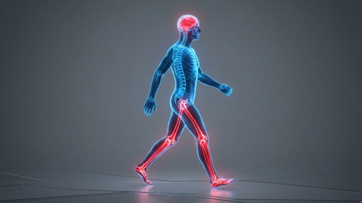
The researchers then used statistical methods to examine how closely these biomarkers overlap with one another. As a composite biomarker, GrimAge2 was strongly related to all of the organ-specific proteomic markers along with many of the other markers. Walking time, a biomarker with similar predictive power as immune and heart proteomics, was strongly correlated with other biomarkers. While organ age gaps were loosely correlated with each other, they were uncorrelated with physical biomarkers as a whole.

The researchers then sought the biomarkers with the most independent predictive power when excluding other biomarkers, aiming to minimize redundancy. Four stood out in this regard: white matter volume, total brain volume, walking time, and the cognitive metric g. Combining these four markers accounted for 19% of the variance in mortality risk, and including the other 17 only raised this by 4%.

One protein stands out among many

Finally, this study took a look at proteins themselves. GDF15, which has been associated with cellular senescence, was found to be the protein most closely related to mortality risk after adjustment for lifestyle factors, and, of all the biomarkers in this study, it was only surpassed by GrimAge2 in overall predictive power. The neuropeptide NPS was found to have the strongest negative association: people who express more NPS are less likely to die of any cause.

As expected, the proteins most associated with mortality were associated with inflammation, including chemokine and interleukin signaling. The proteins that were associated with longer lifespans, on the other hand, were related to the maintenance of genomic stability, such as retroelement regulation and chromatin management.

The researchers acknowledge this study’s limitations, including the fact that this relatively small cohort only includes healthy Scottish people. This study also used time-point measurements and did not consider trajectories. This was also solely an observational study and did not delve into mechanisms. However, its core finding, that brain imaging and lung measurements are more effective than organ-specific proteomic clocks in predicting overall mortality, offers sobering insight.

We would like to ask you a small favor. We are a non-profit foundation, and unlike some other organizations, we have no shareholders and no products to sell you. All our news and educational content is free for everyone to read, but it does mean that we rely on the help of people like you. Every contribution, no matter if it’s big or small, supports independent ethical journalism and sustains our future.

Literature

[1] Salih, A., Nichols, T., Szabo, L., Petersen, S. E., & Raisi-Estabragh, Z. (2023). Conceptual overview of biological age estimation. Aging and disease, 14(3), 583.

[2] Jylhävä, J., Pedersen, N. L., & Hägg, S. (2017). Biological age predictors. EBioMedicine, 21, 29-36.

[3] de Souza, A. F., de Oliveira, D. C., Ramírez, P. C., de Oliveira Máximo, R., Luiz, M. M., Delinocente, M. L. B., … & da Silva Alexandre, T. (2025). Low gait speed is better than frailty and sarcopenia at identifying the risk of disability in older adults. Age and Ageing, 54(4), afaf104.

[4] Fletcher, E., Gavett, B., Harvey, D., Farias, S. T., Olichney, J., Beckett, L., … & Mungas, D. (2018). Brain volume change and cognitive trajectories in aging. Neuropsychology, 32(4), 436.

[5] Wang, Q., Zhan, Y., Pedersen, N. L., Fang, F., & Hägg, S. (2018). Telomere length and all-cause mortality: a meta-analysis. Ageing research reviews, 48, 11-20.

[6] Lu, A. T., Binder, A. M., Zhang, J., Yan, Q., Reiner, A. P., Cox, S. R., … & Horvath, S. (2022). DNA methylation GrimAge version 2. Aging (Albany NY), 14(23), 9484.

[7] Oh, H. S. H., Rutledge, J., Nachun, D., Pálovics, R., Abiose, O., Moran-Losada, P., … & Wyss-Coray, T. (2023). Organ aging signatures in the plasma proteome track health and disease. Nature, 624(7990), 164-172.

FDA at ARDD

FDA Leaders Name Longevity a Priority at ARDD

At a panel in Boston, senior officials announced plans to include aging in the agency’s regulatory science agenda and discussed how therapies could move toward approval.

Longevity has a partner

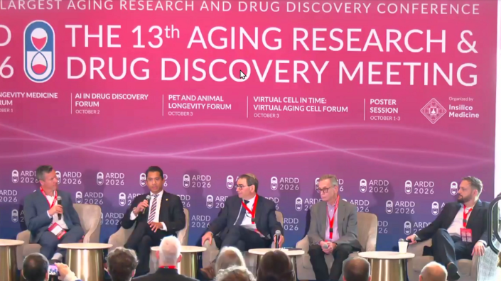
The 2026 edition of the Aging Research and Drug Discovery Meeting (ARDD) kicked off yesterday. This is one of the biggest and longest-running longevity conferences, and it has been traditionally held in Copenhagen. This year, it arrived in Boston instead, with a slightly truncated schedule (three days versus the usual five) but with an equally impressive lineup and program.

Day 1 was all about longevity going mainstream, with discussions centered on policy, regulation, and clinical translation. Gone are the days when longevity events were largely ignored by the people calling the shots. This time, a panel titled “Matching Clinical Trials of Therapeutics & Regulatory Mandates” boasted four FDA heavyweights. Prompted by moderator Andrew Brack, Program Manager at ARPA-H, they described the agency’s growing interest in aging and longevity and discussed how gerotherapeutics could progress toward approval.

“This is an important topic for the agency and HHS at large,” said Lowell Zeta, Deputy Commissioner for Strategic Initiatives. “It’s a defining moment, an inflection point for the future of FDA and how we adapt to the rapidly evolving science in this space.”

The most tangible announcement was that aging and longevity will feature in the forthcoming update to FDA’s Focus Areas of Regulatory Science (FARS). Drawing applause from the crowd, Steven Kozlowski, the agency’s Chief Scientist, described FARS as “high-level principles on areas that are very, very important to us; aging and longevity will be one of those topics.” Kozlowski gave a tentative release target of the 2027 fiscal year, which also began yesterday. The document’s previous version was released several years ago.

Seeking approval

Beyond that announcement, the panelists drilled down into what an approval process for longevity therapies could look like. Jeffrey Siegel, Director of the Office of Drug Evaluation Sciences, outlined two possible approaches to demonstrating broader effects on aging. One would involve accumulating evidence that a treatment benefits several age-related conditions; another, drawing on the concept of intrinsic capacity, would measure deterioration across multiple physical and mental capacities in a defined population and show that treatment slows it.

Justin Penzenstadler, Acting Associate Director of the Office of Cardiology, Hematology, Endocrinology, and Nephrology, expects early trials that could support approval to focus on age-related comorbidities or mortality. Collecting functional assessments alongside those outcomes could help establish additional metrics for later trials.

One obvious caveat is that a broader aging claim needs evidence beyond a drug’s established benefits. If a cardiometabolic treatment extends survival through its known cardiovascular effects, that alone does not demonstrate broader geroprotective activity, Penzenstadler noted.

Safety also shapes the choice of trial population. Decades of treatment, potentially modest benefits, and relatively low baseline risk make for a demanding benefit–risk calculation. As Penzenstadler put it, “We’re thinking about potentially treating somebody for thirty years to derive a couple-year benefit.” Consequently, he favors initial trials in older, higher-risk populations, where the calculation “is a bit more straightforward.”

Biomarkers could help shorten trials by serving as proxies for clinical outcomes – but only with evidence that they reliably reflect benefit. The panel discussed collecting both clinical outcomes and candidate biomarkers in trials to build that evidence.

Siegel distinguished prognostic biomarkers, which predict future risk, from surrogate endpoints. “A surrogate endpoint biomarker may also be prognostic, but it has the additional feature that it changes with treatment, and that change reflects the clinical benefit downstream,” he said.

Collaboration and convergence

Asked how the longevity community could advance regulatory progress, Penzenstadler’s main recommendation was to establish a pre-competitive consortium to work out “a cookbook of biomarkers” to include in trials. Agreeing on compatible trial designs up front would allow different companies to pool their results and build evidence for biomarker validation. “I think that’s probably the number one thing that is actionable that will push the field,” he said.

His second recommendation was to hold regular, structured meetings between regulators, researchers, and industry, helping both sides understand emerging evidence and the challenges of developing therapies.

Several recommendations voiced during the panel echoed initiatives that are already underway. For instance, the proposed Targeting Aging with Metformin (TAME) trial would examine multiple age-related diseases, while XPRIZE Healthspan focuses on muscle, cognitive, and immune function. These correspond to the two approaches described by Siegel. Likewise, the Biomarkers of Aging Consortium reflects the field’s recognition that developing better measurements requires collaboration.

This convergence comes amid growing engagement between FDA officials and the longevity field, which seems to have intensified further this year. FDA participation in events such as A4LI’s June DC summit provided ample opportunities for cross-pollination. What we heard at ARDD suggests that this dialogue may be beginning to bear fruit.

The panel also gave longevity sponsors a concrete address: Penzenstadler identified his clinical review office as being responsible for aging as an indication. He encouraged sponsors to submit actual protocols and supporting evidence through the Investigational New Drug (IND) process, giving FDA something specific to assess and respond to.

We would like to ask you a small favor. We are a non-profit foundation, and unlike some other organizations, we have no shareholders and no products to sell you. All our news and educational content is free for everyone to read, but it does mean that we rely on the help of people like you. Every contribution, no matter if it’s big or small, supports independent ethical journalism and sustains our future.
Biomarkers of Aging Consortium

Global Leaders in Geroscience Convene for Biomarkers of Aging

The Biomarkers of Aging Consortium will host its fourth annual Biomarkers of Aging Conference on October 5 and 6, 2026, at the Joseph B. Martin Conference Center at Harvard Medical School. The meeting brings together academic, clinical, industry, regulatory, and philanthropic leaders working to develop, validate, and apply biomarkers of aging.

Therapies that target aging itself are approaching the clinic. Their progress depends on reliable, standardized ways to measure biological aging and to show whether an intervention changes it. The conference is built around closing that gap.

Program Highlights

The 2026 program features nearly 30 speakers from leading universities, research institutes, companies, and funding and regulatory agencies. Invited speakers include:

  • Epigenetic clocks and pace of aging: Steve Horvath (UCLA), developer of the first pan-tissue epigenetic clock, and Dan Belsky (Columbia University), coinventor of the DunedinPACE clock
  • Quantitative and systems views of aging: Uri Alon (Weizmann Institute of Science), Albert-László Barabási (Northeastern University), and Michael Levin (Tufts University), bringing mathematical, network medicine, and bioelectric perspectives Mechanisms of aging and longevity: Vera Gorbunova (University of Rochester), Thomas Rando (UCLA), and Mike Snyder (Stanford), spanning exceptionally long-lived species, stem cell aging, and longitudinal multi-omic profiling of people
  • AI and causal biology in industry: Morgan Levine (stealth-stage company) and Martin Borch Jensen (Gordian Biotechnology), building AI models of biology and causal atlases of age-related disease
  • Funding, regulation, and translation: Andrew Brack (ARPA-H), Cynthia Grossman (U.S. Food and Drug Administration), Jamie Justice (XPRIZE Healthspan), and Lynne Cox (Wellcome Leap), speaking to how aging biomarkers move toward trials and approved endpoints

Sessions span novel biomarker discovery, multi-omic and AI-based approaches, immune and cellular aging, and the use of biomarkers to evaluate longevity interventions. Selected poster presenters will give short talks, and an expert jury will award poster prizes.

The conference is cochaired by Vadim Gladyshev, Chiara Herzog, Andrea Cipriano, Jesse Poganik, and Mahdi Moqri The full program is available here.

From the Conference Chairs

“The field of aging biomarkers is constantly evolving and growing, and this is our most diverse year yet. We’re excited to bring experts and stakeholders in areas such as molecular screening to population science and the ethics of longevity together – across academia, biotech, pharma and policy,” said Chiara Herzog, Ph.D., conference co-chair, executive committee member, and Assistant Professor at the University of Cambridge.

“The science of measuring aging has moved remarkably fast, but speed is not the same as trust,” said Jesse Poganik, Ph.D., conference co-chair, co-director of the Biomarkers of Aging Consortium, and Investigator at Brigham and Women’s Hospital and Harvard Medical School. “Before an aging biomarker can guide a clinical trial or a treatment decision, the field needs to agree on how it is validated and what it actually tells us. This meeting puts the people building these tools in the same room as the people who fund, regulate, and depend on them.”

New for 2026: Satellite Programs

The conference anchors a week of related programming at Harvard Medical School. A full-day Replacement in Aging Session, focused on replacement and regenerative strategies, runs as part of the conference, and conference tickets also include access to the Gerophysics Session. On October 7, the Consortium will co-host two satellite symposia in the Pechet Room of the Joseph B. Martin Conference Center: the Brain Aging Symposium from 8:00 am to 2:00 pm, followed by the Reproductive Aging Symposium from 2:00 pm to 6:30 pm.

About the Biomarkers of Aging Consortium

The Biomarkers of Aging Consortium is a collaborative initiative spanning more than 50 institutions. It supports the global geroscience community by developing standardized, clinically validated ways to measure aging and assess the outcomes of interventions. Its programs include the Biolearn open-source platform, the Biomarkers of Aging Challenge, The Longevity Study, and an annual conference. The Consortium is fiscally sponsored by the Methuselah Foundation.

Media Contact

Jesse Poganik, Biomarkers of Aging Consortium: community@agingconsortium.org

We would like to ask you a small favor. We are a non-profit foundation, and unlike some other organizations, we have no shareholders and no products to sell you. All our news and educational content is free for everyone to read, but it does mean that we rely on the help of people like you. Every contribution, no matter if it’s big or small, supports independent ethical journalism and sustains our future.
Rejuvenation Roundup September 2026

Rejuvenation Roundup September 2026

Which generation will be considered “the last to die” of age-related diseases is ultimately a question of technological development and deployment. Here’s what’s been done on that front in September.

Interviews

Last Generation to DieThe Last Generation to Die: Interview With Tim Maupin: Filmmaker Tim Maupin has spent a number of years developing The Last Generation to Die, a near-future film about what happens when the first true rejuvenation technologies arrive and who gets left behind.

Research Roundup

Mice With Growth Hormone Halted in Middle Age Live Longer: Researchers publishing in Aging Cell have described how stopping growth hormone receptor production in midlife significantly lengthens the lives of mice.

Food-Ignoring MouseLate-Life GLP-1 Treatment Increases Lifespan in Female Mice: In a new study, semaglutide given late in life increased median lifespan in female mice by more than 12% and improved various health markers. Semaglutide’s effect possibly went beyond caloric restriction.

Comparing the Responsiveness of Epigenetic Aging Biomarkers: In a recent study, researchers have evaluated how responsive several DNA methylation (DNAm) biomarkers are to a range of longevity interventions.

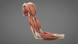
Weak musclesCalcium Mishandling Contributes to Muscle Loss: Researchers have discovered and explained how calcium mishandling in muscle cells leads to long-term muscle weakness in older people.

Lung Fibrosis Drug Lowers Biological Age Estimates: Six different aging clocks detected younger blood protein profiles in patients taking the experimental lung-fibrosis drug rentosertib. This suggests possible anti-aging effects, but separating those from the narrower benefits of treating fibrosis remains a challenge.

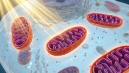
NeuronsMitochondrial Performance May Strongly Influence Learning: A team of researchers has discovered that mitochondrial health and location may be critical to why cognitive plasticity decreases with age.

An Entirely New Target for Fighting Senescence: Researchers have established how a protein that is nearly absent in the literature, PTCHD4, is linked to cellular senescence.

Sunlight on mitochondriaClearing Damaged Mitochondria to Fight Skin Photoaging: A cocktail of molecules released by human umbilical cord mesenchymal stem cells can mitigate sun-induced skin aging in mice by restoring their cells’ ability to get rid of their own damaged mitochondria.

Thymus Grafts Grow in Mouse Spleens, Restoring Immunity: In a new study, thymic tissue implanted in the spleen improved immune responses against viruses and tumors and reversed age-related thymic involution in mice.

Time-restricted eatingIntermittent Fasting Shows Promise in a Huntington’s Trial: In a pilot study, patients engaging in intermittent fasting demonstrated improvements in Huntington’s disease, a neurodegenerative condition currently considered untreatable.

Transplanted Immune Cells Donate Mitochondria to Neighbors: According to a new study using a mouse model of a rare disease, transplanted immune cells donate their mitochondria to neighboring donor cells, improving energy production and rescuing function.

Mouse treadmillPhysical Activity Delays Ovarian Aging in Mice: Results from a recent study suggest a delay in ovarian aging in mice following treadmill training; this effect was mediated by adiponectin. Pharmacologically activating adiponectin receptors extended reproductive span in mice.

Giving Cells Fresh Mitochondria Helps Clear Damaged Ones: Researchers have discovered that BNIP3, which regulates mitochondrial maintenance, is increased with aging, harming the heart’s ability to process energy.

StrengthCreatine Protects Lean Mass Even Without Exercise: According to a new study, creatine supplementation might help maintain and increase lean mass even without diet or exercise. It also enhanced the effects of a combined diet and exercise intervention.

A New Metric for Overall Senescent Cell Burden: Researchers have created a deep learning-based biomarker, SASP Score, that evaluates the combined effects of the senescence-associated secretory phenotype (SASP) circulating in the bloodstream.

Heart AcclimatizationTransplanted Hearts Shift Toward Recipients’ Molecular Age: A new preprint study suggests that transplanted hearts’ epigenetic age and possibly function are influenced by their recipients’ age.

Rapamycin increases cerebral blood flow and modulates metabolic, inflammatory, and microbiome profiles in healthy middle-aged APOE4 carriers: a pilot single-arm trial: These findings suggest that rapamycin may mitigate early cerebrovascular and systemic dysfunction in APOE4 carriers and support a precision medicine approach in which therapeutic response is influenced by genotype.

Metformin therapy increases leukocyte telomere length, telomerase activity, and longevity gene expression in Asian Indians with prediabetes: Metformin significantly increased all of these metrics compared to placebo.

Metformin Use Is Associated With Better Cognitive Performance in Older Adults With Type 2 Diabetes: Metformin is associated with better cognition in older T2DM adults. The association was not statistically explained by B12 or MMA. The benefit was concentrated in younger-old and cognitively vulnerable subgroups.

Inducible chronic NAD deficiency in mice reveals multi-systemic phenotypical, metabolic, and transcriptional changes: Dietary niacin repletion reversed phenotypic, biochemical, senescence-associated, and transcriptional abnormalities, demonstrating that recovery of tissue NAD levels restored systemic function.

Clearance of p16-positive cardiac cells improves age-related cardiac remodeling in mice: Selective clearance of cardiac cells with a p16-targeted probe reduces age-related signs of diastolic dysfunction, cellular hypertrophy, and fibrosis. These effects appear to be due to the targeting of senescent fibroblasts and cardiomyocytes.

Microfluidic Mechanical Reactivation of Senescent Stem Cells: This platform establishes a non-genetic, mechanobiological approach to functional stem cell reactivation, offering a scalable strategy for restoring stem cell function and providing a foundation for future cellular rejuvenation strategies.

A probiotic Bifidobacterium pseudocatenulatum converts hesperidin to bioactive hesperetin and improves metabolic dysfunction in aging mice: These findings support a microbiome-enabled strategy in which probiotic-assisted hesperetin bioconversion activates the CISD2 longevity pathway to improve metabolic health during aging.

Conditional Senescence and Longevity Mechanisms in Early-Branching Metazoans: Insights from Hydra: These observations suggest that aging in early-branching metazoans is a regulated, context-dependent process characterized by substantial lineage-specific variation rather than an inevitable, universal consequence of cellular senescence.

How to live for centuries: common denominators of organisms with exceptional longevity: Out of the 101 multicellular species with a maximum lifespan of 250+ years, 11 are animals and 90 are plants.

Control theory of aging and longevity for gerotherapeutic drug discovery: state space, vector fields, modalities, safety, and the minimum safe cost of functional restoration: This paper posits a control-theoretic framework that is not merely descriptive but prescriptive: it specifies which intervention, at which dose and sequence, under which safety constraints, will restore a measured biological state to a functional region.

Aging rate indicators and the search for anti-aging drugs: This article presents recent work on anti-aging drugs that are effective in mice, provides evidence for candidate “aging rate indicators” (ARIs), and outlines a roadmap for translating these into clinical research to slow aging in humans.

A demographic perspective on longevity and healthspan: Rethinking population ageing through a positive and prospective lens: In this context, healthspan, the portion of life lived in good health and with preserved functional capacity—and not longevity—becomes a central criterion for assessing ageing.

Enhancing Healthy Longevity: Scoping Review of Practices and Interventions: This scoping review identifies the existing and emerging practices and interventions that promote healthy longevity, identifies the key components of these practices and interventions, and considers how stakeholders contribute to these practices and interventions.

News Nuggets

LSM 1From World Cup Champions to Longevity Champions: Fresh from Spain’s spectacular victory in the 2026 FIFA World Cup, Madrid has prepared for another kind of world-class gathering: one focused not on winning more years of sporting glory, but on winning more healthy years of life.

Invested in Her Campaign Extends Fundraising Deadline: Driven by ongoing support from their community, The Longevity Science Foundation had extended the deadline for the Invested in Her campaign to October 1, 2026, as they work toward a $250,000 fundraising target to support early-stage research into ovarian aging and menopause.

We would like to ask you a small favor. We are a non-profit foundation, and unlike some other organizations, we have no shareholders and no products to sell you. All our news and educational content is free for everyone to read, but it does mean that we rely on the help of people like you. Every contribution, no matter if it’s big or small, supports independent ethical journalism and sustains our future.

Heart Acclimatization

Transplanted Hearts Shift Toward Recipients’ Molecular Age

A new preprint study suggests that transplanted hearts’ epigenetic age and possibly function are influenced by their recipients’ age. The findings can be relevant to both expanding the donor pool and rejuvenation through organ replacement [1].

The heart of the matter

Over the last few decades, organ transplantation has revolutionized treatment of multiple conditions. It is also one of the most promising “corner-cutting” anti-aging strategies: why understand and learn how to alter the insanely complex biology of aging when we can simply replace organs with brand-new spare parts?

Unfortunately, organ availability remains a major bottleneck. In recent years, the demand for organ transplants has grown considerably, in part due to population aging, while supply has failed to keep up. Growing organs in genetically modified animals is a challenging but promising route [2]. However, until this happens on a large scale, human organs remain the only source.

For organs such as the heart, younger donors are generally preferred, although hearts from older but particularly healthy donors are sometimes used. However, previous research suggests that younger tissues transplanted into older bodies can acquire older molecular profiles, and vice versa. A new study from Harvard, published as a preprint and not yet peer-reviewed, provides an intriguing and potentially valuable insight into this phenomenon.

The recipient’s age matters, the heart’s age is less important

First, the researchers transplanted hearts between young, middle-aged, and old mice, while leaving each recipient’s own heart in place. This design connected the graft to the recipient’s circulation while also retaining the native heart for comparison. The mice belonged to the same inbred strain, C57BL/6, to minimize the immune reaction against genetically foreign tissue.

Four to six months later, the researchers collected the graft, native heart, liver, and blood and used three epigenetic clocks to estimate their biological ages. All three showed that the transplanted heart’s biological age shifted toward the recipient’s age: young hearts developed older-looking methylation patterns in older mice, while old hearts showed younger-looking patterns in younger mice. This suggests that the body’s environment can alter aspects of a heart’s biological aging.

“The most striking finding is how strongly the age of the recipient influences the biological age of the transplanted heart,” Vadim Gladyshev, a professor of medicine at Harvard Medical School and a corresponding author of the study, told Lifespan News. “An old heart placed into a young organism becomes molecularly younger, while a young heart placed into an old organism moves in the opposite direction. This tells us that the biological age of an organ is shaped by the systemic environment in which it resides.”

Having found that the recipient’s age influences the graft, the authors tested the reverse. They measured the epigenetic ages of the recipients’ native heart, liver, and blood and found no consistent effect. Thus, at least in this experimental setting, the body had a much clearer effect on the transplanted heart than vice versa.

The authors next examined the transcriptome, comparing gene expression patterns with previously established signatures of normal aging and of lifespan-extending interventions. RNA sequencing showed substantial gene expression changes: young grafts in old recipients shifted toward aging-associated expression patterns, while old grafts in young recipients shifted toward patterns associated with longevity interventions.

The strongest findings involved mitochondrial processes: these energy-related gene programs were downregulated in young hearts transplanted into old bodies and upregulated in old hearts transplanted into young bodies, relative to hearts from donors transplanted into age-matched recipients.

The researchers then went beyond mice and examined human heart transplants using a hospital dataset of 407 transplantations between 2002 and 2022. For the molecular analysis, the researchers took archived heart-muscle biopsies from just 11 recipients. Five had received hearts from older donors, while six had received hearts from younger donors.

Using methylation clocks, they calculated age deviation: clock-estimated age minus donor chronological age. Two of three clocks found significant differences between the older-to-younger and younger-to-older transplant groups. The third moved in the same direction but failed to reach statistical significance.

“What I like about this study is that it straddles both the fundamental biology of aging and its clinical translation,” said Jesse Poganik, an instructor in medicine at Harvard Medical School and a co-first author of the study. “On the one hand, we discovered a phenomenon that we termed biological age assimilation, that appears to be a fundamental and general principle of biological aging. On the other hand, the idea that older organs assimilate the biological age of younger recipients has important potential implications for transplant medicine, including efforts to reduce the organ donor shortage.”

Human data points in the same direction

The researchers also examined clinical records from hundreds of patients about one year after heart transplantation. After accounting for donor age and recipient sex, older recipients had higher heart rates, thinner posterior walls of the heart’s main pumping chamber, and lower exercise capacity, although the proportion of blood pumped out with each beat was not significantly associated with recipient age. These results are consistent with the idea that the recipient’s environment influences the transplanted heart’s condition and performance. However, differences in health, medications, and exercise habits could also help explain the results, and exercise capacity depends on more than the heart.

“For transplantation, this raises the possibility that older donor organs may be more useful than we currently assume,” said Gladyshev, “although we still need to understand long-term outcomes and the effects of irreversible structural damage.”

While old hearts being rejuvenated by younger bodies suggests a possibility of expanding the donor pool with older donors, there is the flip side of the coin: younger hearts acquiring older molecular profiles in older bodies raises the question of how long the rejuvenating effects of organ replacement can last without also rejuvenating the rest of the body.

“The immediate question these findings raise is on the viability of tissue/cell type targeted aging therapeutics,” said Poganik. “If we have a way to rejuvenate a heart, does it assimilate to the age of the old host months-years later? I think this is an important question for the future. Possible implications are that focus should be on systemic interventions, or perhaps we need to identify targeted interventions that can outpace the biological age assimilation effects (should such interventions exist).”

“What I find most interesting here is that the effect goes both ways: not only do old recipients ‘re-age’ young hearts, but young recipients seem to rejuvenate old hearts, at least to some degree,” said Yuri Deigin, CEO of the cellular reprogramming startup YouthBio, who was not involved in this study. “To me, that is a pretty strong indication that biological age is not just a passive record of how much local damage a tissue has accumulated. At least part of it looks like an actively maintained state that the rest of the organism keeps imposing on its tissues. This matters a lot for organ replacement. If you put a young organ into an old body, it seems the body will simply start teaching that organ to be old. Conversely, an older organ placed into a younger body may have much more capacity for rejuvenation than we usually assume.”

We would like to ask you a small favor. We are a non-profit foundation, and unlike some other organizations, we have no shareholders and no products to sell you. All our news and educational content is free for everyone to read, but it does mean that we rely on the help of people like you. Every contribution, no matter if it’s big or small, supports independent ethical journalism and sustains our future.

Literature

[1] Poganik, J. R., Matsunaga, T., Tyshkovskiy, A., Lu, A., Haghani, A., Zhou, H., … & Gladyshev, V. N. (2026). Transplanted hearts assimilate the recipient’s biological age. bioRxiv, 2026-09.

[2] Anand, R. P., Layer, J. V., Heja, D., Hirose, T., Lassiter, G., Firl, D. J., … & Qin, W. (2023). Design and testing of a humanized porcine donor for xenotransplantation. Nature, 622(7982), 393-401.

[3] Ding, R., Chen, X., Wu, D., Wei, R., Hong, Q., Shi, S., … & Xie, Y. (2013). Effects of aging on kidney graft function, oxidative stress and gene expression after kidney transplantation. PLoS One, 8(6), e65613.

Bad blood vessel proteins

A New Metric for Overall Senescent Cell Burden

Researchers have created a deep learning-based biomarker, SASP Score, that evaluates the combined effects of the senescence-associated secretory phenotype (SASP) circulating in the bloodstream.

Measuring an overall burden

Gathering the data needed to evaluate a person’s circulating SASP is as simple as taking a blood draw. However, as bloodstream SASP is a combination of many different proteins secreted by many types of senescent cells located throughout the body, and as these proteins have nonlinear relationships to each other along with various aspects of health [1], measuring the entirety of the SASP as a single, combined biomarker that has any predictive utility is not an easy task.

This led the researchers to employ a deep learning algorithm to analyze this complex data, calling it SASP Score. They hold that their algorithm solves two key problems in SASP analysis: it accounts for these nonlinear relationships, and it can be used across multiple data-gathering platforms without the need for additional data transformation. This algorithm is based on Guided AutoEncoder with Transformer (GAET) architecture, which is particularly useful in determining nonlinear relationships and has been previously used in aging clock development [2].

To develop it, they used data from the well-used UK Biobank, specifically the UK Biobank Pharma Proteomics Project (UKB-PPP). Out of 54,219 participants, the team used data from 50,997. Only three proteins were discarded before initial analysis, as they were not present in most samples. After evaluating the literature, the researchers chose a total of 38 proteins with which to build their biomarker. Many of these selections, such as the CCL family, the CXCL family, and the IL family of inflammatory factors, are well-known as being related to senescence in a large variety of cells. The researchers randomly selected 85% of this data to use for development, reserving the other 15% for validation.

Not a complete aging clock

This paper’s authors note that in developing SASP Score, they used chronological age only to guide model development; chronological age is not part of SASP Score evaluations. They are also careful to say that, as it only measures cellular senescence, it is not a general biological aging clock and is not meant to be used as one. However, it was found to be closely correlated with chronological age when used with UK Biobank data, to roughly the same degree as PhenoAge, BioAge, and proteomic clocks measuring age and healthspan. Unsurprisingly, higher SASP Scores are correlated with markers of functional loss as well, including a composite frailty index, high blood pressure, a decline in lung and heart fitness, slower walking, and reduced grip strength.

A higher SASP Score is also correlated with a significantly greater risk of death and age-related disease after controlling for other health-related factors, including chronological age, smoking, drinking, blood pressure, and BMI. People with high SASP Scores were found to be roughly 1.4 times as likely to die for any reason as people with low SASP Scores. While some cancers were found to be not significantly correlated, or even inversely correlated, with a high SASP Score, conditions such as dementia, stroke, and particularly chronic kidney disease are all much more likely to be present alongside an increase in total SASP Score. SASP Score was found to have stronger predictive power in this regard than evaluations of any particular protein.

Exercise may put the brakes on SASP increase

For further validation, the team used SASP Score on a different data set, this one derived from the MEDEX study, which was developed to gauge the effects of exercise. Because that study recruited healthy older people without specific age-related diseases, a higher SASP Score was not correlated as strongly with chronological age in MEDEX data. In the non-exercise group, the average SASP Score was found to significantly increase over the 18 months of that study. However, the scores of people who engaged in regular exercise for those 18 months were largely flat, suggesting a protective effect.

The creators of SASP Score note that as a blood-based marker, it is systemic and general in nature, and they intentionally chose SASP proteins that are common between highly heterogenous senescent cells. While a consistently elevated SASP Score is statistically likely to signify the presence of potentially deadly long-term conditions, it cannot be used to determine which of those conditions it is. This metric is intended to be used alongside biological aging clocks as a supplementary measurement, as it contains useful information about overall senescent cell burden and can be used to quickly estimate the effectiveness of lifestyle and pharmacological interventions.

We would like to ask you a small favor. We are a non-profit foundation, and unlike some other organizations, we have no shareholders and no products to sell you. All our news and educational content is free for everyone to read, but it does mean that we rely on the help of people like you. Every contribution, no matter if it’s big or small, supports independent ethical journalism and sustains our future.

Literature

[1] Fafián-Labora, J. A., & O’Loghlen, A. (2020). Classical and nonclassical intercellular communication in senescence and ageing. Trends in cell biology, 30(8), 628-639.

[2] Sayed, N., Huang, Y., Nguyen, K., Krejciova-Rajaniemi, Z., Grawe, A. P., Gao, T., … & Furman, D. (2021). An inflammatory aging clock (iAge) based on deep learning tracks multimorbidity, immunosenescence, frailty and cardiovascular aging. Nature aging, 1(7), 598-615.

Strength

Creatine Protects Lean Mass Even Without Exercise

According to a new study, creatine supplementation might help maintain and increase lean mass even without diet or exercise. It also enhanced the effects of a combined diet and exercise intervention [1].

How to lose weight but not muscle

As people age, they often accumulate fat while losing muscle. When these changes occur together, in what is called sarcopenic obesity, they can undermine mobility and metabolic health, contributing to other age-related conditions [2].

Losing weight is a natural solution, but caloric restriction carries the risk of further reducing muscle mass. This creates a practical problem: how can middle-aged and older adults lose fat while preserving or increasing their lean tissue and strength? Resistance training helps address this problem, while creatine might provide additional support because it participates in the regeneration of ATP, the molecule cells use to power their activities.

Creatine’s potential benefits extend beyond muscle. The brain is also energy-hungry, and previous studies have suggested that creatine supplementation might improve cognitive function [3]. One study linked creatine intake to lower blood levels of neurofilament light chain, a marker of neuroaxonal damage [4].

A new study from Texas A&M University, published in the Journal of the International Society of Sports Nutrition, set out to evaluate the effects of creatine monohydrate (CrM) supplementation on body composition and cognition in healthy middle-aged and older adults, both on its own and alongside an exercise and weight-loss program. The study duration was 12 weeks, and the investigated CrM dose was five grams twice a day, higher than typical maintenance supplementation.

Creatine helps preserve lean mass

Participants first chose whether to join the exercise program. Within each category, they were randomly assigned to creatine or placebo. This means that supplement allocation was randomized, but exercise participation was not.

64 participants completed the study and were included in the main analysis. They were aged 45-65, with an average age of approximately 54.5 years. 40 were women and 24 were men. Their average BMI was approximately 30. Participants and staff were blinded to supplement assignment.

The exercise program included resistance and aerobic training three times a week. Resistance exercises included three sets of ten repetitions, with progressively increasing loads. Aerobic exercise lasted about 20 minutes per session. Participants were also asked to clock at least 10,000 steps on non-training days. The intended dietary intervention involved a daily energy deficit of approximately 300 to 500 calories.

Food questionnaires did not show statistically significant differences in dietary changes between groups over time. Average reported protein intake was about 74 grams daily, or 0.94 grams per kilogram of body weight. Hence, this was not a high-protein intervention.

To assess body composition, the researchers used DXA, an X-ray method that estimates fat, lean tissue, and bone mineral. Both creatine groups gained lean tissue, while the placebo groups showed little change.

Exercise and dieting reduced fat, and adding creatine produced an even greater reduction in body-fat percentage. At twelve weeks, body-fat percentage declined by about 3.24% with exercise/diet plus creatine, versus 1.87% with exercise/diet plus placebo.

However, measured lean tissue is not necessarily all muscle. Creatine can increase body water, which affects DXA lean-mass estimates.

Increased strength, inconsistent cognitive results

The researchers next tested maximal strength, muscular endurance, and treadmill performance. Strength increased most consistently in the active intervention groups, with the creatine and exercise group showing the largest gains.

After twelve weeks, leg-press maximum increased by about 34% with exercise plus creatine, versus 18% with exercise plus placebo. Bench-press maximum increased by about 23% with exercise plus creatine, versus 12% with exercise plus placebo. Interestingly, creatine without exercise also improved bench-press strength relative to the non-exercising placebo group.

Treadmill time to exhaustion improved, especially with exercise plus creatine, but creatine did not produce a clear additional improvement in peak oxygen uptake. This is consistent with creatine being mainly beneficial for muscle strength.

Most overall cognitive analyses did not detect statistically significant differences between the groups, although a few individual comparisons did. For instance, on a word recognition test, where participants are asked to identify previously presented words among unfamiliar ones, people on creatine generally performed better. Taken together, however, the cognitive tests did not demonstrate a broad cognitive benefit.

This study’s most intriguing finding is probably that creatine might help preserve or increase muscle mass even when not combined with diet or exercise. However, this study was small and had its fair share of limitations.

We would like to ask you a small favor. We are a non-profit foundation, and unlike some other organizations, we have no shareholders and no products to sell you. All our news and educational content is free for everyone to read, but it does mean that we rely on the help of people like you. Every contribution, no matter if it’s big or small, supports independent ethical journalism and sustains our future.

Literature

[1] Chun, J., Liu, Y., Kibler, G. L., Lee, H., Babakhani, K., Rhoades, N., … & Kreider, R. B. (2026). Effects of creatine supplementation with and without exercise and diet intervention on body composition, cognitive function, and markers of health in middle-aged and older adults. Journal of the International Society of Sports Nutrition, 23(sup1), 2716273.

[2] Zamboni, M., Mazzali, G., Fantin, F., Rossi, A., & Di Francesco, V. (2008). Sarcopenic obesity: a new category of obesity in the elderly. Nutrition, metabolism and cardiovascular diseases, 18(5), 388-395.

[3] Avgerinos, K. I., Spyrou, N., Bougioukas, K. I., & Kapogiannis, D. (2018). Effects of creatine supplementation on cognitive function of healthy individuals: A systematic review of randomized controlled trials. Experimental gerontology, 108, 166-173.

[4] Ostojic, S. M., Grasaas, E., Baltic, S., & Cvejic, J. (2024). Dietary creatine is associated with lower serum neurofilament light chain levels. Applied Physiology, Nutrition, and Metabolism, 49(8), 1121-1123.

Mitochondria for the heart

Giving Cells Fresh Mitochondria Helps Clear Damaged Ones

Researchers have discovered that BNIP3, which regulates mitochondrial maintenance, is increased with aging, harming the heart’s ability to process energy. Mitochondrial transplantation was found to be effective against this increase in a mouse model.

The heart needs energy

Mitophagy, the process of eliminating damaged mitochondria, is necessary for the proper function of organs and staving off mitochondrial dysfunction [1]. If cells can’t begin or finish the process of mitophagy, the damaged mitochondria accumulate within cells, harming energy production and increasing oxidative stress, particularly in hardworking heart muscle cells (cardiomyocytes) [2], which are the focus of this study.

BNIP3, which is located in the membranes of mitochondria, is activated under stress conditions, spurring the formation of mitophagosomes, which conduct mitophagy, by recruiting LC3B in autophagosomes [3]. Normally, this would be beneficial, but BNIP3 is also linked to cellular death by apoptosis, and excessive BNIP3 is linked to heart failure [4].

We have recently reported on research that uses transplanted cells to donate mitochondria in the brain. This team has previously found that the same can occur in the heart [5], but that previous work did not closely investigate energy metabolism nor did it look at BNIP3.

Overfilling the cellular incinerator

Microscopic examination determined that senescent heart cells have more, not fewer, mitophagosomes than younger cells. LC3B is upregulated as well. When chloroquine, which damages mitochondria, is introduced to younger cells, it stimulates increases in LC3B; however, it does nothing to increase mitophagy in senescent cells. Therefore, the mitophagy process is “functionally saturated”: mitophagy initiation is boosted to its maximum, but the damaged mitochondria are not being destroyed quickly enough to keep up, leading to their accumulation. This is referred to as a blockade of mitophagic flux.

Transplantation of mitochondria from mesenchymal stem cells (MSCs) alleviated some of this dysfunction in mice that had been artificially aged through the administration of doxorubicin, which causes cells to become senescent. Compared to a control group that had only been aged in this way, aged mice given these mitochondria experienced less senescence as measured by SA-β-gal, better heart function according to measurements of volume and flow, and reductions of age-related mitophagosome accumulation.

Unsurprisingly, this aging was found to be connected to an increase in murine Bnip3 along with other senescence-related genes, and this result was confirmed by data derived from naturally aged mice as well as data from human cells. The PINK1/Parkin mitophagy pathway was not affected. In the researchers’ mouse model, murine Bnip3 expression was reduced by mitochondrial transplants.

BNIP3 needs to be regulated

Further work with human cardiomyocytes confirmed that BNIP3 is a cause rather than a downstream consequence. Upregulating BNIP3 in these cells led directly to an increase in the senescence marker p16. Creating mice that were both artificially aged and overexpress BNIP3 led to mitochondrial transplantation having no benefit. An examination of LC3B determined that “aberrant BNIP3 expression directly perturbs the process of mitophagy,” meaning that preventing the age-related increase in its overexpression is key to preventing the blockade of mitophagic flux and thus delaying the senescence of heart cells.

Using both animal and human data, the researchers also found that a hypoxia-inducible factor, HIF-3α, is a regulator of BNIP3, which concurs with previous work [6]. Unsurprisingly, HIF-3α was upregulated in aged human and mouse cardiomyocytes. Working with human cardiomyocytes, the researchers found that overexpressing HIF-3α overexpresses BNIP3 as well, and BNIP is depleted when HIF-3α is depleted. Knocking down HIF-3α while directly overexpressing BNIP3 reduces some of BNIP3’s negative effects.

By using CCCP, an inducer of mitochondrial damage, the researchers found that HIF-3α is overexpressed when ATP is scarce. Therefore, at least some of the benefits of mitochondrial transplantation stem from its resulting increases in ATP, which diminish HIF-3α and thus BNIP3, clearing the blockage of mitophagic flux.

However, the biochemical relationship between HIF-3α and ATP scarcity was not determined, and the researchers note that they did not analyze how native mitochondria and transplanted mitochondria interact. Furthermore, this was only a murine and cellular study, and it is not yet clear if this approach is safe for human beings.

We would like to ask you a small favor. We are a non-profit foundation, and unlike some other organizations, we have no shareholders and no products to sell you. All our news and educational content is free for everyone to read, but it does mean that we rely on the help of people like you. Every contribution, no matter if it’s big or small, supports independent ethical journalism and sustains our future.

Literature

[1] Lin, J., Chen, X., Du, Y., Li, J., Guo, T., & Luo, S. (2024). Mitophagy in cell death regulation: insights into mechanisms and disease implications. Biomolecules, 14(10), 1270.

[2] Xu, X., Pang, Y., & Fan, X. (2025). Mitochondria in oxidative stress, inflammation and aging: from mechanisms to therapeutic advances. Signal transduction and targeted therapy, 10(1), 190.

[3] Lu, Y., Li, Z., Zhang, S., Zhang, T., Liu, Y., & Zhang, L. (2023). Cellular mitophagy: mechanism, roles in diseases and small molecule pharmacological regulation. Theranostics, 13(2), 736.

[4] Wu, Q. Q., Yao, Q., Hu, T. T., Wan, Y., Xie, Q. W., Zhao, J. H., … & Tang, Q. Z. (2022). Tax1 banding protein 1 exacerbates heart failure in mice by activating ITCH-P73-BNIP3-mediated cardiomyocyte apoptosis. Acta Pharmacologica Sinica, 43(10), 2562-2572.

[5] Jin, N., Zhang, M., Zhou, L., Jin, S., Cheng, H., Li, X., … & Xie, J. (2024). Mitochondria transplantation alleviates cardiomyocytes apoptosis through inhibiting AMPKα‐mTOR mediated excessive autophagy. The FASEB Journal, 38(10), e23655.

[6] Huang, L., Wang, L., Yuan, D., Xu, Y., Wang, Y., Yao, K., … & Liu, D. (2025). Overexpression of BNIP3 in renal carcinoma cells can promote apoptosis of renal carcinoma cells through HIF-1α-BNIP3-mediated autophagy. Frontiers in Oncology, 15, 1614378.