The Blog

Building a Future Free of Age-Related Disease

Public Longevity Group

Lifespan Research Institute Launches Public Longevity Group

[Mountain View, September 17, 2025] — Lifespan Research Institute (LRI) today announced the launch of the Public Longevity Group (PLG), a new initiative focused on bridging the cultural gap between scientific breakthroughs in aging and their public acceptance. To kickstart its work, PLG has opened a crowdfunding campaign to develop tools that measure and strengthen public trust in longevity science.

While the science of longevity biotechnology continues to advance, skepticism and cultural resistance limit progress, with some studies showing that more than half of Americans would reject a safe, proven therapy to extend life. This hesitation poses risks of raising costs, delaying health-promoting regulation, and slowing the delivery of treatments that could combat age-related diseases and extend healthy lifespan.

“The breakthrough that unlocks all other breakthroughs is public trust,” said Sho Joseph Ozaki Tan, Founder of PLG. “Without it, even the most promising therapies may never reach the people they’re meant to help. PLG exists to change that.”

“Persuasion is a science too,” said Keith Comito, CEO of Lifespan Research Institute. “To bring health-extending technologies to the public as quickly as possible, we must approach advocacy with the same rigor as our research. With PLG, we’ll be able to systematically measure and increase social receptivity, making the public’s appetite for credible longevity therapies unmistakable to policymakers, investors, and the public itself.”

PLG is developing the first data-driven cultural intelligence system for longevity—a platform designed to track real-time sentiment, test narratives, and identify which messages resonate and which backfire. Early tools include:

  • The Longevity Cultural Clock: a cultural barometer mapping readiness and resistance across demographics and regions.
  • Sentiment Dashboards: real-time monitoring of public, investor, and policymaker perceptions.
  • Narrative Testing Tools: data-driven analysis that will enable robust pathways to public support.

The crowdfunding campaign will provide the initial $100,000 needed to launch these tools, creating the cultural foundation required for healthier, longer lives.

With a lean, data-driven team, the group aims to provide open-access cultural insights for advocates and policymakers while offering advanced analytics to mission-aligned partners.

Campaign Timeline:

  • Campaign completion: November 2, 2025
  • Dashboard development: Dec 2025 – Feb 2026
  • First survey deployment: Feb – Apr 2026
  • Beta dashboard launch: May 2026
  • First public insight report: June 2026

Supporters can contribute directly at: https://lifespan.io/campaigns/public-longevity-group/

The PLG campaign is sponsored by the members of LRI’s Lifespan Alliance, a consortium of mission-aligned organizations that believe in the promise of extending healthy human lifespan. Newly-joined members include OpenCures, AgelessRx, and Lento Bio.

About Lifespan Research Institute

Lifespan Research Institute accelerates the science and systems needed for longer, healthier lives by uniting researchers, investors, and the public to drive lasting impact. LRI advances breakthrough science, builds high-impact ecosystems, and connects the global longevity community.

Media Contact:

Christie Sacco

Marketing Director

Lifespan Research Institute

christie.sacco@lifespan.io

(650) 336-1780

We would like to ask you a small favor. We are a non-profit foundation, and unlike some other organizations, we have no shareholders and no products to sell you. All our news and educational content is free for everyone to read, but it does mean that we rely on the help of people like you. Every contribution, no matter if it’s big or small, supports independent ethical journalism and sustains our future.
Thymus and tumor

Why Male Mice Lose Anti-Cancer Immunity Earlier

A new study found that shrinking reserves of naive T cells weaken tumor recognition in male mice by middle age more than in females. Restoring thymic activity improved protection [1].

Do men lose anti-cancer defenses with age?

CD8+ T cells are immune cells that can kill infected or cancerous cells. However, an individual T cell cannot recognize every threat: its T cell receptor only recognizes particular antigens. Therefore, broad immune protection requires a diverse T cell population.

Naive T cells are those that have not yet been activated by their matching antigen. Their initial activation (priming) usually occurs in lymph nodes and depends on the appropriate T cell bumping into an antigen-presenting cell that displays its matching antigen.

This creates a problem: even if the body maintains a diverse collection of T cell receptors overall, a particular lymph node might contain too few cells to reliably include those able to recognize a nearby tumor. This might be one of the reasons why cancer incidence rises with age.

Interestingly, men have higher rates of many cancers, with the gap becoming more pronounced around middle age [2]. A new study from Massachusetts General Hospital, published in Nature Aging, investigates whether differences in how the immune system ages could help explain this disparity – in particular, whether T cell responses against tumors become less efficient in men.

Sex differences emerge

When the researchers implanted melanoma into young and middle-aged mice, tumor growth was similar between young males and females, but middle-aged males controlled it less effectively than middle-aged females did. Depleting CD8+ T cells accelerated tumor growth in both sexes, erasing the female advantage. This established that the difference depended on this particular type of immune cell.

Middle-aged female mice had more tumor-specific and activated CD8+ T cells within their tumors. The next question was where the difference in activation originated. Removing the spleen did not eliminate the female advantage, but using mice without functional lymph nodes did. This implicated lymph nodes as responsible for the stronger female response.

Female tumor-draining nodes, which receive fluid and antigens from the tumor’s location, contained more activated and tumor-specific CD8+ T cells, but there was no evidence of faster proliferation of activated cells. Antigen uptake and presentation by antigen-presenting cells also did not differ significantly between sexes. That pointed toward a different issue: the supply of T cells capable of recognizing the displayed antigen.

Counting specific cell populations indeed showed a substantial absolute loss of naive CD8+ T cells with age. Importantly, male mice started losing these cells earlier than females. Published human blood datasets that the researchers used also suggested an age-related, sex-biased depletion of naive CD8+ cells.

Human CT scans showed smaller lymph nodes in men. Mouse experiments revealed that lymph node size and cell numbers declined earlier in males. Interestingly, receptor diversity was high in both sexes, but a smaller node could retain high relative diversity while still having fewer distinct receptors in absolute terms.

Memory issues

The researchers also found an early male bias toward “virtual memory cells” [3]. When a naive T cell encounters its antigen, it undergoes rapid expansion, with some of its clones becoming cytotoxic CD8+ T cells and some becoming memory T cells. The latter linger in the body, ready to mount a fast immune response against the now familiar threat. Virtual memory cells resemble regular ones but arise from naive T cells that had never encountered an antigen that matches their receptor.

Naive T cells may be slower to respond because they require activation, but they maintain their ability to rapidly expand, while memory T cells gradually lose it with age. The paper’s overarching idea is that the male-specific bias towards naive T cells becoming virtual memory T cells begins to dampen anti-tumor responses later in life.

Indeed, the researchers found that virtual memory cells produced more immune cytokines (signaling molecules) after stimulation but proliferated less effectively. They also showed more senescence-associated gene expression.

The researchers treated young mice with an interleukin-15 (IL-15) complex. In this setting, this cytokine facilitates virtual memory formation. A brief treatment increased the number of virtual memory cells in young females but also caused them to have fewer naive CD8+ T cells seven months later. At this point, but not early after treatment, tumor control was significantly impaired.

The thymus and the testes

The team also investigated the role of the thymus, where T cells mature. With age, this crucial organ shrinks and turns into fat (thymic involution) [4], and this process is known to happen faster in males.

Human imaging and blood measurements linked poorer thymic preservation, which is more pronounced in men, with fewer naive CD8+ T cells. Markers identifying cells recently released from the thymus suggested a relationship between thymic output and the reserve of naive T cells.

The researchers then experimentally depleted immune cells in middle-aged mice. Females rebuilt their naive CD8+ population faster and had more cells that recently originated in the thymus. Thymus removal at six weeks of age eliminated the female advantage.

When challenged with melanoma in middle age, thymectomized females also lost their advantage in tumor control. This suggests that continued thymic function after early life contributes to later protection.

The final experiments asked whether the deficit could be reversed. Knowing that androgens, including testosterone, suppress thymic activity, the researchers reduced androgen signaling through surgical removal of the testes or treatment with degarelix, a drug that suppresses gonadal hormone production.

Androgen suppression increased thymic cellularity, repopulated lymph nodes, and restored naive CD8+ T cell numbers in middle-aged male mice. They had stronger tumor-specific responses and controlled melanoma more effectively, substantially reducing the sex difference.

We would like to ask you a small favor. We are a non-profit foundation, and unlike some other organizations, we have no shareholders and no products to sell you. All our news and educational content is free for everyone to read, but it does mean that we rely on the help of people like you. Every contribution, no matter if it’s big or small, supports independent ethical journalism and sustains our future.

Literature

[1] Menzel, L., Zschummel, M., O’Melia, M. J., Sandstedt, M., Zhou, H., Lei, P. J., … & Padera, T. P. (2026). Lymph node contraction links sex-biased naive CD8+ T cell decline to compromised antigen recognition during middle age Nature Aging, 1-17.

[2] Cook, M. B., Dawsey, S. M., Freedman, N. D., Inskip, P. D., Wichner, S. M., Quraishi, S. M., … & McGlynn, K. A. (2009). Sex disparities in cancer incidence by period and age Cancer Epidemiology Biomarkers & Prevention, 18(4), 1174-1182.

[3] White, J. T., Cross, E. W., Burchill, M. A., Danhorn, T., McCarter, M. D., Rosen, H. R., … & Kedl, R. M. (2016). Virtual memory T cells develop and mediate bystander protective immunity in an IL-15-dependent manner. Nature communications, 7(1), 11291.

[4] Araki, T., Nishino, M., Gao, W., Dupuis, J., Hunninghake, G. M., Murakami, T., … & Hatabu, H. (2016). Normal thymus in adults: appearance on CT and associations with age, sex, BMI and smoking. European radiology, 26(1), 15-24.

Rejuvenation Roundup October 2026

Rejuvenation Roundup October 2026

As the weather cools down, the race for functional interventions heats up, and members of our field have been looking for ways to push through bottlenecks both regulatory and scientific. Here’s what we’ve reported last month.

Interviews

Laura Minquini InterviewLaura Minquini on the Quest for Female Longevity: Laura Minquini is not a scientist, but that hasn’t stopped her from joining the front lines of the war on aging: specifically, female reproductive aging.

Advocacy and Analysis

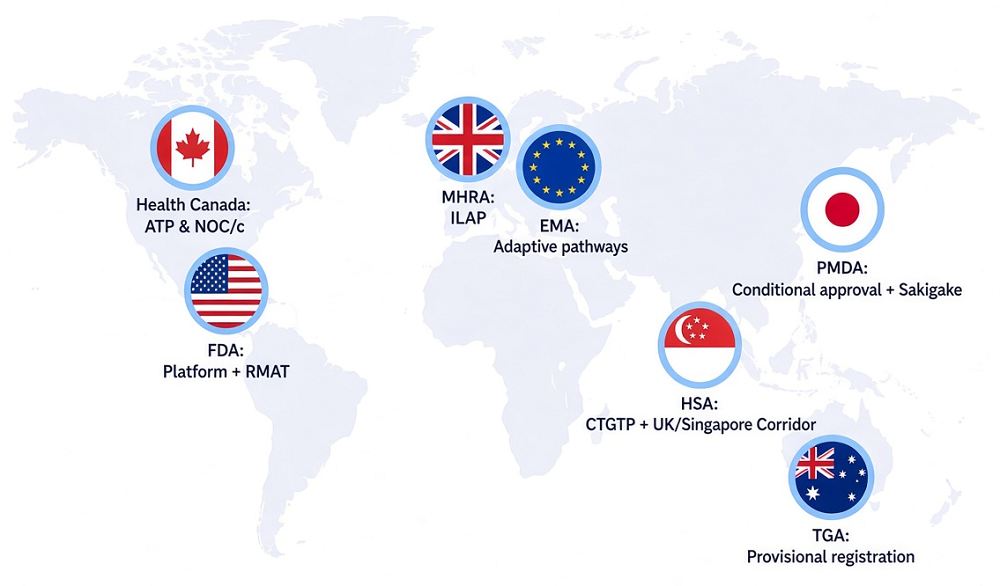
FDA Leaders Name Longevity a Priority at ARDD: At a panel in Boston, senior officials announced plans to include aging in the agency’s regulatory science agenda and discussed how therapies could move toward approval.

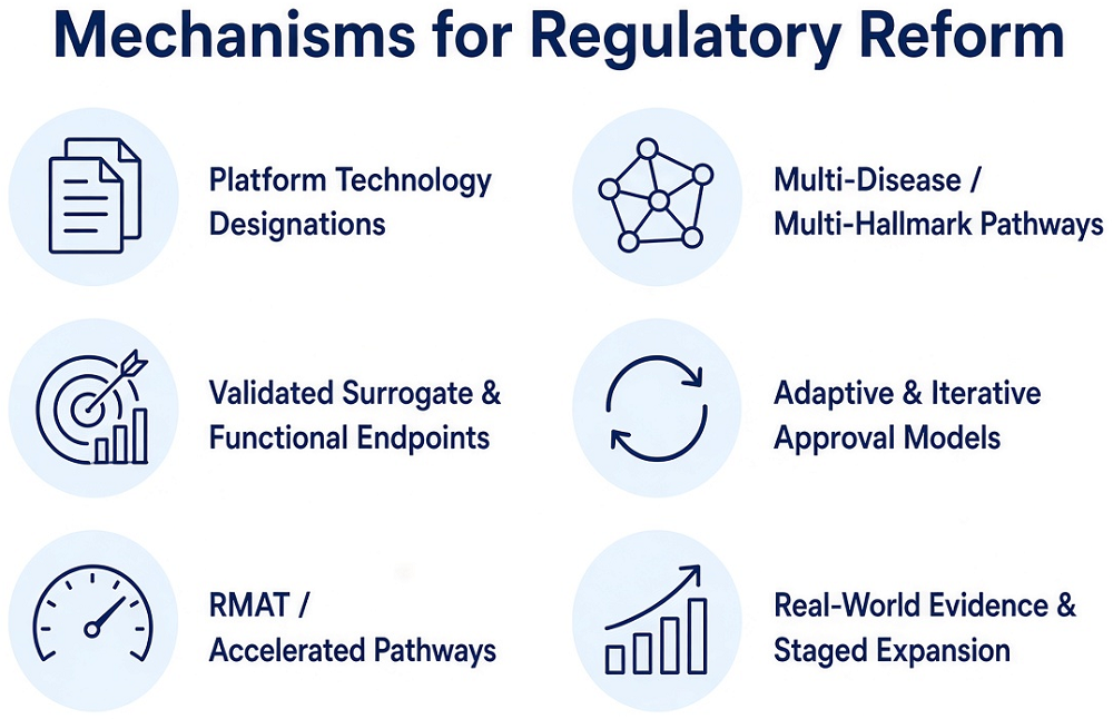
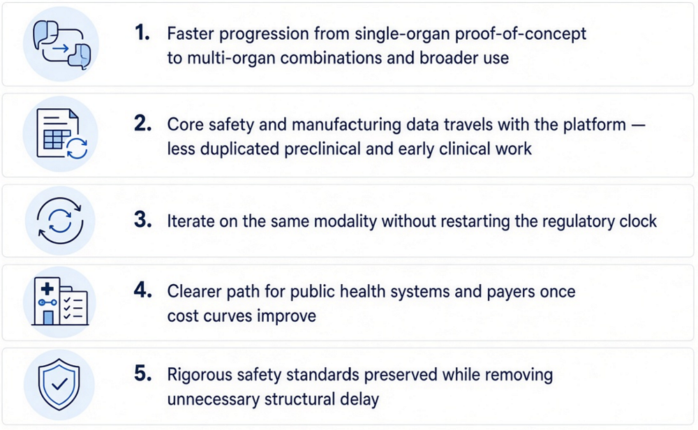
Regulatory Bottleneck 1The Regulatory Bottleneck in Aging Biology: Researchers are developing biomedical repair therapies to address multiple age-related diseases at once, yet they are forced to put them through regulatory pathways built for single-disease drugs. The result is delay, duplication, and unnecessary cost precisely when speed matters the most.

ARDD 2026: Traditional Biology to AI-Driven Breakthroughs: One of the oldest and biggest longevity conferences moved this year across the Atlantic, from Copenhagen to Boston. The new location brought ARDD close to several important longevity hubs and might have contributed to this edition’s practical, entrepreneurial, and forward-oriented spirit.

Research Roundup

Brain and walkingThe Biomarkers That Predict Mortality Risk: Researchers have discovered key biomarkers that are strongly linked to all-cause mortality and identified four measurements with independent predictive power.

Declining Autophagy Impairs Senescent Cell Clearance: A new study links an age-related decline in cellular maintenance to impaired immune clearance. Activating the pathway reduced signs of senescence in aged mice.

Proteins or extracellular vesiclesUntangling Differences in Senescent Cellular Signaling: In Aging, researchers have described the differences between the secretions of cells driven senescent through a cancer gene and cells that stopped proliferating due to p16 expression.

Gut Bacteria Molecule Slows Atherosclerosis in Mice: A new study highlights a fatty acid produced by a particular genus of bacteria as a “natural statin,” suggesting a new way of treating atherosclerosis.

Inflamed lower back discsAn Anti-Senescence Gene May Fight Spinal Disc Deterioration: A team of researchers has described how a little-known gene, ZNF865, has a significant influence on back pain through the regulation of cellular senescence.

Low-Fat Vegan Diet Shows Benefits in Type 2 Diabetes: A human trial found that even without caloric restriction, a low-fat, plant-based diet improved blood sugar control, reduced liver fat, and lowered the quantity of advanced glycation end products (AGEs) in the skin.

Proteins in the brainMenopausal Serum Protein Changes May Predict Brain Health: A recent analysis of serum protein changes identified 16 proteins related to inflammatory, synaptic, metabolic, and Alzheimer’s disease processes whose levels change during the menopausal transition.

Making SIRT1 and NAD+ Therapies Work in People: Reviewers publishing in Biology have elucidated SIRT1 functional reserve as an aspect of aging, describing how its depletion is an upstream cause of age-related disorders at the cellular level and explaining why many prospective therapies that increase SIRT1 and NAD+ have not had promising results.

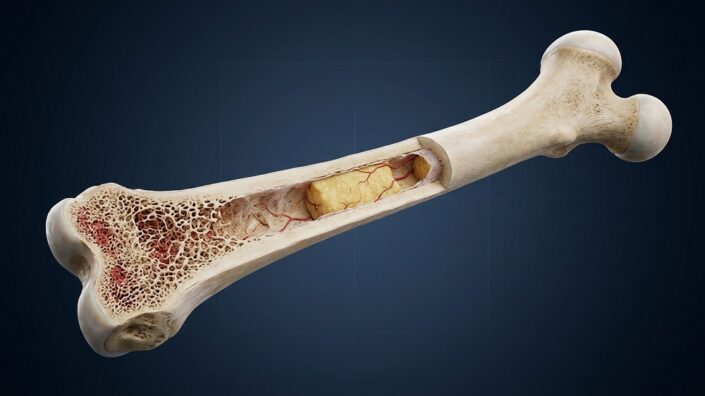
Bone structureBlocking STING Helps Preserve Bone in Aging Mice: A new study links STING-mediated signals from marrow fat cells to inflammation and declining NAD⁺ levels in bone-forming cells. Interrupting this pathway helped preserve aging bone.

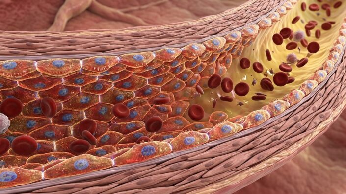
An RNA Regulator Mitigates Senescence in Blood Vessels: Researchers publishing in Aging Cell have found that the regulatory protein MTR4 fights DNA damage in the blood vessel lining but decreases with aging.

CT Scan RobotAI Extracts Broader Health Insights From Routine CT Scans: A new study suggests that CT scans can provide information about a wide range of conditions, including some not typically diagnosed through imaging.

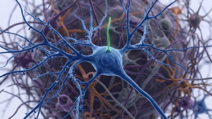
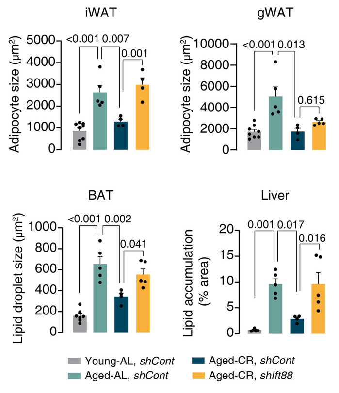
How Some Neuronal Cilia May Affect the Whole Body: A team of researchers has written a paper with a surprising finding: that many of the benefits of caloric restriction and increased NAD+ may be due to their effects on the cilia of hypothalamic neurons.

RNA DNA InteractionRejuvenation Factor Extends Lifespan and Healthspan in Mice: A recent study identified SRSF1, an RNA-processing and splicing factor, as a molecule with geroprotective potential.

An Astrocytic Factor Slows Whole-Body Aging in Mice: In Aging Cell, researchers have described how increasing fibroblast growth factor 13 (FGF13) in astrocytes reduces aging throughout the bodies of mice.

College for T CellsAn Anti-Senescence Vaccine Increases Mouse Lifespan: Late-life treatment with a three-element senolytic vaccine led to lower senescence markers and a 12% increase in median lifespan in female B6 mice.

Midlife time-restricted feeding preserves skeletal muscle insulin responsiveness and function during aging: Midlife time-restricted feeding preserved lean mass, muscle performance, glucose tolerance, and insulin-stimulated glucose uptake in male mice.

Vitamin D Deficiency, Supplementation, and Aging-Related Morbidity and Mortality Across Two Longitudinal Population Cohorts: These findings support severe vitamin D deficiency as an important and potentially modifiable determinant of systemic aging vulnerability and age-related multimorbidity in human populations.

The Effects of Blueberry Consumption on Cerebral Blood Flow and Brain Glutathione Levels in Older Adults: A Double-Blind Randomized, Placebo-Controlled Trial: Compared with placebo, blueberry consumption increased blood flow in the anterior cingulate cortex, a brain region relevant to age-related cognitive decline.

Resistance Training Improves Respiratory Muscle Strength and Immune Response in Older Adults: A Randomized Clinical Trial: The concomitant increase in circulating Klotho suggests that resistance training may promote biological mechanisms associated with healthy aging, supporting its implementation as a non-pharmacological strategy to preserve functional independence and respiratory health in the elderly.

When to Trust Epigenetic Clocks: Avoiding False Positives in Aging Interventions: These findings provide a framework for interpreting epigenetic clock results in anti-aging intervention studies, and suggest that some previously published single-clock findings may warrant reexamination.

Investigating the efficacy and toxicity of partial in vivo reprogramming as a rejuvenation intervention: These findings highlight tissue-specific responses to partial reprogramming and emphasize the need for larger, controlled studies to clarify physiological outcomes and safety.

H3K9me3 inhibition reverses Alzheimer’s progression by restoring synaptic and immune proteostasis across the brain–retina axis: This study identifies H3K9me3 accumulation as a shared, reversible epigenetic contributor to Alzheimer’s disease.

Synergistic reversal of mesenchymal stem cell senescence by engineered exosomal delivery of a dual miR-195/miR-188 sponge: Exosome-mediated delivery of a dual miRNA sponge represents a mechanism-driven strategy to reprogram senescence-associated gene networks and restore MSC function.

Chronic Senolytic Treatment and/or Aerobic Exercise Reduce Senescence and Improve Myocardial Function During Aging in the Heart: These findings demonstrate that long-term exercise or senolytics limit senescence at the cellular level within the myocardium and improve ventricular function.

Aged microenvironment-triggered nanoreactors rejuvenate senescent LepR+ cells to remodel aged bone marrow: Collectively, these findings suggest that the CLPM nanoreactor offers a promising approach for rejuvenation of the aging skeletal system.

Dynamic force sensing by bone delays aging and extends lifespan: This could potentially be manipulated to prevent aging, extend health, and aid those unable to exercise or with decreased mobility.

Elamipretide Rejuvenates Oocyte Quality and Restores Female Fertility During Reproductive Aging: Collectively, this study identifies a novel peptide therapeutic candidate for aging-related infertility, showing that elamipretide restores the quality of aged oocytes for fertility.

Heat-treated Lactiplantibacillus plantarum MYO Improves Muscle Strength and Modulates Gut Microbiota in Older Adults with Reduced Muscle Strength: This approach significantly improved muscle strength and muscle quality, and these muscle strength–enhancing effects may be associated with alterations in the gut microbiota.

The polycentric aging framework: epigenetic regulation as a dominant lifespan-limiting center in human aging: This paper holds that clock shifts should be interpreted against tissue-specific function, healthspan, morbidity, and mortality rather than as stand-alone evidence of rejuvenation.

Seven ways not to think carefully about aging: Accumulating data from mouse studies suggest that many or all varieties of slow-aging, long-lived mice, even as young adults, share multiple physiological and molecular traits whose effects may shed light on the pathogenesis of late-life disease, and whose origins may point to new anti-aging interventions.

Lysergic acid diethylamide extends lifespan in Caenorhabditis elegans: Together, these findings identify LSD as a tool to investigate how serotonergic signaling interfaces with conserved longevity-related pathways.

News Nuggets

Biomarkers of Aging ConsortiumGlobal Leaders in Geroscience Convene for Biomarkers of Aging: The Biomarkers of Aging Consortium has hosted its fourth annual Biomarkers of Aging Conference on October 5 and 6, 2026, at the Joseph B. Martin Conference Center at Harvard Medical School.

Evipedia Goes Hugging Face: Evipedia is a comprehensive, continuously updated online encyclopedia that provides much-needed, accurate, and up-to-date information on a wide range of health and longevity-related interventions.

Life BiosciencesLife Biosciences Announces First-in-Human Phase 1 Trial Data: Administration of ER-100 was well-tolerated through Day 56 in three participants with glaucoma, supporting dose escalation in the ongoing trial.

We would like to ask you a small favor. We are a non-profit foundation, and unlike some other organizations, we have no shareholders and no products to sell you. All our news and educational content is free for everyone to read, but it does mean that we rely on the help of people like you. Every contribution, no matter if it’s big or small, supports independent ethical journalism and sustains our future.

College for T Cells

An Anti-Senescence Vaccine Increases Mouse Lifespan

In a new study, late-life treatment with a three-element senolytic vaccine led to lower senescence markers and a 12% increase in median lifespan in female B6 mice. However, short-lived controls complicate the picture [1].

Helping the immune system kill “zombie cells”

Cellular senescence is a double-edged sword. It plays important roles in early development, wound healing, and cancer surveillance, but when senescent cells are not cleared by the immune system and instead start to accumulate in tissues, they contribute to aging and disease.

To make the aging immune system more effective against senescent cells, scientists have experimented with anti-senescence (senolytic) vaccines [2]. The idea is simple: teach the immune system to recognize a protein associated with these cells and eliminate cells displaying it. Execution, however, has been trickier.

A new study from a group of Chinese scientists, published in Advanced Science, describes a senolytic vaccine consisting of three main elements that is aimed at dendritic cells, the immune cells that collect proteins, process them, and display fragments to T cells. The antigen is Gpnmb, a protein previously used in experimental senolytic vaccination. A special piece of CpG DNA acts as an adjuvant via the receptor TLR9, which recognizes this DNA as a microbial-like alarm signal and promotes T cell activation against the antigen. Finally, the CK5 protein scaffold packages the ingredients into nanoparticles that get absorbed by the cells.

The researchers assembled the three components into nanoparticles approximately 90 nanometers in diameter. They then generated dendritic cells from the bone marrow of young mice. Along with CK5, the team used a different scaffolding protein, K36.

Improved immune responses and senescence markers

Both nanoparticle vaccines improved antigen uptake and dendritic-cell activation compared with simpler formulations. Dendritic cells exposed to the nanoparticle vaccines stimulated greater CD8 T-cell proliferation. However, the CK5-based vaccine released Gpnmb into the dendritic cells’ cytosol more effectively than its K36-based counterpart.

This led the researchers to investigate CK5 separately. A gene expression analysis showed that treating dendritic cells with CK5 reduced expression of Aurora kinases A and B, proteins best known for regulating cell division.

Previous research has linked Aurora-kinase inhibition to reduced activity of PI3K/AKT signaling – a pathway associated with senescence [3]. This led the scientists to investigate whether CK5 can actually alleviate cellular senescence. To test the connection, they compared CK5 with K36 and danusertib, an Aurora-kinase inhibitor. D-galactose was used to produce senescence-like stress in cultured dendritic cells.

When the researchers compared the two vaccines, the CK5-based formulation reduced activation of PI3K/AKT and the senescence-associated markers p16, p21, and p53 while preserving cell viability. The treated cells also showed signs of improved metabolism: increased oxygen consumption and extracellular acidification.

The AI model AlphaFold3 predicted interactions between CK5 and each of the two Aurora kinases. However, this in itself does not prove the proposed mechanism.

A lifespan increase that comes with a caveat

The researchers first tested this vaccine’s health effects in mice fed a high-fat diet from four weeks of age, who were then vaccinated at 60 weeks. The CK5 vaccine elicited stronger responses from dendritic cells and CD8 T cells than the comparator groups, which included vaccines lacking one of the elements, and a K36-based vaccine. More CD8 T cells expressed interferon-γ and granzyme B, molecules associated with immune activation and cytotoxic capacity. The researchers then asked whether those T cells could effectively kill targets. Indeed, T cells collected from CK5-vaccinated mice killed more senescent Gpnmb-high cells in culture.

In the mice, vaccination reduced senescence markers in visceral fat and improved liver fat accumulation. The authors report approximately 50% lower senescence-associated β-galactosidase staining relative to the comparator groups (this experiment did not include untreated controls).

Importantly, glucose tolerance and insulin responses also improved. Body weight and food intake did not differ significantly between groups. This makes reduced eating or weight loss less likely explanations. Median lifespan reached 91 weeks, compared with 79-83 weeks in the comparator groups.

Then came the most important test: effect on lifespan in naturally aged mice. Normally fed mice were vaccinated at 75 weeks of age. The CK5 vaccine and the K36-plus-danusertib combination both strengthened immune responses, reduced senescence markers in fat tissue, and improved glucose regulation. Neither significantly changed food intake or body weight.

Mice that received the CK5 vaccine had a 12% longer median lifespan than untreated controls. This is a strong result considering how late in life the intervention started. However, two important caveats apply: the lifespan experiments were fairly small, with 10 mice per group, and the controls were relatively short-lived for this mouse strain (C57BL/6). Short-lived controls are a known issue in mouse lifespan experiments [4], although this does not necessarily invalidate the results. More rigorous studies are needed to test this intriguing proof of concept.

We would like to ask you a small favor. We are a non-profit foundation, and unlike some other organizations, we have no shareholders and no products to sell you. All our news and educational content is free for everyone to read, but it does mean that we rely on the help of people like you. Every contribution, no matter if it’s big or small, supports independent ethical journalism and sustains our future.

Literature

[1] Q. Song, X. Du, W. Dong, Y. Wang, X. Wang, and Q. Yin, A Bi-Adjuvant Senolytic Nanovaccine Modulating Dendritic Cell Senescence Ameliorates Aging-Related Metabolic Dysfunction. (2026). Advanced Science

[2] Wu, R., Sun, F., Zhang, W., Ren, J., & Liu, G. H. (2024). Targeting aging and age-related diseases with vaccines. Nature Aging, 4(4), 464-482.

[3] Astle, M. V., Hannan, K. M., Ng, P. Y., Lee, R. S., George, A. J., Hsu, A. K., … & Pearson, R. B. (2012). AKT induces senescence in human cells via mTORC1 and p53 in the absence of DNA damage: implications for targeting mTOR during malignancy. Oncogene, 31(15), 1949-1962.

[4] Pabis, K., Barardo, D., Gruber, J., Sirbu, O., Malavolta, M., Selvarajoo, K., … & Kennedy, B. K. (2024). The impact of short-lived controls on the interpretation of lifespan experiments and progress in geroscience–Through the lens of the “900-day rule”. Ageing Research Reviews, 101, 102512.

Astrocytes

An Astrocytic Factor Slows Whole-Body Aging in Mice

In Aging Cell, researchers have described how increasing fibroblast growth factor 13 (FGF13) in astrocytes reduces aging throughout the bodies of mice.

A factor from a well-known family

FGFs are signaling molecules that share structural and functional commonalities and play crucial roles in biology, including neural function [1]. Some of these molecules are secreted, and others are not; one of the secreted molecules, FGF21, has been thoroughly researched as a regulator of aging processes.

However, the subject of this study, FGF13, is a non-secreted molecule with functions that are not related to the FGF receptor [2]. Previous work has found that it has beneficial effects in a mouse model of Parkinson’s [3], while its expression in endothelial cells is detrimental in a mouse model of diabetes [4]. Unlike FGF21, its role in age-related diseases has not been thoroughly explored.

Brain inflammation may spread to the body

The researchers first examined public mouse datasets and confirmed that FGF13 indeed decreases in the brains of mice; while it does not decrease in neurons, it decreases in other cells, including oligodendrocytes, astrocytes, and microglia. Interestingly, while FGF13 smoothly declined with age in the hippocampus, it increased in the cortex at 6 months of age before decreasing. While macaque data was less comprehensive, it was similar to that of mice, with a particularly strong effect in the astrocytes. Therefore, the researchers chose those cells for further experimentation.

Knocking out FGF13 in the astrocytes of aging mice exacerbated their aging. The affected mice performed even more poorly on the Morris water maze test than the aged control group did. Additionally, the affected mice had more cellular senescence in the heart, skin, liver, spleen, and kidney, even though the knockout was targeted to astrocytes, and they also had higher levels of circulating inflammatory cytokines. This led the researchers to conclude that these mice’s brains were “amplifying and transmitting aging signals to other tissues via the blood to accelerate systemic aging.”

The researchers then performed the opposite experiment, using a similar technique to increase FGF13 in the astrocytes of older mice instead. In the Morris water maze test, the treated mice performed similarly to young mice, having a substantial advantage over the untreated mice. In related spatial reasoning tests, the elevated plus maze test and the open field test, the treated mice also performed significantly better than the untreated mice, although not quite to the level of young mice.

Similar benefits were found in other tissues as well, despite this still being an astrocyte-specific intervention. Inflammatory cytokines in the bloodstream were substantially decreased, particularly IFN-γ and IL-6. Most other benefits were significant but modest, with decreases in skin, liver, spleen, and kidney senescence (although not in the heart). FGF13 overexpression in astrocytes also decreased age-related weight gain in mice.

The researchers then performed cellular experiments. They found that complement protein C3, which marks the presence of neurodegenerative diseases, is significantly increased in FGF13-deficient astrocytes. Cx43, which marks age-related neuronal injury, is increased with FGF13 deficiency. Co-culturing these deficient astrocytes with neurons found that they impaired the neurons’ synapses, and co-culturing them with microglia revealed that they induced the microglia to release inflammatory factors. FGF13 overexpression in neurons revealed benefits in all of these areas, restoring neuronal synaptic proteins, reducing microglial expression of cytokines, and reducing levels of proteins associated with injury and degeneration.

Autophagy may be the crux

RNA sequencing revealed some of the mechanisms of action behind these effects. 625 genes were upregulated and 195 were downregulated in astrocytes with the overexpression of FGF13. These genes affected multiple pathways, including lysosomal transport, inflammatory factor production, metabolism, and various signals.

FGF13 was specifically found to have a physical interaction with the protein Phafin2, a protein that is related to the cellular maintenance process of autophagy [5]. Like FGF13, Phafin2 decreases with age in mice, while increasing FGF13 also increases Phafin2. An analysis of the mice’s brain tissue found that the FGF13-overexpressing animals had increases in autophagy, as evidenced by increases in autophagosomes and autolysosomes along with the autophagy markers P62 and LC3. Similar results were found in cellular experiments.

These researchers have provided evidence that an upstream regulator in one particular brain cell has downstream ramifications throughout the body, suggesting that a treatment to increase FGF13 in the astrocytes of older people may improve multiple age-related conditions. However, this paper does not suggest any potential methods of doing so, and further work will need to be done to confirm these results and determine whether this approach is valid in non-human primates and then in people.

We would like to ask you a small favor. We are a non-profit foundation, and unlike some other organizations, we have no shareholders and no products to sell you. All our news and educational content is free for everyone to read, but it does mean that we rely on the help of people like you. Every contribution, no matter if it’s big or small, supports independent ethical journalism and sustains our future.

Literature

[1] Bollenbecker, S., Barnes, J. W., & Krick, S. (2023). Fibroblast growth factor signaling in development and disease. International Journal of Molecular Sciences, 24(11), 9734.

[2] Naderi, J., Johnson, A. K., Thakkar, H., Chandravanshi, B., Ksiazek, A., Anand, A., … & Chaurasia, B. (2025). Ceramide-induced FGF13 impairs systemic metabolic health. Cell metabolism, 37(5), 1206-1222.

[3] Song, N., Wang, X., Ha, L., Hu, L., Mei, S., Liang, Y., … & Lu, M. (2025). Neuronal FGF13 Inhibits Mitochondria‐Derived Damage Signals to Prevent Neuroinflammation and Neurodegeneration in a Mouse Model of Parkinson’s Disease. Advanced science, 12(28), 2503579.

[4] Sun, J., Guan, X., Niu, C., Chen, P., Li, Y., Wang, X., … & Cong, W. (2023). FGF13-Sensitive alteration of parkin safeguards mitochondrial homeostasis in endothelium of diabetic nephropathy. Diabetes, 72(1), 97-111.

[5] Tang, T. X., Jo, A., Deng, J., Ellena, J. F., Lazar, I. M., Davis, R. M., & Capelluto, D. G. (2017). Structural, thermodynamic, and phosphatidylinositol 3‐phosphate binding properties of Phafin2. Protein Science, 26(4), 814-823.

RNA DNA Interaction

Rejuvenation Factor Extends Lifespan and Healthspan in Mice

A recent study identified SRSF1, an RNA-processing and splicing factor, as a molecule with geroprotective potential. Inducing SRSF1 expression rejuvenated human cells in vitro, extended lifespan, and significantly reduced frailty in mice [1].

Alternative approaches

Differentiated cells have the capacity to rejuvenate if an appropriate molecular environment is provided. The best-known factors that enable cellular rejuvenation are the Yamanaka factors, which can reprogram differentiated cells into induced pluripotent stem (iPS) cells. While cellular reprogramming with Yamanaka factors can reverse aging signatures, it can cause cells to lose their identity. This has prompted researchers to seek alternative approaches, especially since rejuvenation and dedifferentiation processes can be separated [2-4].

“In reprogramming, there is a fundamental tradeoff between rejuvenation and cell identity; achieving one without losing the other requires walking a fine line,” said Alex Plesa, Ph.D., the study’s first co-author.

These researchers searched for an alternative approach to cell rejuvenation by investigating age-associated shifts in gene expression, since the genes that a cell expresses at a given time (the transcriptome) defines the cell’s identity and changes with age.

Building a model

This team started by building a transcriptomic aging model. They trained a machine-learning predictor on gene expression data from primary normal human dermal fibroblasts from people aged 1 to 96 years, teaching it to assess cellular age. They grouped the gene expression data by its associated cellular processes, allowing them to identify modules that best predict chronological age. The model also identified interventions and cellular states that affect cellular age faster than a commonly used DNAm clock, suggesting that gene expression changes are much faster and more sensitive than more stable DNA methylation changes.

“The most prominently used ‘aging clock,’ the epigenetic aging clock, assesses chemical modifications to the genome that occur with age as one of the hallmarks of aging, but it doesn’t scale well to screening approaches because of high costs and challenges with interpreting its widespread changes,” said Plesa. “The idea of predicting the age of cells based on their gene expression made a lot of sense, since every rejuvenation process inevitably has to manifest itself in altered gene expression programs that lead to the recovery of more youthful cell functionalities,” said Plesa.

Rejuvenating cells and body

The developed models were used in the screen that identified the age-reversal factor SRSF1, an RNA-processing and splicing factor whose expression declines during fibroblast aging.

The authors built a library of preselected genes previously linked to human-aging-associated networks. They overexpressed those genes in human dermal fibroblasts and used their transcriptomic age predictor to evaluate how each gene affected the cells’ transcriptome. SRSF1 was the most robust hit for transcriptomic rejuvenation (comparable to OSKM). Further cell culture experiments showed that SRSF1 induction reverses multiple hallmarks of aging: it reduced senescence markers and senescence-associated secretory phenotype programs, proteostatic stress, and inflammatory signaling, while inducing DNA repair pathways.

Unlike OSKM-driven rejuvenation, SRSF1-mediated rejuvenation does not cause cells to lose their cellular identity in terms of transcriptional state and morphology, despite reducing transcriptomic and epigenetic age.

At the whole-body level, SRSF1 induction extended healthspan and lifespan in aged mice. Specifically, acute SRSF1 induction reduced frailty by 44%, accelerated wound closure, reduced mortality risk, and extended median lifespan by 14% in aged mice. This was achieved despite using a viral vector with a tropism for the liver, limiting its systemic effects.

“It is also very likely that primarily targeting the liver can reverse aging processes in other organs and tissues and that rejuvenation programs can be communicated and thus amplified within the body,” said Plesa.

Based on these results, the authors suggested that delayed physiological deterioration is a key contributor to lifespan extension, since they observed an association between frailty burden and survival. Additionally, comparing gene expression in mouse liver with previously analyzed human fibroblasts pointed to downregulation of similar molecular pathways: age-associated stress responses, inflammatory pathways, and cell-cycle programs following SRSF1 induction. However, these observations came from a small-scale experiment with only 9-10 female mice per group.

Constitutive SRSF1 induction produced higher SRSF1 levels, and while it didn’t cause toxicity, it did not improve wound repair or muscle regeneration. These observations point to the importance of treatment timing and dosing. As the authors suggest, SRSF1 might not be a simple ‘on switch’ for regenerative processes but a precisely regulated molecule. Therefore, designing a future therapy that would use SRSF1 induction would require precise dosing and timing regimens.

Alternatively spliced

The DNA code for a gene is not a direct, straight line of code; it’s made of exons (coding parts) interspaced by introns (non-coding). When the gene is transcribed into RNA (an intermediary between a gene and a protein), some exons can be included, and some can be removed, allowing multiple versions of a given protein and adding another layer of gene expression regulation and protein functions. SRSF1 induction affected these processes and created a “consistent splicing landscape dominated by alternative first exon (~45%) and skipped exon (~25%) events.”

Analyzing this splicing data alongside data on differentially expressed genes pointed to dual regulation of key genes in epigenetic and endoplasmic reticulum pathways, offering insights into SRSF1’s rejuvenation effects; however, future studies are required to understand the mechanistic links between those molecular processes and observed cellular effects, such as tissue rejuvenation.

“We observed a strong trend of alternatively spliced mRNAs, but we are only just starting to understand how these changes bring about the multi-faceted rejuvenation program in tissues,” said Plesa.

A geroprotective therapeutic target

This study identified SRSF1 as a key player in regulating aging processes and a potential geroprotective target. The authors see it as a potential target for future anti-aging therapies.

“We were working on the premise that over-expressing a single gene with validated rejuvenation outcomes could eventually be advanced into an organ or tissue-specific gene therapy if combined with the right delivery system,” said Plesa.

“It has been a long-standing goal of my lab to devise clinically useful de-aging strategies by identifying genes that, when engineered in the genome for under or over-expression in aging target tissues, extend human healthspan. Our platform and findings on SRSF1 are an important step on that quest,” said Church, who is a Professor of Genetics at Harvard Medical School. “Aging research is transiting an inflection point where the exponential progress of biotech is impacting the core aging processes which would otherwise kill 90% of us.”

We would like to ask you a small favor. We are a non-profit foundation, and unlike some other organizations, we have no shareholders and no products to sell you. All our news and educational content is free for everyone to read, but it does mean that we rely on the help of people like you. Every contribution, no matter if it’s big or small, supports independent ethical journalism and sustains our future.

Literature

[1] Plesa, A. M., Jung, S., Wang, H. H., Omar, F., Shadpour, M., Choy Buentello, D., Perez-Matos, M. C., Horwitz, N., Cai, G., Ngian, Z. K., Magalhaes, C., Wagers, A. J., Mair, W. B., Del Sol, A., & Church, G. M. (2026). Transcriptomic reprogramming screen identifies SRSF1 as a regulator of aging. Nature aging, 6(10), 2057–2073.

[2] Rando, T. A., & Chang, H. Y. (2012). Aging, rejuvenation, and epigenetic reprogramming: resetting the aging clock. Cell, 148(1-2), 46–57.

[3] Olova, N., Simpson, D. J., Marioni, R. E., & Chandra, T. (2019). Partial reprogramming induces a steady decline in epigenetic age before loss of somatic identity. Aging cell, 18(1), e12877.

[4] Roux, A. E., Zhang, C., Paw, J., Zavala-Solorio, J., Malahias, E., Vijay, T., Kolumam, G., Kenyon, C., & Kimmel, J. C. (2022). Diverse partial reprogramming strategies restore youthful gene expression and transiently suppress cell identity. Cell systems, 13(7), 574–587.e11.

ARDD 2026: Traditional Biology to AI-Driven Breakthroughs

One of the oldest and biggest longevity conferences moved this year across the Atlantic, from Copenhagen to Boston. The new location brought ARDD close to several important longevity hubs and might have contributed to this edition’s practical, entrepreneurial, and forward-oriented spirit. We have already reported on the unprecedented panel where FDA named longevity a priority. Now, we bring you a curated selection of talks spanning areas from calorie restriction to creating “digital twins” for human patients and achieving longevity escape velocity in the foreseeable future. We apologize for not being able to include many other equally exciting and informative talks.

Vishwa Deep Dixit on Metabolic Checkpoints in Inflammaging

Vishwa Deep Dixit of Yale presented cysteine and taurine metabolism as two checkpoints that link nutrient availability to the aging-related inflammation known as inflammaging.

The NLRP3 inflammasome is a protein complex that drives inflammatory responses in tissue-resident macrophages. According to Dixit, increased inflammasome activity is associated with biological age and mortality, while animals with lower NLRP3 activity are protected from multiple degenerative changes.

To understand what regulates this complex, Dixit’s team started by analyzing results from the CALERIE calorie restriction trial in humans. Interestingly, the target was 25% calorie restriction, but participants achieved only about 14%. Dixit suggested this might be closer to what people can sustain long-term in real-life conditions.

Longitudinal adipose-tissue metabolomics showed lower cysteine and glutathione but higher taurine. Dixit interpreted this as a shift in sulfur-amino-acid metabolism toward taurine production. The transsulfuration pathway connects methionine to cysteine, which, in turn, supplies both glutathione synthesis and the taurine pathway.

To test cysteine’s role, Dixit’s team combined deletion of a cysteine-producing enzyme with dietary restriction in mice. The animals lost about 30% of their weight within six days. This was accompanied by the conversion of white fat into thermogenic brown/beige fat. The effect was so severe that the mice had to be euthanized.

Dixit reminded the audience that restricting methionine, another amino acid, has extended lifespan in multiple animal studies. However, historical methionine-restricted diets also lacked cystine, cysteine’s oxidized dimer. Dixit suggested that cysteine depletion is what actually drives the benefits attributed to methionine restriction.

In C. elegans experiments reported in a preprint, Dixit’s team recorded longer lifespans and improved survival after heat stress with cysteine restriction. Methionine restriction did not produce those benefits. Adding back cysteine reversed the heat-stress survival advantage.

According to Dixit, humans in calorie-restricted states prioritize making taurine, of which cysteine is a precursor. Giving taurine to old mice reduced inflammatory cytokine levels. Conversely, experiments showed that less taurine in the system led to stronger inflammasome activation.

In activated macrophages, intracellular taurine levels decline as taurine leaves the cell. Restoring taurine suppresses NLRP3 inflammasome activity and rescues inflammatory cell death.

But which comes first? Taurine loss persisted when NLRP3, ASC, or gasdermin D was knocked out. Dixit therefore places taurine efflux upstream of inflammasome assembly. His overarching idea is that taurine acts as a metabolic damage sensor: its efflux from the cell triggers inflammasome activation. During a Q&A period, he said that his team had not identified the exact export mechanism yet.

Dixit closed by asking whether NLRP3 inhibitors could be useful against inflammaging. His findings also suggest another way to approach the problem: keeping taurine inside macrophages. In these experiments, restoring it helped shut down the inflammatory response.

Kevin Duffin on How Tirzepatide Changes Biological Aging Clocks

The conference saw a significant presence of Big Pharma representatives. Kevin Duffin from Eli Lilly reported on a recent trial of the effect of tirzepatide on a battery of biological age clocks. Tirzepatide is a dual agonist against GLP-1 and GIP. It is the active ingredient in Mounjaro, which is branded for type 2 diabetes, and in Zepbound, which is branded for overweight and obesity.

Duffin first reminded that his company has run tens of clinical trials on tirzepatide and other GLP-1-based drugs and has seen significant improvements in weight loss, obesity, and other parameters, such as liver, kidney, and cardiovascular disease. He mentioned significant reductions in hospitalization in heart failure as well as in all-cause mortality.

“Yesterday, we talked about what is a good aging drug,” Duffin said. “One of the definitions was a drug that shows improvements in multiple organ diseases. Well, I think we checked the box there.”

Finally, the company decided to look at biological age clocks, including organ-specific clocks. Duffin presented data specifically about epigenetic (methylation-based) clocks. An impetus to do the study, he noted, came in part from Novo Nordisk, Lilly’s competitor in the GLP-1 field, presenting data on a small cohort at the last-year ARDD.

The trial, named SURMOUNT-5, is one in a group of trials in patients with obesity. It was done on a relatively young population, overweight but not diabetic. However, for the clocks study, older patients, aged from 50 to about 80, were picked out.

The trial, according to Duffin, produced good outcomes. It looked at tirzepatide versus semaglutide, with both drugs producing significant weight loss, reductions in blood pressure, fasting insulin, triglycerides, and LDL cholesterol. However, in his talk, Duffin only gave details on the tirzepatide results.

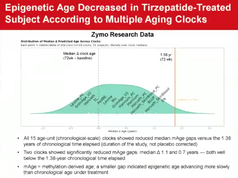
Since it was a 72-week study, the participants’ chronological age increased by 1.38 years. Some of the clocks showed a slight increase versus baseline but smaller than the chronological age increase. A minority of the clocks showed decreases even compared to baseline – in other words, rejuvenation rather than slower aging.

Duffin mentioned that, as a rule, second-generation clocks showed stronger age decreases. Pace-of-aging clocks were also used (DunedinPACE and FraminghamPACE), showing a reduction in the pace of aging with the drug.

The team sent blood samples to two companies: Zymo and TruDiagnostic. The latter also measured organ-specific aging, recording biological age decreases in multiple organs and systems, including the heart, immune system, kidney, and liver.

“I point those out specifically because we saw good clinical endpoints in those disease areas in our clinical trials,” Duffin said. He noted that this consistency between clinical endpoint data and biomarker data is exactly what we want to see in longevity trials. Epigenetics were also used to predict multiple protein biomarker changes. Many, but not all, changed concordantly.

Can the effect be separated from weight loss? When asked this during a Q&A period, Duffin said that the benefits probably come mostly from weight loss and improved glucose regulation.
ARDD Presentations 1

Alex Zhavoronkov on AI-driven Drug Discovery for Longevity

One of the conference’s founders, Alex Zhavoronkov, CEO of Insilico, presented results from a 12-week Phase 2a trial of rentosertib (an AI-discovered inhibitor of TNIK developed in-house by Insilico for idiopathic pulmonary fibrosis) and a subsequent proteomic analysis published last month in Nature Biotechnology. However, before diving into the data, he discussed at length the wider topic of drug discovery for longevity.

Although today, no drugs or therapies exist that significantly prolong human lifespan, adding even three years of life to everyone on the planet “is more lifetimes than we ever lost in all wars ever fought,” Zhavoronkov said. This makes increasing longevity one of humanity’s most important goals.

Unfortunately, most current strategies for identifying longevity therapeutics work slowly. Insilico claims to offer a sustainable business model of making money and reinvesting it back into science with the use of AI, which profoundly speeds up preclinical discovery.

As much as the company and its founder are passionate about longevity, aging research is currently more of a byproduct, according to Zhavoronkov. The idea is to find therapeutic targets that are present in aging and disease at the same time, approve candidates for a specific indication, expand the number of indications, and then try to go after aging. About 70% of Insilico’s pipeline consists of dual-purpose approaches.

“The reason we chose IPF as the first indication is that, if you are developing a longevity therapeutic, you need to outperform the standard of care,” explained Zhavoronkov. “In some diseases, it’s almost impossible with a longevity therapeutic, because it is likely to work very mildly. In our case, we wanted to show it in an indication where nothing works.”

Zhavoronkov then moved to the results, which he called very early and preliminary. Rentosertib showed a 98 ml increase in the amount of air a person can forcibly exhale (forced vital capacity) with the higher once-daily dose, which he described as “a very promising trend.”

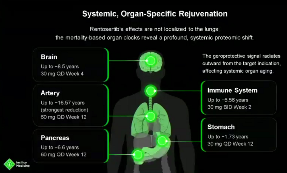
Switching to the newest analysis, Zhavoronkov reported using six proteomic biological age clocks on 42 participants, including models trained to predict chronological age or mortality risk. The clocks indicated reductions in estimated biological age of roughly three to six years after four weeks. The estimates then partly rebounded, with the extent varying between clocks.

Organ-specific estimates also shifted. The most striking was a reported 16.57-year reduction in an arterial clock. To understand whether the drug only affects the disease itself or aging in general, the team compared its protein effects with age-associated patterns in UK Biobank. The drug reversed the trajectories of some of the proteins identified in UK Biobank as responsible for aging. According to Zhavoronkov, the effect had a strong senomorphic component (IPF is thought to be driven in large part by cellular senescence).

Interesting dose-dependent effects emerged. “If you administer it at high dose once a day, it looks more disease-relevant,” he noted. “If you give it twice a day in smaller doses, you see more of an anti-aging effect.”
ARDD Presentations 2

Vadim Gladyshev on Measuring Aging to Understand and Target It

In a sprawling talk, Harvard professor Vadim Gladyshev shared his vision on measuring, targeting and understanding aging, along with some exciting results from his prolific team.

This team, he said, is interested in how function is built with age, from early development to late life. Gladyshev was one of the first scientists to investigate “embryonic reset” – the mysterious disappearance of the signs of aging that occurs in early embryonic development and allows young organisms to be born from old organisms, permitting the continuation of life. The damage levels – what Gladyshev calls the “deleteriome” – begin to increase from this point on all the way until the end of life, even though function follows a different path; we are at our best when we are young adults.

Gladyshev’s team is also interested in organ- and system-specific aging. Not everything in our body fails at once: rather, certain organs and systems age faster in different people, which opens the door to early interventions.

Targeting these “weak links” is one of three major ways of controlling lifespan, he said. Remodeling physiology is another one, including interventions like calorie restriction and rapamycin. This is the most direct and immediate approach, but the expected gains in lifespan are modest.

In this view, achieving radical lifespan extension requires overcoming species-level constraints: increasing maximum human lifespan by drastically slowing down aging. In the future, this can potentially be done with approaches like rejuvenation, replacement, and complex coordinated intervention combinations.

“As you realize, our main goal is to understand aging,” Gladyshev said. “Everything else is an application of that.”

Long-lived species can provide many useful insights. Gladyshev noted the immense differences in lifespan between closely related animals, which proves that tweaking biology can affect aging way stronger than currently known interventions. A similar phenomenon occurs across cell types: despite having the same genome, some cells live only a few days, while others can survive for decades. Gladyshev called their research into these topics “discovering the rules of aging.”

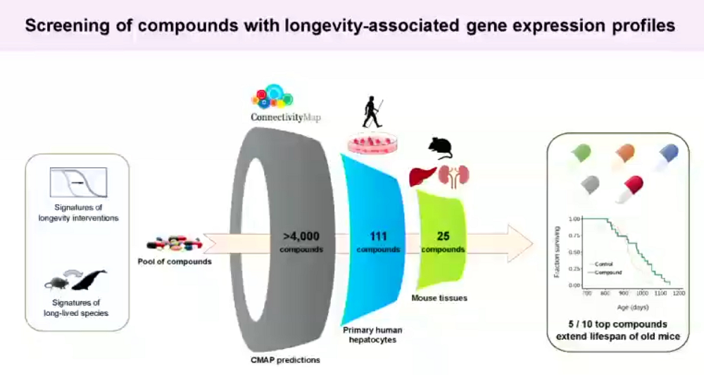
One approach involves longevity-associated expression patterns. The team computationally screened more than 4,000 compounds, then narrowed the list of candidates through tests in human liver cells and mouse tissues. Of ten compounds selected for lifespan experiments, five extended survival in old mice.

Gladyshev explained that while epigenetic clocks are more widely used, transcriptomic data is more widely available, so the researchers built transcriptomics-based aging clocks that strongly correlate with mouse chronological age and mortality. These clocks can be used to pre-screen compounds before committing to lifespan experiments. According to these clocks, Gladyshev said, “diseased tissue is always older,” suggesting a profound connection between aging and disease. The team built a free online tool called Transcriptomic Age Calculator Online (TACO), which allows scientists to analyze their RNA-sequencing data in the context of aging.

Gladyshev touted the team’s recent work on heterochronic heart transplantation. It showed that young hearts in old mice aged rapidly, while old hearts in young mice were rejuvenated. Gladyshev suggested that older donor hearts might therefore be more useful than assumed. However, there were no significant effects on the ages of the transplant recipients’ native organs.

Finally, Gladyshev’s broader proposal was to combine molecular data, imaging, physiology, and longitudinal observations to track biological states. Measuring how the therapy’s molecular target changes is not enough, he argued.
ARDD Presentations 3

James Peyer on Reversing Metabolic Decline Through AMPK Activation

Cambrian Bio has been around for a while, and its founder James Peyer has presented at many conferences. This time, he had exciting new study results to talk about, but not before describing the company’s philosophy: “Cambrian has developed a reputation in this aging biology space for doing the obvious thing, which is to take… pathways that come up again and again in our study of aging biology, but we haven’t been able to drug successfully.”

Two concrete pathways Cambrian is working on are MTOR and AMPK, and this talk was focused on the latter. AMPK is a major regulator of metabolism. It mobilizes energy from the liver and fat tissue, delivers it into the cell, and coordinates it use in the mitochondria to increase energy reserves. As such, it also mediates response to exercise.

Peyer first mentioned metformin as an imperfect AMPK activator. Metformin, he said, gets AMPK activated by inhibiting mitochondrial complex I. Essentially, AMPK activation is paid for by stifling mitochondrial energy production, which might explain metformin’s interference with exercise. Cambrian has been looking for an AMPK activator that escapes such trade-offs.

Peyer noted that some of the previous attempts to develop AMPK activators, such as by Merck with their MK-8722 candidate, led to increased glucose and fatty acid uptake by muscles with concomitant upregulation of mitochondrial function, which caused glycogen accumulation, specifically in cardiac muscle. The opposite approach – stimulating mitochondrial energy production without bringing in extra fuel – turned out to be cytotoxic.

According to Peyer, Cambrian’s candidate ATX-304 successfully activates both sides of the pathway; oxidative phosphorylation “increases in a dose-dependent way in the same concentration ranges that you’re increasing glucose uptake,” leading to an actual decrease in muscle glycogen. This increase in metabolic rate changes cells’ composition, driving up the levels of all five mitochondrial complexes, which suggests large-scale mitochondrial biogenesis.

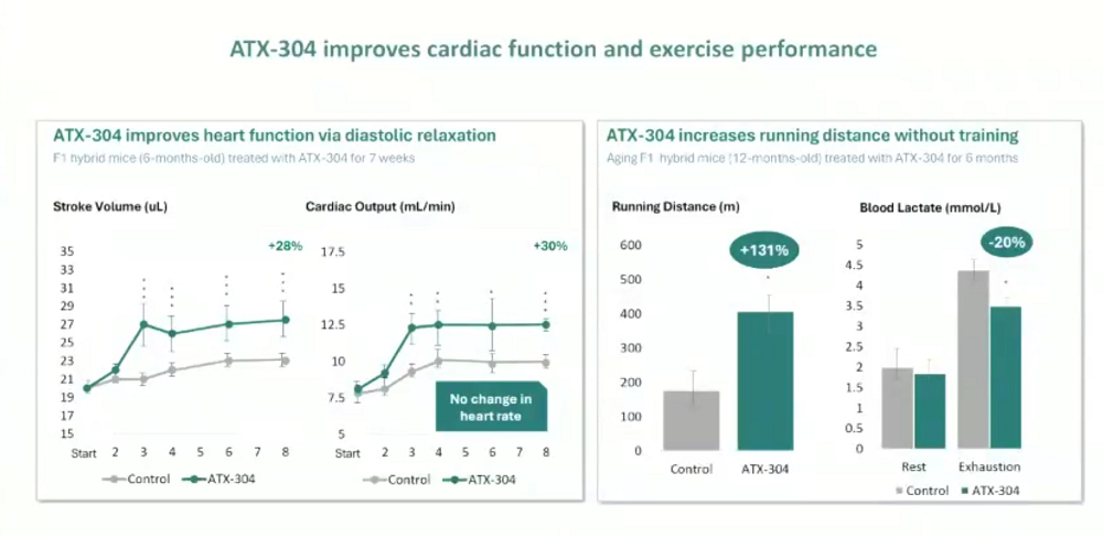
The next question is: can it mimic exercise effects? Indeed, a seven-week trial in mice led to increased stroke volume and cardiac output without affecting heart rate, an effect seen in athletes. Despite a 131% increase in running distance, blood lactate levels in treated mice were actually lower, hinting at superior mitochondrial function.

Cambrian found its indication in obesity. Weight loss in obese mice was comparable with semaglutide, but, importantly, there was no change in food consumption or loss of lean mass, which is a major concern in GLP-1-based therapies. Combination therapy with semaglutide increased weight loss, still without lean mass loss.

Weight gain after stopping GLP-1-based therapy is a known problem. However, mice that had lost weight on semaglutide did not regain it when ATX-304 was used as a maintenance drug.

The next big test would be translating these results into humans. Peyer reported on a small Phase I trial with 23 obese, pre-diabetic participants. The slides were marked confidential, but the company has previously released some data. According to these press materials, ATX-304 produced statistically significant, time-dependent improvements in lipid metabolism and body composition.

ATX-304 increased resting metabolic rate by 8%, consistent with the drug’s mechanism of action. Importantly, no mitochondrial failure or increase in body temperature and 24-hour heart rate were observed. More robust human studies that could prove significant weight loss and muscle preservation are on the way.
ARDD Presentations 4

David Sinclair on NAD Metabolism and Epigenetic Restoration

Harvard professor David Sinclair, one of the most recognizable names in the longevity field, reported on the various directions that his lab has been pursuing. One of the less flashy ones involves boosting the levels of NAD, a molecule that facilitates numerous crucial cellular processes, including DNA repair. “My lab and others have shown that NAD boosting and sirtuin activation as a downstream target are beneficial in mammals,” he said.

The results Sinclair presented come from a ten-year-long project and have not been previously disclosed. His team, he said, discovered “a new type of biology and a new structure in mammalian cells that forms with aging.” They call these structures “inflammules.”

The research was done in yeast cells, a traditional aging model. Inflammules, Sinclair said, are a major reason that NAD is consumed increasingly fast in senescent cells and in older tissues. Even though some research suggests that NAD blood levels don’t decline with age, in many tissues, such as in the brain, they do.

Inflammules consume NAD through PARP14 and promote inflammatory signaling through TRAF6 and NF-κB, contributing to the senescence-associated secretory phenotype (SASP). Disrupting inflammules increased NAD levels in aged fibroblasts. The idea is to address a source of NAD consumption, rather than simply increase its supply.

Sinclair then moved to his Information Theory of Aging, which postulates that much of aging stems from gradual loss of information on how cells should behave (exdifferentiation, or dedifferentiation). A crucial element of the theory is the existence of a hypothetical back-up copy of this epigenetic information in every cell. Sinclair’s lab is currently trying to understand the structure of this information repository. Sinclair signaled that some results might be coming.

The lab does a lot of work on the phenomenon that this theory attempts to explain: epigenetic restoration. From the beginning, Sinclair’s team decided to forgo the classic Yamanaka reprogramming factor, because the full cocktail (OSKM) had already been known to be toxic. Eventually, the researchers discovered that taking out one factor (c-Myc, a known oncogene) reduces the risk while preserving the reprogramming effect. Even lifelong treatment in mice proved to be safe. The team was able to restore vision in mice and non-human primates. Today, Life Biosciences, co-founded by Sinclair, is running the first ever human reprogramming trial aimed at glaucoma and NAION (“the stroke of the eye”).

Sinclair then shared more previously undisclosed data. His team tried putting OSK into cancer cells and tumors in mice, expecting it to either reinforce lineage programs (the cell reacquires its identity), cause further epigenome destabilization, or not have any effect. They found that OSK-treated cancer cells slow down and die. Single-cell multi-omics confirmed that many of the cancer cells regained their cell type identity.

Another project is creating a “chemical reprogramming pill” that could treat the whole body, unlike the viral vectors that are used in Life Bio’s trial. The team screened a total of 80 billion molecules using AI to get down to a small number of possible epigenetic restorers.

The one they are focused on the most is SL-100. In vitro, SL-100 protects ALS motor neurons from stress, preserving neurite length. The treatment benefits even healthy neurons. Molecular assays showed signs of rejuvenation in the treated cells. The scientists ran a pilot lifespan and frailty trial in progeroid mice (LAKI) and detected strong effects on frailty and lifespan, with a 25% mean and a 33% maximum lifespan increase.
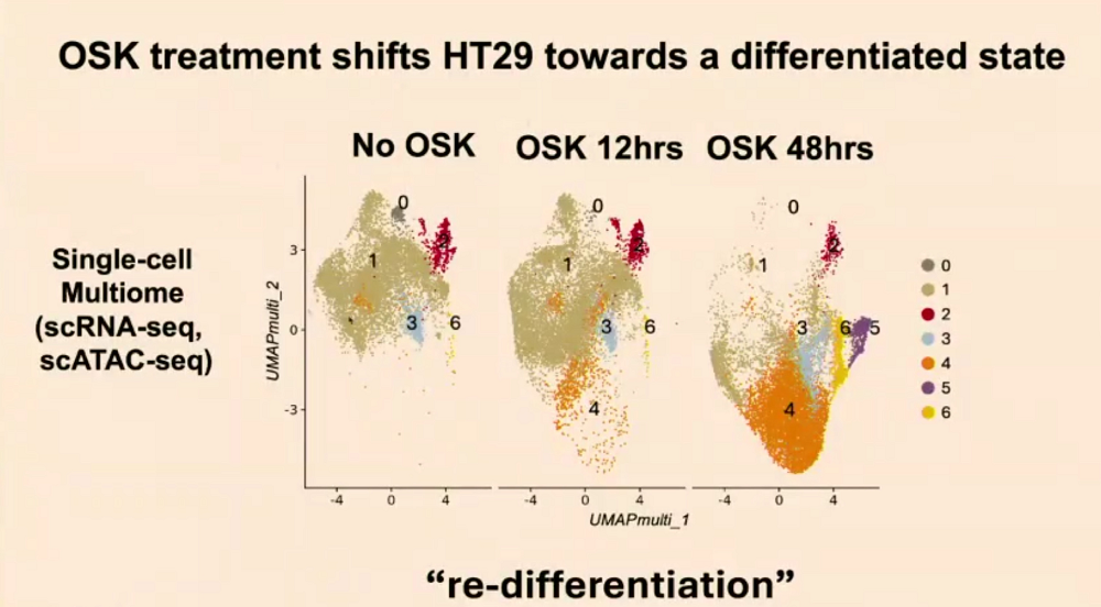
ARDD Presentations 5

Morgan Levine on Why Virtual Cells Alone Won’t Solve Aging

Morgan Levine, former Yale professor and principal investigator at Altos Labs, revealed that she left the company about two months ago to lead a stealth startup, granting the audience a rare glance into what that startup might be up to.

Her talk, like several others at the conference, revolved around virtual cells – computer models that can simulate various aspects of living cells. Levine started with a cold shower, stating that these models “are probably not enough to solve aging.”

The problem, she said, is biology being extremely complex and multidimensional. Innumerable events happen in a cell simultaneously, creating a dynamic and constantly changing state.

The number of experiments needed to understand a cell is immense. Testing whether each of roughly 20,000 protein-coding genes should be turned down or up already requires about 40,000 conditions. Add different cell types, environments, genetic backgrounds, time points, and measurements, and the number reaches billions. Combinations of targets make the problem worse, and there are about 1060 possible drug-like small molecules. “You can imagine, as we start to get up to this multidimensional complexity, this is really infeasible to do experimentally,” Levine said.

This complexity problem can theoretically be solved by in silico experimentation. Morgan, however, is interested in more than just doing a screen. The question virtual cells should be able to answer, through reasoning or other mechanisms, is how to move a cell from the starting state to the desired end state.

Most current virtual cells are built around single-cell RNA-sequencing data. However, RNA is not a cell’s state, but just one of its many dimensions. Capturing the complete data on every such dimension is an immense task, but much of that data also might be redundant. How do we know what we actually need to capture?

We need good benchmarks, said Levin, bringing up the Virtual Cell Challenge by Arc Institute. Its benchmarks test whether models can predict changes in gene expression and distinguish one perturbation from another. However, one metric was sensitive to the scale of predictions, which created opportunities for competing teams to increase their scores without actual biological improvement. For Levine, this illustrated the importance of benchmark design and correct incentives.

“Perturbation-response prediction, particularly RNA-seq, is not the right benchmark for virtual cells, in my opinion,” she noted. “I don’t think it’s that useful for us to be generating raw data, because you’re still lacking interpretability.” Biological data is often very noisy, and when models generate raw data, that includes the noise. Therefore, generative AI may not be the best choice for virtual-cell models.

“The way I think about every type of modality is just an incomplete reflection of this latent state that we’re actually trying to learn,” Levine said. “That’s the thing we care about: how do you actually perceive the latent state and make interpretations of what that means, not how do you perfectly generate the data?”

She argued that we need to build more general models of biology, not just cell models, that capture all the different states of biology and modalities. She likened those different modalities to senses that supply raw data to the brain, which in turn produces perception and reasoning.

“My new endeavor is trying to build this perception layer of biology,” Levine said. She envisions world models of biology that are fed those “sensory inputs” and task prompts and are capable of reasoning their way to solutions.
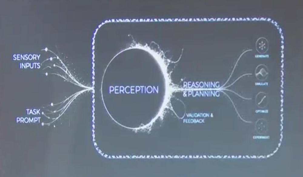
ARDD Presentations 6

Derya Unutmaz on How AI Could Help Defeat Aging

In contrast with more technical and detail-laden talks, Derya Unutmaz, professor at the Jackson Laboratory, unfurled an optimistic general vision of AI helping humanity defeat aging. The groundwork for this breakthrough has been laid by the new tools that have appeared in recent years and contributed to accelerated data generation, such as high-throughput single cell sequencing and CRISPR. Where there is a lot of data, AI can really show its strength.

However, the important part is to use AI, in particular AI-controlled robotic labs, to scale data generation up. Unutmaz expects this virtuous circle to transform biological science and healthcare. Biomedical AI, he said, is already making a tremendous impact in at least three different areas: data representation, prediction, and generation.

Unutmaz told the audience about last year’s collaboration between his lab and OpenAI. A while ago, his team hit a snag with an experiment involving glucose uptake by T cells, which influences their differentiation into subsets. Something in the results did not make sense, and the team did not know how to proceed to figuring out the mechanism.

They then fed the data and the experiment design to ChatGPT Pro when it came out last year. After deliberating for 18 minutes, it proposed an idea that immediately explained the data and an experimental way to validate it. AI scientists that are able to reason over the data, suggest hypotheses and experiments, will soon be developed and paired with smart robotic labs, Unutmaz predicted.

The ultimate goal would be to create the “probabilistic digital twin” – a computer simulation of a particular body’s biology. This would allow early diagnostics, prediction of future conditions, meaningful N-of-1 trials, and development of personalized therapies. A set of such “digital twins” encompassing most common human phenotypes may drastically reduce the need for large-scale clinical trials.

“That’s continuous data generation,” Unutmaz said. “You have this discovery flywheel. You have AI generating the hypothesis, analyzing the data, and coming up with the mechanisms. Then you have screening in silico, which generates more data, determines toxicity, and so on. Then you have the robotic labs, which try the experiments and try to optimize whatever molecule has been designed. Then you also have some clinical feedback from real-life data that feeds into that. It just keeps going, and it gets better and better. The more data it generates, the better it’s going to get.”

This future revolution would be highly relevant to aging because aging is both individual (everyone ages somewhat differently) and begins much earlier than conventionally assumed, as Vadim Gladyshev also pointed in his talk. “We have the resilience to fix those things until a certain time point, because if you didn’t, you would start aging right away,” Unutmaz said.

What prevents most people from aging early – unlike progeria patients – is the mechanisms that are fixing various types of damage fast enough. AI-driven personalized medicine would allow us to keep these mechanisms in good shape for much longer. In fact, Unutmaz predicted that today’s 30-40-year-olds will never age, essentially hitting longevity escape velocity (LEV).

We would like to ask you a small favor. We are a non-profit foundation, and unlike some other organizations, we have no shareholders and no products to sell you. All our news and educational content is free for everyone to read, but it does mean that we rely on the help of people like you. Every contribution, no matter if it’s big or small, supports independent ethical journalism and sustains our future.

Life Biosciences

Life Biosciences Announces First-in-Human Phase 1 Trial Data

Life Biosciences (“Life Bio”), a biotechnology company pioneering cellular rejuvenation medicines to reverse diseases of aging, announced interim data from its Phase 1 clinical trial of ER-100 in three participants with open angle glaucoma (OAG). The data demonstrated that a single intravitreal administration along with daily oral doxycycline was well-tolerated through Day 56 in all three participants and showed preliminary signals of improvement in visual field testing in two participants.

“These initial results provide early information regarding the tolerability of ER-100 and epigenetic restoration in people with open angle glaucoma. We are encouraged by the Data Safety Monitoring Board’s recommendation to escalate to Dose Level 2, a decision that allows us to keep moving this trial forward on behalf of people living with optic neuropathies like glaucoma, a disease where vision loss is often considered irreversible,” said Sharon Rosenzweig-Lipson, Ph.D., Chief Scientific Officer of Life Bio. “While this Phase 1 study is designed to evaluate safety and tolerability, the preliminary observations of visual field improvements in two participants are encouraging as we continue to advance the ER-100 program. This milestone marks an important step in Life Bio’s progress translating epigenetic restoration into a new class of medicines for age-related diseases.”

Presentation highlights:

Safety and Tolerability Observations

  • A single intravitreal administration of Dose Level 1 ER-100 (2×10¹¹ vg) along with daily oral doxycycline to induce OSK expression was well-tolerated in 3 participants with open angle glaucoma (OAG) through Day 56
  • There were no reported dose-limiting toxicities, adverse events of special interest or serious adverse events
  • All reported adverse events were related to procedure, steroid, or pre-existing conditions and were not directly associated with ER-100
  • Data Safety Monitoring Board recommendation was to escalate to Dose Level 2 (6×10¹¹ vg) in OAG participants, with the trial currently ongoing

Visual Field Observations

  • Improvements in Humphrey Visual Fields measurements, a standard eye exam that measures a participant’s ability to detect light across many points in their field of vision, scored in decibels (dB), were observed in 2 of 3 OAG participants at Day 56
  • In each of these 2 participants, ≥ 28% of points in the visual fields showed a > 4 dB improvement

This first-in-human, Phase 1, open-label clinical trial (NCT07290244) is evaluating the safety and tolerability of ER-100 in adults with optic neuropathies. ER-100 uses controlled expression of three transcription factors, OSK (OCT4, SOX2, KLF4), delivered via a single intravitreal injection, with OSK expression induced by systemic doxycycline administered for 8 weeks (56 days). An oral prednisolone taper is administered to minimize risk of inflammation due to AAV-2 immune response. Multiple study visits assessing tolerability and visual endpoints were included in this first 56 day evaluation.

About Optic Neuropathies

Optic neuropathies are a group of disorders characterized by damage to retinal ganglion cells (RGCs), the primary neurons connecting the eye to the brain. Because RGCs do not naturally regenerate, damage results in permanent vision impairment. One such optic neuropathy, open-angle glaucoma (OAG) is a chronic neurodegenerative disease and a leading cause of blindness in older adults. While often associated with elevated intraocular pressure, disease progression frequently continues despite treatment, and some patients suffer from OAG despite normal intraocular pressure. Non-arteritic anterior ischemic optic neuropathy (NAION) is the most common acute optic neuropathy in adults over fifty. It involves sudden, painless vision loss due to insufficient blood flow, for which there are currently no approved treatments.

About ER-100

ER‑100 is an investigational therapy in clinical development for the treatment of optic neuropathies including OAG and NAION. ER‑100 is designed to restore function in retinal ganglion cells using Life Biosciences’ Epigenetic Restoration platform, which utilizes controlled expression of three transcription factors, OCT4, SOX2 and KLF4 (OSK), to reset cellular gene expression patterns and restore cells to a more youthful and functional state. ER‑100 is currently being evaluated in a Phase 1 clinical trial. More information can be found at clinicaltrials.gov (NCT07290244): https://clinicaltrials.gov/study/NCT07290244

About Life Biosciences

Life Biosciences is a biotechnology company pioneering cellular rejuvenation using epigenetic restoration to reverse diseases of aging. The company’s proprietary Epigenetic Restoration platform utilizes three transcription factors, OCT4, SOX2, and KLF4 (OSK), to restore older and damaged cells to a younger and healthier state. This innovative approach targets a root cause of aging at the epigenetic level, and has the potential to address a wide range of serious age-related diseases. Life Bio’s lead program, ER-100, is in development for optic neuropathies, including open-angle glaucoma (OAG) and non-arteritic anterior ischemic optic neuropathy (NAION), with a Phase 1 clinical trial initiated in the first quarter of 2026. Beyond ER-100, the company is strategically broadening its therapeutic pipeline to address additional age-related diseases, underscoring the platform’s versatility and transformative potential. For more information, visit www.lifebiosciences.com or follow Life Bio on Twitter (X) and LinkedIn.

Media Contact

Madelin Hawtin

LifeSci Communications

lifebio@lifescicomms.com

Regulatory Bottleneck 1

The Regulatory Bottleneck in Aging Biology

Researchers are developing biomedical repair therapies to address multiple age-related diseases at once, yet they are forced to put them through regulatory pathways built for single-disease drugs. The result is delay, duplication, and unnecessary cost precisely when speed matters the most.

The single-disease-indication trap

Current regulation still treats age-related diseases as if they were a collection of isolated conditions. The research, however, shows they are deeply interconnected and driven by the same underlying processes, which have been outlined in the Hallmarks of Aging framework.

Regulatory Bottleneck 2

Biomedical repair technologies seek to rejuvenate aged organs and tissues by directly targeting these hallmarks. This means that they have the potential to address multiple age-related diseases at once, rather than treating one disease at a time.

From scientific theory to clinical reality

The biology of aging and the biomedical repair technologies designed to address it are increasingly being seen as an engineering challenge rather than a purely biological one. Advances in tools such as artificial intelligence are accelerating this shift.

Progress has now reached the point at which concepts developed over decades are entering the clinic. Several biomedical repair approaches are either in clinical trials or approaching them.

Regulatory Bottleneck 3

By their nature, biomedical repair approaches are inherently multi-disease therapies. Rather than treating the downstream diseases of aging in isolation, they target the upstream biological drivers that give rise to many of them at once.

Unfortunately, while the technology and tools have improved dramatically, the rules have not.

The regulatory model has not kept pace

Current regulation remains a significant barrier to progress. Legacy frameworks are optimized for single-pathway, single-disease-indication drugs. When applied to interventions that target multiple diseases or aging hallmarks, they create substantial friction.

Several specific bottlenecks currently block progress for multiple-disease or hallmark-based therapies. Each new indication still tends to require largely separate clinical packages. Safety data cannot easily travel across related uses of the same platform.

Surrogate and functional endpoints that reflect biological age or multi-organ improvement still remain poorly accepted. Regulators, including the FDA, have so far been cautious about accepting aging biomarkers and other proxies of organ rejuvenation as clinical endpoints.

Successive improvements to the same platform are slowed by rules that often treat each new version like a brand-new drug. Even modest refinements to delivery, safety switches, or manufacturing can trigger requirements that should not have to be repeated from scratch.

Differing requirements between the FDA and other regulators slow progress further and make cross-border cooperation more difficult and costly.

Today, organ-specific clinical programs are already advancing. However, how quickly those early successes can expand into multiple-organ applications and wider adoption will depend less on new biological insights and more on whether regulations permit platform data sharing, learning across indications, and suitable surrogate or functional endpoints.

The Longevity Dividend

Until that changes, technologies capable of addressing multiple conditions simultaneously will continue to be evaluated and reimbursed as if they were narrow specialty drugs. The cost of this mismatch will be measured in slower clinical gains and a delayed Longevity Dividend.

The Longevity Dividend is a term that describes the vast economic, social, and healthcare benefits that societies gain by extending the period of time people live in good health, rather than just extending life expectancy at all costs [1].

Regulatory Bottleneck 4

More recent economic analyses continue to support the scale of potential gains from healthier and longer lives [2, 3]. A broader look at the fiscal impact on national social security and healthcare systems can be found in our companion piece, Why Democratizing Rejuvenation Is an Economic Imperative.

Improving legacy frameworks and creating new ones that support the development of multiple-disease or hallmark-based therapies is a critical step in realizing that dividend.

Breaking the single-disease trap

These bottlenecks are not the unavoidable cost of rigorous regulation and safety standards. They are the result of frameworks built for single-disease drugs. Updating these frameworks now is essential if interventions that target multiple diseases or hallmarks are to progress from narrow, organ-specific uses into broader applications.

Partial cellular reprogramming is one example of such potential. Human clinical trials are already underway for glaucoma and nonarteritic anterior ischemic optic neuropathy (NAION). Under the current rules, expanding that same platform from these initial ocular indications to other organs and age-related conditions, such as sarcopenia, type 2 diabetes, fatty liver disease, or heart failure, would typically require largely separate clinical packages, repeated safety work, and new endpoint negotiations at each step.

Each of these extra cycles costs years of additional time and massive expenditure. For patients with progressive, age-related diseases, these years represent lost function, declining independence, and in many cases a preventable loss of life.

An effective platform-based regulatory pathway for multiple-disease or hallmark-based therapies would allow safety and manufacturing data to travel with the technology, enabling faster, more coherent expansion while still ensuring appropriate oversight.

Emerging and proposed solutions

Fortunately, several of the needed mechanisms already partially exist or have been formally proposed. These could reduce duplication, support iteration, and better align regulatory evaluation with the biology of aging.

Regulatory Bottleneck 5

Platform technology designations: These would allow core safety, manufacturing, and quality data from one modality to be taken into account when the same platform is applied to other disease indications. This reduces duplicated work and allows drug developers to build on existing evidence instead of repeating the same regulatory steps for every new application of the platform.

Multi-disease/hallmark pathways: This would give explicit recognition that a single intervention may legitimately target shared upstream biology across several conditions. Rather than putting each disease into a separate silo, this approach would allow a more coherent development strategy for therapies whose primary targets are the hallmarks of aging.

Validated surrogate and functional endpoints: Greater acceptance of validated surrogate, multiomic, and functional endpoints, including epigenetic clocks, organ specific functional biomarkers, tissue imaging, and composite scores, would better capture the actual biological effects of these therapies. Current reliance on traditional single-disease clinical endpoints fails to reflect improvements in multiple systems. It also impedes the evaluation of interventions that act on upstream causes.

Adaptive and iterative approval models: These could support successive improvements to the same platform without treating each refinement as a new product. Changes in delivery, safety switches, manufacturing, or factor design could then be evaluated more efficiently, allowing beneficial iterations to reach patients faster while maintaining appropriate oversight.

RMAT and accelerated pathways: More consistent application of RMAT and other accelerated pathways for biomedical repair interventions that address serious unmet needs would provide a clearer route for promising multi-disease/hallmark therapies. Used appropriately, these pathways could reduce unnecessary delay without compromising safety standards.

Real-world evidence and staged expansion: A staged approach could begin with high-need organ-specific indications and then expand outwards, supported by real-world evidence, allowing regulators and developers to build confidence progressively. This staged expansion matches how biomedical repair technologies are actually advancing in the clinic and offers a practical bridge from early, focused approvals to broader multi-organ use.

Ultimately, these reforms are not regulatory leniency; they are the exact modernization required to match the biological interventions of which we are now capable, while still preserving rigorous safety standards.

The door is open for biomedical repair

Recent statements from the agency itself indicate that this shift is already beginning. At the 2026 ARDD conference, senior FDA officials described aging and longevity as a high-priority area of regulatory science and confirmed its inclusion in the forthcoming Focus Areas of Regulatory Science (FARS) document [4]. They also discussed practical pathways for evaluating therapies that target shared aging biology rather than single diseases.

Parallel efforts at ARPA-H, including the PROSPR programme focused on aging biomarkers and healthspan trials, further reinforce the institutional priority now being placed on measurement tools and surrogate endpoints [5].

This signals growing institutional willingness to engage with the core premise of developing multiple-disease or hallmark-based therapies: that interventions acting on upstream aging processes can legitimately be evaluated for effects that cut across traditional disease categories.

While significant work lies ahead for endpoints, evidence standards, and staged expansion, the agency’s public prioritization of aging and longevity is a positive development. It reduces the risk that these areas of focus will be treated as regulatory outliers. It also creates a more constructive environment for developing the tools, platform data, multi-domain measures, and iterative development that these therapies require.

Fortunately, useful precedents already exist in other jurisdictions, offering working examples of how these reforms might be implemented.

International landscape and coordination opportunities

On the global stage, the potential for collaboration could be a massive force multiplier when it comes to developing biomedical repair therapies. A number of international regulatory systems already have their own frameworks to support biomedical repair technologies. This makes for fertile ground when it comes to information sharing and collaborative development.

Regulatory Bottleneck 6

United States: FDA platform technology designations, Regenerative Medicine Advanced Therapy (RMAT) designation, and potential PDUFA-related or legislative updates that could support multi-disease/hallmark development programs. Specifically, our sister organization, the Alliance for Longevity Initiatives (A4LI), has proposed a policy solution for inclusion in the upcoming PDUFA VIII Reauthorization package. Its proposed Multi-Disease Therapeutic Designation (MDTD) framework provides the explicit statutory vehicle needed to execute cross-divisional FDA review teams, operationalize Master Protocols for multi-morbidity, and integrate emerging measurement frameworks like ARPA-H Intrinsic Capacity scores.

United Kingdom: MHRA Innovative Licensing and Access Pathway (ILAP) and related programs designed to accelerate promising innovative medicines, including enhanced regulatory support and coordinated advice.

European Union: EMA adaptive pathways and evolving guidance on advanced therapies, including opportunities for iterative development, real-world evidence, and more flexible evidence packages for complex products.

Japan: Conditional and time-limited approval pathway for regenerative medicine products under the PMD Act, allowing earlier market access when safety is established and efficacy can be reasonably predicted, combined with the Sakigake (pathfinder/pioneer) designation for innovative products.

Canada: Advanced Therapeutic Products (ATP) framework: flexible, risk-based, tailored requirements for complex products that do not fit standard drug/device rules, alongside the Notice of Compliance with Conditions (NOC/c) pathway.

Australia: Provisional registration pathway (and provisional determination for biologicals): time-limited approval based on preliminary clinical data for serious conditions, with confirmatory data required later.

Singapore: Dedicated Cell, Tissue and Gene Therapy Products (CTGTP) regulations with risk stratification (Class 1/2), conditional registration options, and the newer UK–Singapore Regulatory Innovation Corridor for coordinated advice and faster pathways.

Japan currently offers the most relevant existing model

Japan’s conditional and time-limited approval pathway for regenerative medicine products (under the PMD Act) is arguably the most relevant existing model for biomedical repair technologies. It allows earlier marketing authorization when safety is established and efficacy can be reasonably predicted. Additionally, it has a clear time limit (up to 7 years) and mandatory post-release data collection.

The Sakigake designation provides additional acceleration for innovative products. In 2026, Japan granted the first conditional approvals for iPS cell-based products (for severe heart failure and Parkinson’s). This is closer to the practical needs of partial reprogramming and related biomedical repair approaches than many Western regulatory pathways.

This demonstrates that earlier, conditional access with robust post-market obligations is already operationally feasible for complex regenerative products. Japan is essentially offering a blueprint for biomedical repair technology approval frameworks.

Regulatory convergence vs. fragmentation

The existence of these varied but complementary frameworks creates a real opportunity for regulatory convergence. If international agencies coordinate, this could transform the current patchwork of national rules into a unified global pipeline.

Achieving this requires standardisation across four key areas:

  • Platform data packages: Allowing core safety, toxicity, and trial data validated by one agency to be mutually recognized by others could reduce duplication and speed development across borders.
  • Manufacturing standards: Agreeing upon global standards for manufacturing biological therapies.
  • Harmonised biomarkers: Establishing a shared, cross-border lexicon for validating multi-omic clocks, tissue imaging, and functional surrogate endpoints.
  • Joint scientific advice: Expanding frameworks like the UK–Singapore Regulatory Innovation Corridor to allow developers to brief multiple agencies simultaneously, cutting out years of redundant consultation.

By aligning these mechanisms, a clinical win in Tokyo or London could rapidly provide a regulatory springboard in Washington, significantly reducing development costs and time to market.

However, there is a potential risk of fragmentation. If major jurisdictions are unable to come to regulatory agreements, the field will splinter. This fragmentation would act like a stealth tax on innovation, threatening progress in several predictable ways:

  • Jurisdictional arbitrage: If Japan’s conditional PMD Act approval remains isolated, or if the FDA refuses to accept surrogate endpoints validated in Europe, biotech capital will flee to the path of least resistance.
  • Inconsistent standards: If major jurisdictions move at significantly different speeds or apply inconsistent evidentiary standards, developers will face a patchwork of requirements that slows rather than accelerates progress.
  • Trial duplication: Developers will be forced to run separate clinical trials to satisfy the varied procedural quirks of individual nations, inflating the price of final therapies and wasting precious time.
  • Unequal access: Instead of global democratization, we risk creating a world where geographic lotteries dictate access to biomedical repair. In such a world, a patient in one nation could legally access tissue-rejuvenating therapies that remain tied up in administrative gridlock in another.

A coordinated path forward

The bottlenecks standing in the way are no longer challenges of pure biology; they are artifacts of legacy administrative architecture. The inclusion of aging and longevity in the FDA’s 2027 Focus Areas of Regulatory Science (FARS) document proves that institutional willingness exists.

However, goodwill within individual national agencies is not enough, and coordinated dialogue between international agencies is therefore critical. In particular, clear agreement as to what constitutes sufficient evidence for multiple-disease or hallmark-based therapies and platforms will be essential if the field is to make scientific advances an accessible reality for patients.

Without proactive international dialogue to bridge these frameworks, the organ-specific breakthroughs of the 2030s will likely remain trapped in regulatory silos. True modernization requires regulators to actively collaborate before the flood of multiple-disease or hallmark-based therapies arrives in the near future.

What success looks like in practice

If the following reforms are adopted, they would lead to a system capable of evaluating multiple-disease or hallmark-based interventions with the same efficiency it currently applies to single-disease drugs.

Regulatory Bottleneck 7

Therapies could move faster from single-organ, proof-of-concept studies into multi-organ combinations and eventually into broader label or reimbursed use. Under the current system, each new organ or indication usually needs a largely separate clinical package. This is the case even when the underlying modality, manufacturing process, and core safety data remain the same.

A platform-based regulatory pathway for multiple-disease or hallmark-based therapies would allow developers to expand from initial organ-specific evidence rather than starting from scratch. Because core safety, manufacturing, and quality data would travel with the platform rather than having to be repeated for every new indication, much of the duplicated preclinical and early clinical work that currently slows expansion would be avoided.

Developers would also be able to iterate on the same base modality. This would allow developers to adjust delivery systems, refine therapeutic factors, or improve manufacturing without repeating the regulatory process each time. Under the current system, even minor refinements are often treated as entirely new products, triggering fresh safety packages and endpoint negotiations.

An iterative pathway would allow successive improvements to be evaluated against the existing evidence for that platform, enabling faster optimization but ensuring changes do not introduce new risks.

For healthcare systems, the route from approval to actual patient access would become clearer and more practical once manufacturing costs fall. Regulators and payers would have better evidence on which to evaluate these new technologies, rather than a fragmented collection of narrow, disease-specific packages.

Throughout this process, rigorous safety standards would also remain in place. The pivotal change would be the removal of the structural delay that no longer serves a protective purpose. The result would be a reduction in long-term care burden and a gain of healthy, productive, and independent years. This is the very core of the Longevity Dividend.

In short, success would mean a regulatory system that finally matches the pioneering biology it is asked to govern. It would allow multiple-disease or hallmark-based therapies to advance in a staged, coherent way while fully preserving strict safety standards.

Policy recommendations and calls to action

These reforms will only matter if the right institutions and decision makers implement them. These recommendations are therefore directed at legislators, regulators, funders, health systems, and international bodies best placed to reduce that friction.

Legislators

Existing laws and guidance were mainly designed for single-disease therapies, which is a barrier. Adopting clear and enabling language that supports platform-driven and multi-disease/hallmark-based development routes will help to address this. Revising current statutes and guidance to recognize that one modality can target several related disorders through common upstream biology would streamline development without compromising safety standards.

Regulators

For regulators, issuing clear guidance in three operational areas is key: Acceptance and qualification of multiomic, functional, and composite endpoints that better capture the effects of interventions acting on aging biology; rules for cross-referencing core safety, manufacturing, and quality data across indications within the same platform; and the use of adaptive and iterative designs so that successive improvements to an established modality do not mean starting over.

The recent inclusion of aging and longevity in the FDA’s Focus Areas of Regulatory Science (FARS) document is a constructive signal. However, translating that priority into concrete guidance is the crucial next step.

Funders and healthcare systems

Support the pre-competitive work required to validate new endpoints and build a real-world evidence infrastructure that is capable of tracking multiple-system outcomes over time. Without reliable measurement tools and longitudinal data systems, even well-designed regulatory pathways will struggle to generate the evidence needed for broader adoption and reimbursement.

International bodies

Promote structured dialogue on convergence of approaches to platform technologies, adaptive pathways, and multi-domain endpoints. Differing national requirements increase development costs, hinder collaboration, and delay access. Greater alignment and building on existing initiatives such as ILAP, RMAT, conditional approval mechanisms, and reciprocal recognition arrangements would accelerate responsible progress while preserving rigorous oversight.

These steps are modest in regulatory terms yet could have a potentially high impact in their practical effect. They do not require new science, but they do require the frameworks that evaluate science to catch up with the biology now entering the clinic.

Aligning regulation with the biology of aging

Scientific progress in biomedical repair is accelerating. Partial reprogramming, targeted clearance of extracellular damage, senolytic and senomorphic approaches, and related multi-hallmark strategies are moving from laboratory concepts into clinical development. Our understanding of aging biology and the technology to address it are no longer the primary bottlenecks.

The binding constraint is now regulatory and institutional. Frameworks designed for single disease indication products still force unnecessary duplication of effort, slow iteration, and delay the translation of organ-level advances into broader patient benefit. This is not a failure of scientific ambition; it is a mismatch between the tools we now have and the rules used to evaluate them.

Modernizing these rules is the greatest leverage available to us. Platform designations, multiple-disease or hallmark-based regulatory pathways, better validated surrogate and functional endpoints, adaptive designs, and staged expansion supported by real-world evidence would allow the field to move at the pace that the science already supports. Done properly, this preserves rigorous safety standards while removing structural delay that no longer serves a protective purpose.

The result would be faster, more coherent progress from early clinical validation to a scalable, economically viable population benefit. This is the practical foundation for a serious attempt to partake of the Longevity Dividend. The science is ready, and now the remaining task is for the regulatory system to catch up.

We would like to ask you a small favor. We are a non-profit foundation, and unlike some other organizations, we have no shareholders and no products to sell you. All our news and educational content is free for everyone to read, but it does mean that we rely on the help of people like you. Every contribution, no matter if it’s big or small, supports independent ethical journalism and sustains our future.

Literature

[1] Olshansky, S. J., Perry, D., Miller, R. A., & Butler, R. N. (2007). Pursuing the longevity dividend: scientific goals for an aging world. Annals of the New York Academy of Sciences, 1114(1), 11–13.

[2] Scott, A. (2025). “The Longevity Dividend”. IMF Finance & Development, June 2025.

[3] 2026 World Economic Forum / Marsh report “The Longevity Dividend: The Business Case for Linking Health and Wealth”.

[4] Lifespan Research Institute (2026). “FDA Leaders Name Longevity a Priority at ARDD”.

[5] ARPA-H (2026). “Research teams to add more healthy years to Americans’ lives as they age”.

Laura Minquini Interview

Laura Minquini on the Quest for Female Longevity

Laura Minquini is not a scientist, but that hasn’t stopped her from joining the front lines of the war on aging: specifically, female reproductive aging. Several years ago, after a successful stint in consumer technology, Laura discovered the longevity field when decentralized science was all the rage. Inspired by VitaDAO’s success, she founded AthenaDAO, a decentralized autonomous organization devoted to female health and longevity. Now comes the next step: AthenaBio, a more conventional 501(c)(3) nonprofit with the same broad focus. We talked with Laura about her journey and why female longevity requires special attention (and more female billionaires).

Tell me about your journey into longevity and your personal connection to the field.

First, I’m not a scientist. I got into longevity after we sold my other company. I was looking for a mission, and by pure fluke, while volunteering with my dog, who was still a puppy, at retirement homes, I discovered how we age. I wanted to do something about that.

Let’s take a step back. What was the company?

I started as a fashion editor. When I moved from Canada to Paris, I went into the business side, doing trend forecasting and qualitative research for companies like LVMH. This was before everything was available on Instagram and Google. You had to be part of the industry to have access.

Then I had an opportunity to start a consumer tech company. My business partner and I were the first to do licensed accessories for Apple in Europe. We did design, manufacturing, distribution, everything in between. I call it my MBA in business. We were in Apple stores, Amazon, and fashion distribution, selling across the globe.

It was super interesting and super fun, but it wasn’t what I thought my life would be. My partner wanted to focus on his other business, and I didn’t want to keep doing this. We said, “Let’s call it a day.” Because of how the market was, it was okay to do that, but then you have this moment of, “What do I want to do next?” I looked for a while before I found it.

After we sold it, I wanted to do something interesting that had impact and would make some money. Volunteering with my dog at retirement homes made me think, “Oh my God, this needs help.”

I did a market study and went to Aging 2.0 in San Francisco, thinking, “Tech and aging, that’s how we’ll figure it out.” I hated it. We were talking about diapers and data systems. I thought, “This is not how we’re going to change how we age.”

Then I met my first longevity biotech founder and started reading about the biology of longevity. I thought, “Okay, this is it. Rejuvenation and stopping aging.” Not everybody wants to live forever, but everybody would love to age better and be healthy, whether their goal is 80, 100, or 150.

People should have a choice.

Yes. If you want to call it a day at 80, call it a day at 80. If you want to live forever, live forever. The big question is having health or not having health. For that, we need to figure out the biology.

I got very passionate about it around the pandemic. You probably remember this app called Clubhouse, where you could talk to people you would never otherwise reach. Nathan Cheng and I started panels called The Future of Longevity. We had George Church, David Sinclair, Matt Kaeberlein – everybody who is someone in longevity. We never would’ve had access to these people if not for that app. People were bored at home, so they joined. It was a crazy time, but that was the interesting thing about it: you could have conversations that wouldn’t otherwise happen.

A friend introduced me to Jennifer Garrison, who was working on ovarian aging. Meeting her changed everything. I realized what a huge change menopause is for women, and that became my focus.

I don’t think women are going to have longevity unless we figure out that part of the equation. Women live longer, but they live longer postmenopausal, which is in many ways a compromised state of health.

Four years ago, we started AthenaDAO to fund translational research for women’s health. As a non-scientist, my mission is to bring more capital and attention to scientific research.

I don’t have the temperament to be an influencer. I don’t want to sell supplements. I get that humans want to know what to buy and do today, and that’s why all this placebo works so well, but to live to a thousand years, or whatever people want – or, more basically, for women not to spend a third of their lives in a compromised state – we need to understand biology and stop some of its processes.

That means science and research. Not supplements, meditation, red light, or protocols. Literally therapies.

I agree. Those are Band-Aids.

Think of it: for how many years did Americans battle with weight gain?

Until the GLP-1 drugs came.

Yes. One drug, and everything changed. You can take all the supplements and do all the protocols you want. The only thing that will ever make a difference is when we start having therapies for these things.

I know you as the leader of AthenaDAO, which started during the DAO craze. I understand it’s now morphing into AthenaBio, a more traditional nonprofit.

AthenaDAO is still alive. We’re advancing the assets we funded and functioning as an advocacy resource and network for women and researchers interested in female health.

AthenaBio is about getting more capital allocated. When I started AthenaDAO, they asked, “Why don’t you make this a foundation?” I went against it, and I think we did the right thing then. Now is the right time for a foundation, a 501(c)(3) nonprofit.

These are separate entities with different mandates. AthenaBio is based on a thesis that took four years to form: what’s the biggest unlock in this space? I fully believe it’s data.

We’ll get to AthenaBio, but first, for people who are not familiar with this term, what is a DAO? Why did you choose it, what has AthenaDAO accomplished, and why create a more traditional structure?

Clubhouse plays a role again. During the pandemic, we had this huge crypto rally and the NFT craze. People were awash with cash, I guess because of those government checks. They were trading JPEGs of rocks for millions of dollars.

I’m curious about new technology, and I thought, “This is insane. These rocks are so useless.” I understood the people selling them and pitching them as the biggest game changer in the world. I could see how people were ascribing value to them, and it was interesting to watch that mania and hype. I got the sellers’ side. The consumer side was more interesting: “Wow, these people are totally going for it.”

Through longevity, I discovered VitaDAO – the first longevity DAO [decentralized autonomous organization] – as it was launching. The idea was to use blockchain to fund science. You create a token, and people buy it for governance rights, to vote on the science and join the community.

I joined early, stepped away for my own project, then came back. I got more involved because I was talking to Jennifer Garrison about funding research for her organization. I tried presenting the project within VitaDAO.

VitaDAO had a very successful launch. They had what I considered the god of longevity, Aubrey de Grey, and the god of crypto, Vitalik Buterin, together on YouTube. Everybody was at home watching.

All the stars aligned. Literal stars.

Oh my God, all the stars aligned. The token auction brought in millions and millions of dollars.

But no one was interested in funding women’s health or ovarian aging. Then I was asked to MC a DeSci conference. I met the team behind Molecule, who launched VitaDAO, who wanted to do other DAOs, and other women and technologists interested in the space.

Coming back to what a DAO is: you can say it’s a group with a shared mission. You form an organization, figure out how to run it, and go into the market with a governance token. We have an ATH token. Our mission was funding translational research in women’s health: something from a lab that could become a diagnostic, drug, or therapeutic.

We funded projects from $50,000 up to, I guess, $300,000. We ran four cohorts, all around ovarian and reproductive health. Eventually, we could address every kind of condition in women’s health, but those calls for submissions need to be focused.

We funded $1.5 million in research across seven labs, twenty fellowships, and five or six academic conferences, alongside initiatives bringing attention to female researchers and women in biotech. Our big validation moment came this year when we were featured in TIME’s longevity issue.

We supported a company spinning out of an academic lab. Three IP research projects we funded are moving through the regular biotech pipeline and have successfully proven their thesis and theory.

But science takes time and money, and crypto’s timelines don’t work with science’s timelines. With AI, there’s an overemphasis on engineering problems that can be proven fast. You can generate data quickly for those engineering problems. But researchers doing experiments can’t just produce results on demand. Hard scientific discovery for a new molecule or target validation takes time and a lot of capital.

So, what is AthenaBio all about?

Our five-year mandate is to model, measure, and aggregate ovarian aging data, creating robust datasets for the era of AI. It’s a specific end goal: what we’re going to achieve with this capital within a certain period, rather than, “Give me the money and let me figure out what to do.” That distinction matters to me.

AthenaDAO was great schooling: building a science and deal-flow team, doing due diligence, managing projects by milestones to get IP. AthenaBio, on the other hand, is a new type of research organization, closer to an Arc Institute or an FRO, a focused research organization.

The difference is that I don’t think moving labs into a centralized location makes sense here. Most focused research organizations want to build the structure themselves and bring researchers into one place. But when you imagine the capital allocation, moving labs, setting everything up, and only then getting the work done, we’re going to waste a lot of time and capital.

We’d work with labs across the US and the globe, developing protocols and ways to measure things. A big issue is batch integration and batch correction: figuring out what we can take from each lab and finding standardized endpoints to make the datasets valuable.

In five years, we would have new datasets for research and open science. So, again, t’s not, “Give me the money, and it’ll be fine whether we get something done or not.”

So, data collection that’s decentralized but standardized?

Yes, though I don’t want “decentralized” to make it sound like crypto. It’s open science.

Multiple labs working with the same standards and endpoints?

One thing I know is lacking – and here I’ll go back to my fashion life – is something like Pantone. It was our biggest license when we made cases and cables.

I met the founder’s daughter. Her father used to be a paint salesman, I think, and met people who had developed a way of coding colors. You didn’t have to say, “The red that’s a little more white.”

Imagine working with China, LA, New York, and Paris and saying, “Give me the red,” without a code. That’s how the world of paint and color was. Pantone was a genius idea. Now everybody in the creative field has these Pantone books so you can specify exactly what you mean.

We haven’t standardized data as well for our field. We need to know which decisions can’t be made today in ovarian biology, and what’s the failure point. There’s also a crucial lack of sex-specific information. We need to collect more of it and make it available for research.

How has female health been overlooked by medicine and healthcare systems, and where are we today?

It’s only been 30 years since women were mandated into clinical trials. We’re missing a lot of sex-specific information.

We have to look beyond discrimination, too. There are horrible drug development stories where information wasn’t taken into account, causing deaths or birth defects.

Because of female reproduction, clinical research in women became more expensive and difficult. Excluding women optimized the budgets. It starts with mice: female mice are more expensive because they have fertility cycles. With women, you have to be mindful of repercussions like infertility or birth defects.

That created a bias toward excluding females, but women are not men who are 25% smaller. Having ovaries changes everything. It’s also an organ that stops working within our lifetime.

And it orchestrates much of female biology. I don’t think men have anything comparable in its overall impact.

No. Women’s bodies are still a battlefield. Women don’t have full bodily autonomy, to different degrees across the globe. Our reproductive system is weirdly overregulated or not regulated. It’s taboo. People confuse the ovaries’ function with reproduction, when they regulate so much else.

Then there’s who makes decisions in academia. It’s only been maybe 30 or 40 years that academics have become more varied. I’m not saying men don’t study female health, but decision makers have mostly been men. If it’s not a problem you face, why think of it?

Your thesis describes the ovary as a uniquely high-leverage organ and one of the least understood. Could studying its earlier aging give us insights into aging in both sexes?

Five or six years ago, I think, Jennifer Garrison started the Global Consortium for Reproductive Longevity and Equality (GCRLE), which she’s since left. She was the first person I heard advocating so concretely for the ovary as a way to study aging. She did the rounds in longevity many years ago.

For certain aspects of human aging, a longitudinal study would take 70 or 80 years. With the ovary, you have a lifespan of 15, 25, 30, 35 years. It’s a great organ to study longitudinally. Maybe others said it before, but Jennifer was the first I heard really pushing this message.

Studying ovaries would have benefits beyond female health. It could also ultimately reveal things about males. That would be an unlock for the longevity space. Take cellular reprogramming: why don’t we try ovaries first? We could figure it out faster.

That’s a good candidate, I’d say.

In the last New York Times article about Altos Labs, the CEO, Hal Barron, mentioned ovaries. He didn’t say they’re doing it, but I posted the quote, thinking, “Oh my God, I hope they are.” Vittorio Sebastiano and I have talked a lot about this. I’m always trying to get funding so he can do the research.

Sexual dimorphism is the elephant in the room in longevity. Male and female aging are so different, including how geroprotectors work, yet that often gets swept under the rug. What’s your view?

Alzheimer’s is a clear example. More women have it than men, but we’re probably not studying it in women as much as we should.

Then, there are hormones. This factory works within you for years, shaping how your body works. When those hormones aren’t there, it causes havoc. Men have their own problems with testosterone and andropause, but there’s a bigger loss o the female side.

Women also metabolize many drugs differently. Clinical trials have to account for that. Not studying sex-specific differences is constantly to the detriment of bringing things to market.

Turning to AI and advocacy: your whitepaper leans heavily on AI. We probably need it to solve aging’s immense complexity. Given public attitudes ranging from legitimate concerns to demonization, could longevity do more to make that case?

I think AI will do great things. I also think we’re overestimating what it can do, both bad and good.

A lot is at play over who wins: the US versus China, new companies versus the two huge companies that already exist. I think AI is a net positive, and the train has left the station.

I like regulation within limits. This is why I don’t think the FDA sucks, by the way. Could it be better? Yes, but it isn’t solely responsible for where we are in drug discovery. With AI, if we regulate new companies coming in, how will we get innovation? There has to be give and take.

That’s why we’re building our thesis around AI. How will you cure ovarian cancer without researching the fundamental biology, or even having datasets to run models that could find treatments?

My point is that saying AI could develop drugs doesn’t sound convincing enough when the survival of the species is on the other side of the argument. Saying that probably only AI could cure aging might be more convincing. Should we be more vocal about it?

I don’t know if that will work. They say AI will cure cancer because people care about cancer. They don’t care about aging or ovaries.

I don’t make the rules. We need to be realistic. Cancer touches everyone: men, women, dogs. It’s multispecies, genderless. We’ve all lost friends or family to it; some of us have experienced it.

Dementia scares us too, but that’s later in life. Some of us have experienced cancer ourselves, some haven’t, but it touches all our lives. People know it can happen at any stage, from childhood onward. But when it comes to aging, many people say, “Oh my God, no, don’t cure aging.”

That’s basically longevity’s central PR problem.

You have to meet people where they are. Sometimes our field goes to a place people can’t relate to.

In my almost seven years in longevity advocacy, I don’t think we’ve moved the needle much on that.

If anything, it’s moved to wellness, which I find even more dangerous.

Working for a longevity nonprofit, I know how hard fundraising is. Any insights into doing it right? Has the situation improved or worsened?

Ask me in six months. I’ve worked in other fields. I feel like I’ve sold things my entire life. I’ve never found a more complicated, challenging area than women’s health.

The barriers to capital are fascinating. People throw money at other areas of biotech, including philanthropic money. Our job is figuring out what will make someone give you money. For me now, it’s more of a numbers game: trying to meet everyone and anyone who could help with this mission. Every day, we’re figuring out what those levers are.

For reasons that I don’t entirely understand, longevity in general is hard to fundraise for.

And yet, women’s health is a thousand times harder. Longevity has the Brian Armstrongs, people putting their capital or image into it. Vitalik gave a lot. Nobody in crypto has given that kind of money to female health.

You could say we keep hitting the glass ceiling. It illustrates the lack of women among the richest and most powerful people.

People always say, “There’s Melinda Gates.” That’s one woman. Longevity has Jeff Bezos with Altos, Peter Thiel, so many billionaires. Jeff Bezos’s ex-wife gave millions to many things, but nothing I could concretely point to in research.

My call to action is to please let people know we are doing concrete work to address the problem, we have a successful proof of concept on deploying capital and due diligence, we are not just writing or figuring out what is needed, we know and only need the partners and capital that can support this.

We would like to ask you a small favor. We are a non-profit foundation, and unlike some other organizations, we have no shareholders and no products to sell you. All our news and educational content is free for everyone to read, but it does mean that we rely on the help of people like you. Every contribution, no matter if it’s big or small, supports independent ethical journalism and sustains our future.
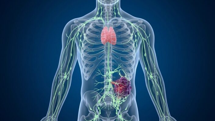
Cilia on a neuron

How Some Neuronal Cilia May Affect the Whole Body

A team of researchers has written a paper with a surprising finding: that many of the benefits of caloric restriction and increased NAD+ may be due to their effects on the cilia of hypothalamic neurons.

Neuronal cilia have a relationship to overall health

To interact with their environment and maintain internal balance, many cells, including hypothalamic neurons, have primary cilia on their external surfaces [1]. Genetic disorders that lead to diabetes and other metabolic issues include ciliary dysfunction among their symptoms, and this dysfunction appears to play a causative role [2]. Impairing ciliary function in adult mice leads to similar metabolic disorders [3].

As cilia and the maintenance process of autophagy are connected [4], and long-lived neurons require functional autophagy to stay alive [5], ciliary health in these cells is critical. Unfortunately, the cilia of neurons shorten with age [6]. This research focuses on the previously undocumented relationship of cilia to NAD+, which has been linked to autophagy through its connection to SIRT1 [7].

SIRT1, NAD+, and cilia

This paper’s first finding was unsurprising: both cilia length and NAD+ decline in the hypothalamus between 3 and 24 months of age in mice. A further experiment found that there is a causative effect, as culturing hypothalamic neurons in vitro and depleting NAD+ from them using FK866 significantly shortened the length of these cells’ cilia, although it did not affect the generation of cilia. Increasing NAD+ in hypothalamic neurons in vitro directly through its precursors lengthened cilia, and suppressing the NAD+ consumer CD38 was even more effective in this. Further experiments found that this affects retinal pigment epithelial cells, and experiments with depleting and repleting NAD+ in 3-month-old mice confirmed its causative relationship to ciliary length in hypothalamic neurons in vivo.

The researchers then looked into the impact of SIRT1. Overexpressing normal SIRT1 in hypothalamic neurons had similar effects to suppressing CD38 in terms of lengthening cilia, and suppressing SIRT1 significantly decreased the effects of increased NAD+. This was found to be due to a specific deacetylase in SIRT1; abnormal SIRT1 that lacked this deacetylase had no effect, and suppressing SIRT1 deacetylase activity also neutralized the effects of increasing NAD+.

An RNA analysis provided more information about these effects. Upregulating SIRT1 also upregulated several genes that are related to ciliary length, and increasing NAD+ through NMN upregulated a few of these genes as well. Roughly a decade ago, this team found that leptin increases RFX1, which is linked to ciliary growth; in this study, the team found that SIRT1 increases the effectiveness on RFX1 through deacetylation. The team also found a connection to autophagy; knocking down a key autophagy-related gene blunted the effects of SIRT1 on ciliary length.

A core causative role

The researchers found that the cilia play a fundamental, causative role in this relationship. Directly suppressing genes that are related to ciliary growth through silencing RNA also suppressed the beneficial effects of NAD+ on autophagy. A protein that appears in the cilia, CHMP4B, was found to be a core part of this relationship; when NAD+ was depleted, this protein stopped appearing in the cilia where it normally does and instead travels to the lysosomes. Using a toxin to injure lysosomal membranes caused CHMP4B to appear there as well. Further work confirmed this conclusion: cilia contain CHMP4B, which returns to lysosomes to maintain their integrity, but NAD+ depletion harms lysosomes and depletes cilia of this protein.

Caloric restriction, which increases NAD+, was found to have similar effects as direct NAD+ interventions in vivo. 24-month-old mice were put on a caloric restriction regimen, and some of them were given silencing RNA against a key ciliary growth gene in the hypothalamus. While calorically restricted aged mice without this silencing RNA had metabolic measurements that were more akin to those of young mice, aged mice with this silencing RNA had some adipose tissue measurements that suggested that they had never been given caloric restriction even though they were. This, therefore, demonstrates that “hypothalamic primary cilia are required for the CR–induced improvements in aging–associated metabolic phenotypes.”

Cilia metabolic effects

This study’s findings are rather striking, as they point to very specific parts of very specific neurons as being fundamental, causative actors in a commonplace longevity intervention. Further work should be done to confirm these findings and determine whether or not there may be interventions that can directly engage the cilia in the hypothalamus in order to treat metabolic disorders.

We would like to ask you a small favor. We are a non-profit foundation, and unlike some other organizations, we have no shareholders and no products to sell you. All our news and educational content is free for everyone to read, but it does mean that we rely on the help of people like you. Every contribution, no matter if it’s big or small, supports independent ethical journalism and sustains our future.

Literature

[1] Singla, V., & Reiter, J. F. (2006). The primary cilium as the cell’s antenna: signaling at a sensory organelle. science, 313(5787), 629-633.

[2] Lee, C. H., Kang, G. M., & Kim, M. S. (2022). Mechanisms of weight control by primary cilia. Molecules and cells, 45(4), 169-176.

[3] Davenport, J. R., Watts, A. J., Roper, V. C., Croyle, M. J., van Groen, T., Wyss, J. M., … & Yoder, B. K. (2007). Disruption of intraflagellar transport in adult mice leads to obesity and slow-onset cystic kidney disease. Current Biology, 17(18), 1586-1594.

[4] Orhon, I., Dupont, N., Pampliega, O., Cuervo, A. M., & Codogno, P. (2015). Autophagy and regulation of cilia function and assembly. Cell Death & Differentiation, 22(3), 389-397.

[5] Rubinsztein, D. C., DiFiglia, M., Heintz, N., Nixon, R. A., Qin, Z. H., Ravikumar, B., … & Tolkovsky, A. (2005). Autophagy and its possible roles in nervous system diseases, damage and repair. Autophagy, 1(1), 11-22.

[6] Oya, M., Miyasaka, Y., Nakamura, Y., Tanaka, M., Suganami, T., Mashimo, T., & Nakamura, K. (2024). Age-related ciliopathy: Obesogenic shortening of melanocortin-4 receptor-bearing neuronal primary cilia. Cell Metabolism, 36(5), 1044-1058.

[7] Lee, I. H., Cao, L., Mostoslavsky, R., Lombard, D. B., Liu, J., Bruns, N. E., … & Finkel, T. (2008). A role for the NAD-dependent deacetylase Sirt1 in the regulation of autophagy. Proceedings of the National Academy of Sciences, 105(9), 3374-3379.

[8] Kang, G. M., Han, Y. M., Ko, H. W., Kim, J., Oh, B. C., Kwon, I., & Kim, M. S. (2015). Leptin elongates hypothalamic neuronal cilia via transcriptional regulation and actin destabilization. Journal of Biological Chemistry, 290(29), 18146-18155.

CT Scan Robot

AI Extracts Broader Health Insights From Routine CT Scans

A new study suggests that CT scans can provide information about a wide range of conditions, including some not typically diagnosed through imaging. Direct comparison with human radiologists is the next step [1].

Getting more from a scan

A CT scan’s goal is usually to answer a concrete clinical question, such as what is causing abdominal pain or whether a suspected tumor is there. However, the images also capture other tissues and organs. The amount of information in a scan greatly exceeds what any radiologist can possibly register and may point to unrelated and yet undiagnosed conditions. A new study from the University of Pennsylvania, published in npj Digital Medicine, describes an AI model that can glean such clues, raise alarms, and assess future risks.

The authors developed Percival, a vision-language model (VLM) trained on CT images and their accompanying radiology reports. The model was trained on more than 400,000 CT image volumes paired with radiology reports from 52,000 participants. These came from the Penn Medicine BioBank and included different anatomical regions, scanner manufacturers, and imaging protocols, with and without contrast.

The model, which uses 86 million parameters, was trained to match images to their corresponding reports. It produces a numerical representation of each scan (an embedding) that encodes patterns it learned from the images.

Signal detected

Principal component analysis showed that the embeddings reflected differences in patients’ age, sex, and body size. This served as a preliminary check that the model captures meaningful patient characteristics. However, associations with blood pressure, pulse, oxygen saturation, and respiratory rate were weak.

The researchers then tested associations between the embeddings and dozens of types of laboratory measurements, taken within 60 days of imaging. They found significant associations involving 35 measurements (corrected for multiple comparisons), including markers related to kidney function, liver function, glucose and lipid metabolism, blood-cell counts, and inflammation.

This suggested that the scans contain broader physiological information. However, when the researchers tried estimating lab values directly, performance was largely weak. Only some measurements showed predictive power, including albumin, hemoglobin, and creatinine.

The researchers then asked whether the embeddings corresponded to diagnoses across multiple organ systems. They found associations with 351 diagnosis categories, spanning cardiovascular, respiratory, cancer, metabolic, and other conditions, documented or diagnosed around the time of the scan. They also found that some image patterns were associated with a greater number of diagnoses recorded after imaging (“future disease burden”).

After establishing these broad associations, the researchers trained disease-specific classifiers to distinguish people with and without recorded diagnoses. They compared Percival with CT-FM, an AI model trained using images without paired reports, which Percival outperformed. The results were the strongest for atrial fibrillation, coronary atherosclerosis, heart failure, and pancreatic cancer. For some of those conditions, such as atrial fibrillation, primary diagnosis does not ordinarily rely on CT.

Future disease prediction

The ultimate test was predicting later diagnoses. Percival competed with an image-only AI model, as well as with predictions that used organ measurements or age and sex, outperforming all of them. Top results were achieved for heart failure, cirrhosis, and acute kidney failure.

“Percival can help identify unrecognized disease, predict future progression, and anticipate major cardiovascular and liver complications,” said co-author Charles Kahn, a professor of radiology. “Because many patients already have substantial heart or metabolic disease when they enter specialty care, routine imaging may provide an opportunity for earlier recognition and intervention.”

However, a later recorded diagnosis does not necessarily mean that a disease developed after imaging. The researchers did not exclude diagnoses made shortly afterwards. Some predictions may therefore relate to an existing condition that had not yet been formally documented. It is a valuable capability but different from forecasting disease years before onset.

“Our teams have developed something that learns broadly from imaging data and uncovers connections between scans, lab values, people’s physiology, and clinical outcomes to potentially give more accurate assessments of health or even ring alarm bells early,” said senior author Walter Witschey, an associate professor of radiology and the associate vice chair of research IT. “This is work that points toward a future in which medical imaging becomes a foundation for more personalized and proactive health care.”

The study suggests that there is potential value in vision-language models that can combine several types of data from the patient’s health records. However, no direct comparison with human radiologists was made, so the study does not establish that Percival is better at diagnosing the conditions a CT scan was ordered to investigate.

We would like to ask you a small favor. We are a non-profit foundation, and unlike some other organizations, we have no shareholders and no products to sell you. All our news and educational content is free for everyone to read, but it does mean that we rely on the help of people like you. Every contribution, no matter if it’s big or small, supports independent ethical journalism and sustains our future.

Literature

[1] Beeche, C. A., Kim, J., Tavolinejad, H., Zhao, B., Dong, J., Sharma, R., … & Penn Medicine Biobank. (2026). Generalizable CT vision-language modeling for population health and disease risk. npj Digital Medicine.

Endothelial cells

An RNA Regulator Mitigates Senescence in Blood Vessels

Researchers publishing in Aging Cell have found that the regulatory protein MTR4 fights DNA damage in the blood vessel lining but decreases with aging.

Vulnerable by nature

This paper begins by noting the properties, and aging, of vascular endothelial cells (VECs), which line the blood vessels and govern their permeability [1]. As these cells are directly exposed to inflammatory factors and other blood-based stresses, they are some of the most rapidly aging cells in the human body [2]. When these cells become senescent, they excrete harmful senescence-related compounds (the SASP) directly into the bloodstream, and senescent VECs are connected to a wide variety of cardiovascular and other diseases, including hypertension and diabetes [3].

Their connection to multiple diseases, and bloodstream availability, make them an “attractive target” for interventions. However, killing senescent VECs off en masse through senolytics may be a bad idea, as one experiment found that this practice can promote fibrosis and shorten lifespan in mice [4] while another found that it can make hypertension worse [5].

Therefore, these researchers focus on methods to bolster these cells’ resilience, which was found to be a sound approach in previous experiments [6, 7]. Most previous work has focused on well-known factors such as Klotho and sirtuins, but these researchers chose a different target: MTR4, an RNA helicase that plays crucial roles in RNA maintenance [8] and the cell cycle [9]. While impeding part of its function has been found to lead to senescence [10], its true impact as a senescence regulator was not previously examined.

Expected changes with age

These researchers first used VECs derived from the human umbilical cord (HUVECs) and drove them senescent through genotoxins, oxidative stress, or replication. In all of these cells, MTR4 significantly declined. Quiescent stem cells, which had no stimulus to divide but were not senescent, did not exhibit a similar decline in MTR4. Similar results were found in mice; 3-month-old mice exhibited significantly higher levels of MTR4 than 20-month-old mice in multiple organs, including the heart, liver, and aorta.

The study then sought to ascertain a cause-and-effect relationship by using silencing RNA to turn off MTR4 in proliferating HUVECs. Markers of cellular senescence in these cells substantially increased, including SA-β-gal, while SASP factor expression increased as well. The inverse experiment also yielded results: replicatively senescent HUVECs that overexpressed MTR4 partially reversed such senescent phenotypes, including broad reductions in SASP expression.

The first link in a chain

An RNA sequencing analysis confirmed these results, although most of these results were ascertained to be downstream of its true effects. The authors identified 92 potentially interacting proteins, used the GenAge database to discover five particularly notable ones, and found that it physically interacts with BUB3 in a way that suppresses cellular senescence; directly overexpressing BUB3 had similar effects to directly overexpressing MTR4. Generating a mutant version of MTR4 that could not interact with BUB3 had no beneficial effects, and MTR4 was found to physically stabilize BUB3.

The researchers found another link in the chain: BUBR1, which itself is stabilized by BUB3. BUBR1 is a core spindle assembly checkpoint kinase that protects DNA during division. The researchers hold that this protection is the core reason behind the antisenescence effects of MTR4.

However, interestingly, RNA expression of MTR4 is increased rather than decreased in senescent cells. The reason that its protein levels decrease, therefore, is accelerated decomposition. The researchers found that this was being done through the ubiquitin-proteasome system rather than autophagic processes. This ubiquitination was found to be caused by an increase in another protein, SYVN1. The researchers even identified the particular lysine on this protein, K638, that allows for this ubiquitination.

Strong effects on blood vessels

The researchers then turned to mice and found significant results on their physiology and behavior after exposure to adeno-associated viruses (AAVs) that influence Mtr4. Aged mice that overexpressed Mtr4 had increased exploratory behavior and better vascular function, while young mice that underexpressed it had reduced exploratory behavior along with vascular dysfunction. Similar results were even found in C. elegans worms; worms that overexpressed mtr-4 lived considerably longer than wild-type worms, approaching the lifespans of well-known longevity mutants in the species, while underexpressing worms had significantly reduced lifespan.

The authors of this paper hold that MTR4 is “a conserved antiaging factor in the vascular endothelium.” While these results are strong, finding treatments that effectively target MTR4 in people will be its own challenge.

We would like to ask you a small favor. We are a non-profit foundation, and unlike some other organizations, we have no shareholders and no products to sell you. All our news and educational content is free for everyone to read, but it does mean that we rely on the help of people like you. Every contribution, no matter if it’s big or small, supports independent ethical journalism and sustains our future.

Literature

[1] Augustin, H. G., & Koh, G. Y. (2024). A systems view of the vascular endothelium in health and disease. Cell, 187(18), 4833-4858.

[2] Bloom, S. I., Islam, M. T., Lesniewski, L. A., & Donato, A. J. (2023). Mechanisms and consequences of endothelial cell senescence. Nature Reviews Cardiology, 20(1), 38-51.

[3] Han, Y., & Kim, S. Y. (2023). Endothelial senescence in vascular diseases: current understanding and future opportunities in senotherapeutics. Experimental & Molecular Medicine, 55(1), 1-12.

[4] Grosse, L., Wagner, N., Emelyanov, A., Molina, C., Lacas-Gervais, S., Wagner, K. D., & Bulavin, D. V. (2020). Defined p16High senescent cell types are indispensable for mouse healthspan. Cell metabolism, 32(1), 87-99.

[5] Born, E., Lipskaia, L., Breau, M., Houssaini, A., Beaulieu, D., Marcos, E., … & Abid, S. (2023). Eliminating senescent cells can promote pulmonary hypertension development and progression. Circulation, 147(8), 650-666.

[6] Chivite, I., Monelli, E., Munar-Gelabert, M., Gómez-Valadés, A. G., Alvarado-Diaz, A., Pozo, M., … & Claret, M. (2026). Deletion of Mfn2 in endothelial cells triggers a mitohormetic response that improves systemic metabolism and healthspan in mice. Cell Metabolism, 38(3), 546-564.

[7] Pang, J., Pan, L., Shen, W., Yi, G., Kong, X., Zhang, H., … & Zhang, Y. (2025). Endothelial LRRC8A delays vascular ageing in natural and accelerated ageing mouse models. Cardiovascular Research, 121(16), 2549-2564.

[8] Olsen, K. J., & Johnson, S. J. (2021). Mtr4 RNA helicase structures and interactions. Biological chemistry, 402(5), 605-616.

[9] Zhang, L., Wang, J., Tang, Z., Lin, Z., Su, R., Hu, N., … & Cheng, H. (2025). The nuclear exosome co-factor MTR4 shapes the transcriptome for meiotic initiation. Nature Communications, 16(1), 2605.

[10] Han, X., Xing, L., Hong, Y., Zhang, X., Hao, B., Lu, J. Y., … & Shen, X. (2024). Nuclear RNA homeostasis promotes systems-level coordination of cell fate and senescence. Cell Stem Cell, 31(5), 694-716.

Bone structure

Blocking STING Helps Preserve Bone in Aging Mice

A new study links STING-mediated signals from marrow fat cells to inflammation and declining NAD⁺ levels in bone-forming cells. Interrupting this pathway helped preserve aging bone [1].

Making old bones

Our bones live a complex life, as they are constantly remodeled: cells called osteoclasts break down existing bone, while osteoblasts build new bone. Osteoblasts arise from bone marrow mesenchymal stem cells (BMSCs), which can also produce fat cells. With aging, bone marrow accumulates more fat, becomes more inflammatory, and supports bone formation less effectively [2]. However, scientists do not fully understand how this process works yet.

A new study by Chinese researchers, published in Advanced Science, focused on macrophages, a type of immune cell. In particular, they investigated cGAS–STING, an immune signaling pathway activated by DNA appearing in the cell’s cytoplasm outside its usual compartments. This mechanism helps detect viral or bacterial DNA, but with age, it starts to misfire, triggered by stimuli such as leaked mitochondrial DNA [3]. The enzyme cGAS detects this misplaced DNA and produces the messenger molecule cGAMP, which activates STING and downstream inflammatory signaling.

A STING operation

The researchers asked whether STING’s net effect changes with age, what activates it in aging marrow, and how activated macrophages affect neighboring bone-forming cells. First, they compared bone marrow from mice aged 3, 10, 18, and 24 months and found progressively greater activation of the cGAS–STING pathway. This included more DNA in the cytoplasm, more cGAMP, and increased phosphorylation (a sign of activation) of STING and downstream signaling proteins.

They then compared different marrow cell populations. Macrophages had the highest baseline expression of the Sting gene, and their STING activation increased with age. This established macrophages as a plausible intervention target, but it was still unclear whether their STING activity was in itself harmful or a response to other damage.

The authors used a macrophage-specific viral vector to silence Sting from 12 to 18 months of age in mice. This significantly preserved bone density and structure. It also increased measures of new bone deposition and reduced marrow fat-cell numbers.

To understand what in aging marrow activates macrophage STING, the researchers investigated interactions between cells. Old macrophages impaired the ability of young BMSCs to develop into bone-forming cells, whereas old macrophages lacking STING were less harmful.

Next, the team examined potential upstream triggers. Exposure to bacterial lipopolysaccharide (LPS), a component of bacterial membranes, activated STING in macrophages, as expected. Old mice also had more endotoxin in blood and marrow, which can contribute to increased STING levels.

Inflammation and mitochondrial damage, combined

The authors then focused on marrow fat cells. Secretions from old marrow fat cells activated macrophage STING both in culture and when injected into young mice’s marrow. Co-culturing old fat cells with young macrophages produced a similar response.

44 secreted proteins elevated in bone marrow with age. The researchers singled out one of them: DPP4. Of all the tested marrow cell types, fat cells released substantially more soluble DPP4, and old fat cells released more than young ones.

Injecting DPP4 into young mice’s marrow for four weeks worsened bone density and architecture and activated macrophage STING. Conversely, reducing Dpp4 expression in marrow fat cells significantly improved one measure of bone structure health (spacing between trabeculae).

However, DPP4 alone did not significantly activate STING but, rather, strengthened its activation when combined with old marrow fluid. Thus, DPP4 appeared to contribute to an inflammatory environment rather than act as a sufficient trigger by itself.

The authors tested several possible receptors for DPP4 and focused on TLR4, a cell-surface receptor involved in innate immune responses. Blocking TLR4 reduced STING activation, and protein-interaction experiments supported direct binding between DPP4 and TLR4.

Importantly, old macrophages contained more mitochondrial DNA outside their mitochondria. Reducing mitochondrial DNA with low-dose ethidium bromide partially reduced STING activation. Old macrophages also showed more signs of mitochondrial stress.

The researchers suggest that external inflammatory signals, including DPP4-associated TLR4 signaling, “cooperate” with internal mitochondrial damage to sustain STING activity.

The NAD⁺ connection

The researchers also found that macrophage NAD⁺ levels declined with age, while NAD⁺-degrading activity increased. Experimentally activating the pathway lowered macrophage NAD⁺ but suppressing cGAS or STING counteracted these changes. Further experiments suggested that STING signaling increases production of the enzyme CD38, which degrades NAD⁺.

How could an enzyme in macrophages reduce NAD⁺ inside BMSCs? Further experiments suggested that CD38 that sits on the cell surface degrades extracellular NAD⁺ and its precursor NMN, potentially limiting the supply available to nearby cells.

Old BMSCs had impaired mitochondrial function and lower production of the cellular energy currency ATP. NMN supplementation partially rescued these metrics but to a reduced degree in old or artificially activated macrophages. Suppressing cGAS or STING in the macrophages seemed to work as well: macrophages from old mice with Sting or Cd38 knockout had better mitochondrial respiration.

Finally, the team tested a pharmacological intervention: STING inhibitor H-151 combined with NMN supplementation in mice from 12 to 18 months of age. The combination preserved bone density and structure, increased new bone deposition, reduced inflammatory markers, and improved NAD⁺ levels and mitochondrial function in BMSCs.

The researchers used alendronate, an established anti-osteoporosis drug, as a positive control. Improvements in bone structure in the alendronate group were largely similar to the H-151/NMN group.

Among the study’s limitations are its small size and combining two treatments in the intervention experiment. The latter makes it hard to disentangle the effects of STING inhibition from those of NMN supplementation. More research is needed to investigate these interesting findings further.

We would like to ask you a small favor. We are a non-profit foundation, and unlike some other organizations, we have no shareholders and no products to sell you. All our news and educational content is free for everyone to read, but it does mean that we rely on the help of people like you. Every contribution, no matter if it’s big or small, supports independent ethical journalism and sustains our future.

Literature

[1] Li, Z., Gao, K., Wang, M., Liang, S., Li, D., Tan, G., Xue, H., Liu, Y., Zhang, P., Xu, Z., Cheng, W., Wang, Y., & Li, N. (2026). Bone marrow macrophage STING signaling is a direct nexus linking marrow adipose expansion to NAD⁺ depletion and osteogenic decline in skeletal aging. Advanced Science.

[2] Aaron, N., Costa, S., Rosen, C. J., & Qiang, L. (2022). The implications of bone marrow adipose tissue on inflammaging. Frontiers in Endocrinology, 13, 853765.

[3] Gulen, M. F., Samson, N., Keller, A., Schwabenland, M., Liu, C., Glück, S., … & Ablasser, A. (2023). cGAS–STING drives ageing-related inflammation and neurodegeneration. Nature, 620(7973), 374-380.

Mechanical cell

Making SIRT1 and NAD+ Therapies Work in People

Reviewers publishing in Biology have elucidated SIRT1 functional reserve as an aspect of aging, describing how its depletion is an upstream cause of age-related disorders at the cellular level and explaining why many prospective therapies that increase SIRT1 and NAD+ have not had promising results.

Managing a dangerous byproduct

Reactive oxygen species (ROS) are necessarily produced as part of basic cellular biology, and under normal conditions, they serve signaling functions [1]. With aging, however, ROS handling becomes dysregulated, leading to their accumulation and thus to age-related cellular dysfunction [2].

The sirtuin SIRT1, which depends on NAD+ to function [3], is very well-known in its utility in managing ROS. However, the simple presence of SIRT1 within a cell does not necessarily mean that it is active, and even if it is active, it may not be meeting the cell’s fluctuating needs. These reviewers, therefore, introduce the concept of SIRT1 functional reserve, a metric intended to “describe the remaining operational flexibility of SIRT1-dependent regulatory processes under a defined stress demand” and thus measure how well a cell can “engage, coordinate, and recover SIRT1-dependent responses”.

As this is intended to be a measurable and experimentally testable metric rather than simply a broad concept, the reviewers largely focused on SIRT1’s relationship to redox reactions and mitochondria; while SIRT1’s use throughout cells should be measured in its analysis, SIRT1 functional reserve does not reflect every use of SIRT1, nor does it measure cellular aging as a whole.

SIRT1 Functional Reserve

Functional reserve is not availability

The reviewers note that measuring total cellular NAD+ may not reflect its complete relationship to SIRT1 functional reserve. NAD+ is spatially located within various parts of a cell and is used and consumed by a variety of processes and molecules such as CD38 [4]. While the various pools that hold NAD+ may be connected, and mitochondria also hold NAD+ [5], there is no guarantee that a cell that is presumably abundant in total NAD+ is delivering enough of it to SIRT1 in a way that properly handles ROS. Similiarly, NAD+ availability does not necessarily reflect SIRT1 prevalence.

SIRT1 also appears as various isoforms, and the v2 and v3 varieties, which accumulate with age, are less functional than the full-length v1 version [6]. Measuring total SIRT1 in a way that does not account for these isoforms, therefore, lacks usefulness in estimating its functional reserve. Additionally, SIRT1 can be modified as a protein by oxidative and other stresses, some of which degrade its capabilities [7] while others enhance them [8].

SIRT1 is used throughout cells

The reviewers note that aging often impairs proper responses and the recovery from such [9]. SIRT1 is a crucial component of these responses, as it directly interacts with energy-sensing pathways [10]. Measuring this interaction, however, can be counterintuitive; for example, during high-intensity interval exercise, SIRT1 activity increased while its total level decreased [11]. Furthermore, the strength of a stress response may or may not correspond to its resolution; a strong response does not signify full recovery, and a weaker response may signify that the cell has handled the situation with less effort [12].

The reviewers also add that repeated stress, a well-known precursor of cellular decline, may alter SIRT1 dynamics [13] but does not necessarily result in a loss of SIRT1 function. Additionally, SIRT1 helps to regulate the heat shock protein HSF1 [14], which aid in proteostasis, while other heat shock proteins regulate SIRT1 [15]. Disruption of this, therefore, leads to a harmful feedback loop: the very machinery that is supposed to regulate proteostasis is harmed by the lack of proteostasis regulation. SIRT1 also regulates proteostasis in mitochondria [16].

SIRT1 is often part of a greater regulatory framework rather than an independent factor. Therefore, the reviewers warn that “enhancement of SIRT1 activity may yield progressively smaller benefits if other determinants of adaptation become limiting,” which may explain some weaker results seen in studies aimed at SIRT1 enhancement [17]. Such other causes need to be separated in order to properly measure SIRT1 functional reserve.

From function to dysfunction

This review discusses the adaptive-to-maladaptive transition within cells, which is a gradual process that does not have a clear line. During chronic stress, cells compensate in ways that involve either the constant use of compensatory mechanisms or altering their structure. SIRT1 is involved in both cases, although in different ways; it can be engaged in ways that involve stress tolerance, and its function can be affected by cellular changes, particularly senescence. According to the reviewers, therefore, “the same level of SIRT1 protein or activity may not produce equivalent functional output under different cellular conditions.” They propose including stress history as part of estimating the functional reserve of SIRT1.

The species and cell involved also determine the role of SIRT1. Some experiments show a straightforward relationship: increasing oxidative stress in various human cells depletes SIRT1. Others, however, such as caloric restriction and exercise in mice, change SIRT1 in a tissue-specific way. Neural stem cells are particularly vulnerable to perturbations in SIRT1 [18]. Therefore, systemic measurements of SIRT1 and/or NAD+ may not properly reflect its use or influence in cells, and its functional reserve must be measured with cell type in mind.

Towards the clinic

This review provides heavily substantiated explanations of why commonly tested methods, such as increasing SIRT1 and NAD+, along with directly targeting oxidative stress at the mitochondrial level through MitoQ [19], have had mixed results. However, this paper also acknowledges that its own framework requires experimental validation. They propose experiments in which SIRT1 functional reserve is measured by analyzing its forms and locations within the cell along with the locations of NAD+, ROS measurements, and various biomarkers of cellular health and inflammation.

Of course, the “main translational question is whether changes in SIRT1-dependent adaptive capacity can be linked to meaningful functional outcomes in humans.” The amount of work needed to verify and validate this approach, and so move towards SIRT1 and NAD+ therapies with a higher chance of working in people, will be substantial.

We would like to ask you a small favor. We are a non-profit foundation, and unlike some other organizations, we have no shareholders and no products to sell you. All our news and educational content is free for everyone to read, but it does mean that we rely on the help of people like you. Every contribution, no matter if it’s big or small, supports independent ethical journalism and sustains our future.
Literature

[1] Sies, H., Mailloux, R. J., & Jakob, U. (2024). Fundamentals of redox regulation in biology. Nature Reviews Molecular Cell Biology, 25(9), 701-719.

[2] Atayik, M. C., & Çakatay, U. (2023). Redox signaling and modulation in ageing. Biogerontology, 24(5), 603-608.

[3] Covarrubias, A. J., Perrone, R., Grozio, A., & Verdin, E. (2021). NAD+ metabolism and its roles in cellular processes during ageing. Nature reviews Molecular cell biology, 22(2), 119-141.

[4] Chini, C. C., Peclat, T. R., Warner, G. M., Kashyap, S., Espindola-Netto, J. M., de Oliveira, G. C., … & Chini, E. N. (2020). CD38 ecto-enzyme in immune cells is induced during aging and regulates NAD+ and NMN levels. Nature metabolism, 2(11), 1284-1304.

[5] Høyland, L. E., VanLinden, M. R., Niere, M., Strømland, Ø., Sharma, S., Dietze, J., … & Ziegler, M. (2024). Subcellular NAD+ pools are interconnected and buffered by mitochondrial NAD+. Nature Metabolism, 6(12), 2319-2337.

[6] Patyal, P., Ameer, F. S., Verma, A., Zhang, X., Azhar, G., Shrivastava, J., … & Wei, J. Y. (2024). The role of Sirtuin-1 isoforms in regulating mitochondrial function. Current Issues in Molecular Biology, 46(8), 8835-8851.

[7] Caito, S., Rajendrasozhan, S., Cook, S., Chung, S., Yao, H., Friedman, A. E., … & Rahman, I. (2010). SIRT1 is a redox-sensitive deacetylase that is post-translationally modified by oxidants and carbonyl stress. The FASEB Journal, 24(9), 3145.

[8] Han, C., Gu, Y., Shan, H., Mi, W., Sun, J., Shi, M., … & Yu, W. (2017). O-GlcNAcylation of SIRT1 enhances its deacetylase activity and promotes cytoprotection under stress. Nature communications, 8(1), 1491.

[9] Lomeli, N., Bota, D. A., & Davies, K. J. (2017). Diminished stress resistance and defective adaptive homeostasis in age-related diseases. Clinical Science, 131(21), 2573-2599.

[10] Cantó, C., Jiang, L. Q., Deshmukh, A. S., Mataki, C., Coste, A., Lagouge, M., … & Auwerx, J. (2010). Interdependence of AMPK and SIRT1 for metabolic adaptation to fasting and exercise in skeletal muscle. Cell metabolism, 11(3), 213-219.

[11] Gurd, B. J., Perry, C. G., Heigenhauser, G. J., Spriet, L. L., & Bonen, A. (2010). High-intensity interval training increases SIRT1 activity in human skeletal muscle. Applied Physiology, Nutrition, and Metabolism, 35(3), 350-357.

[12] Lamech, L. T., & Haynes, C. M. (2015). The unpredictability of prolonged activation of stress response pathways. The Journal of cell biology, 209(6), 781

[13] Zhang, J., Wang, H., Slotabec, L., Cheng, F., Tan, Y., & Li, J. (2023). Alterations of SIRT1/SIRT3 subcellular distribution in aging undermine cardiometabolic homeostasis during ischemia and reperfusion. Aging Cell, 22(9), e13930.

[14] Westerheide, S. D., Anckar, J., Stevens Jr, S. M., Sistonen, L., & Morimoto, R. I. (2009). Stress-inducible regulation of heat shock factor 1 by the deacetylase SIRT1. Science, 323(5917), 1063-1066.

[15] Białopiotrowicz-Data, E., Noyszewska-Kania, M., Jabłońska, E., Sewastianik, T., Komar, D., Dębek, S., … & Juszczyński, P. (2023). SIRT1 and HSP90α feed-forward circuit safeguards chromosome segregation integrity in diffuse large B cell lymphomas. Cell Death & Disease, 14(10), 667.

[16] Chen, Q., Chang, Y. L., Hung, M. W., Lee, P. L., Hsin, C., & Lin, Y. F. (2026). Sirt1 coordinates the mitochondrial UPR and myocellular proteostasis to preserve muscle integrity during muscle atrophy in zebrafish. Frontiers in Cell and Developmental Biology, 14, 1761278.

[17] Alcendor, R. R., Gao, S., Zhai, P., Zablocki, D., Holle, E., Yu, X., … & Sadoshima, J. (2007). Sirt1 regulates aging and resistance to oxidative stress in the heart. Circulation research, 100(10), 1512-1521.

[18] Wang, G., Wang, F., Ren, J., Qiu, Y., Zhang, W., Gao, S., … & Xu, J. (2018). SIRT1 involved in the regulation of alternative splicing affects the DNA damage response in neural stem cells. Cellular Physiology and Biochemistry, 48(2), 657-669.

[19] Broome, S. C., Whitfield, J., Janssens, K., & Hawley, J. A. (2025). MitoQ supplementation does not impact redox responses to acute exercise in skeletal muscle of older individuals. Redox Biology, 103927.

Proteins in the brain

Menopausal Serum Protein Changes May Predict Brain Health

A recent analysis of serum protein changes identified 16 proteins related to inflammatory, synaptic, metabolic, and Alzheimer’s disease processes whose levels change during the menopausal transition [1]. These changes were linked to cognitive health later in life.

The time for interventions

Menopause is mostly known as a stage that marks the end of female reproduction, but the effects of menopause go beyond the reproductive system and affect many organs and systems, with the impact on the brain being widely discussed and researched. For example, multiple studies have reported that women experience various cognitive difficulties during menopause, such as word-finding or attentional difficulties. A recent review estimated that such problems affect between 26 and 95% of menopausal women [2]. Additionally, different studies suggested that the aging of the hypothalamus plays a causal role in ovarian senescence and menopause induction [3]. These data suggest a strong connection between the brain and menopause, creating a window of opportunity during the menopausal transition for improving brain aging and affecting dementia risk.

“Menopause is a normal physiological process that every person who has ovaries who lives long enough will go through,” said Madeline Wood Alexander, a graduate student at the University of Toronto. “We don’t want it to be viewed as a bad thing, but as an opportunity to better understand and maybe modify the biology of brain aging in women.”

Kaitlin Casaletto, Ph.D., an associate professor at the UCSF Fein Memory and Aging Center and co-senior author of the study, added, “Midlife is a critical window for both men and women where we can really set up the brain to be more resilient or more vulnerable to dementia in later life.” “For women, menopause may be this especially powerful time to intervene.”

Menopause proteomic score

Although many studies suggest a connection between menopause and brain aging, human data remain scarce, limiting the ability to understand the underlying molecular pathways driving these processes.

The researchers of this study began by investigating blood biomarkers associated with spontaneous menopause in healthy midlife women (43-58 years old): 30 premenopausal, 26 perimenopausal, and 24 postmenopausal women, along with 36 age-matched men for comparison.

Comparing serum protein levels (proteomics) showed an increase in 16 proteins in postmenopausal women compared to premenopausal women after adjusting for age. These proteins were related to inflammatory, synaptic, metabolic, and Alzheimer’s disease processes, with inflammatory molecules predominant, consistent with previous studies showing increased systemic inflammation after menopause [4].

Because the cohort used to identify these protein changes was relatively small, they also analyzed data from almost 3,000 women in the UK Biobank. 13 of the 16 proteins identified as showing menopause-stage differences were measured in both cohorts; of those, 9 replicated across cohorts.

The observed changes in synaptic and Alzheimer’s disease blood markers align with previous animal studies showing synaptic plasticity and spine density disruptions [5, 6], along with increases in Aβ and tau positron emission tomography (PET) across menopause [7, 8].

Using those 16 proteins, the researchers created a proteomic score that holistically reflects major menopause-related changes with less data noise. Using this score, they showed that compared to premenopausal women, perimenopausal status was associated with an increase of 1.36 units in menopause scores, and postmenopausal status with a 3.25-unit increase. Importantly, the menopausal stage, rather than chronological age, drove the observed changes.

Hormonal changes and menopausal symptoms

One of the most important changes during menopause is hormonal changes, such as reductions in the ovarian hormones 17β-estradiol and progesterone along with increases in follicle-stimulating hormone (FSH).

In this study, the authors found a positive association between their menopause proteomic score and FSH levels, an inverse association between this score and estradiol levels, and no association with progesterone levels. At a more granular level, they reported that some inflammation-related protein levels were inversely associated with 17β-estradiol, which aligns with the role of estrogens in modulating immune responses [9], while other proteins related to inflammatory, synaptic, metabolic, and Alzheimer’s disease processes were positively associated with FSH.

The authors note that this aligns with previous studies showing that “FSH receptors are expressed abundantly in AD-vulnerable brain regions” and that data from animal models suggest FSH’s role in the development of Alzheimer’s disease, neuroinflammation, and cognitive deficits [10]. In their analysis, the identified proteins were clustered into two separate groups, which suggests that each hormone might be linked to different biological pathways.

Menopause is also widely known as a stage in life when women experience various vasomotor symptoms, such as night sweats or hot flashes, and some of those vasomotor symptoms have been associated with vascular inflammation [11, 12], cardiovascular risk, and Alzheimer’s disease-related brain changes [13]. In this study, the authors analyzed different cohorts for the association between menopausal score and vasomotor symptoms. The results were inconsistent across the cohorts, but some suggested a link between some vasomotor symptoms and menopausal score.

Predicting future cognitive health

Beyond many previously reported links between menopausal changes and brain health, these researchers tested how relevant changes in serum proteins are for later-life cognitive health. They analyzed data from four independent cohorts of older women and found a consistent association between higher (more postmenopausal) baseline menopause scores and worse cognitive health later in life and a higher risk for Alzheimer’s disease dementia, suggesting that changes in certain protein levels during menopause may remain relevant in later decades of life.

“We don’t think that menopause is directly causing dementia,” said Casaletto. “But it’s possible that we may be able to predict a woman’s risk for dementia decades later by the levels of these molecules around menopause.”

While all women will undergo menopause, only a fraction will develop dementia. Molecular signatures during the menopausal transition can provide the earliest signals of future risk of developing the disease.

“We’re identifying molecular shifts in blood, in midlife post-menopausal women, that are also predicting cognition later in life,” said co-senior author Rowan Saloner, Ph.D., assistant professor at the UCSF Fein Memory and Aging Center. “So, we think these molecules are really important markers of both menopause and cognitive aging.”

We would like to ask you a small favor. We are a non-profit foundation, and unlike some other organizations, we have no shareholders and no products to sell you. All our news and educational content is free for everyone to read, but it does mean that we rely on the help of people like you. Every contribution, no matter if it’s big or small, supports independent ethical journalism and sustains our future.

Literature

[1] Wood Alexander, M., Rabin, J. S., Caunca, M., Iadipaolo, A., Cornelis, L., Warrier, R., Walker, K. A., Miolane, N., Bot, V. A., Wood, B., Oh, H. S., Wyss-Coray, T., Pham, A., Borger, J., Diaz, V., Paolillo, E. W., Kramer, J., Pritschet, L., Taylor, C., Panizzon, M. S., … Casaletto, K. B. (2026). Blood proteomics of menopause map to brain aging and dementia risk. Nature medicine, 10.1038/s41591-026-04648-4. Advance online publication.

[2] Gurvich, C., Spector, A., & Hickey, M. (2026). Advances in understanding of cognitive symptoms during menopause. The Lancet Obstetrics, Gynaecology, & Women’s Health, 2(4), e335–e345.
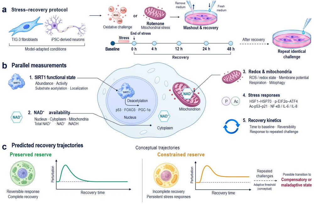
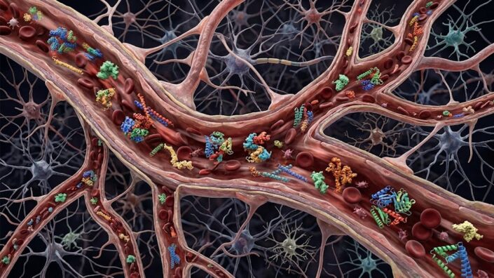
[3] Bacon, E. R., Mishra, A., Wang, Y., Desai, M. K., Yin, F., & Brinton, R. D. (2019). Neuroendocrine aging precedes perimenopause and is regulated by DNA methylation. Neurobiology of aging, 74, 213–224.

[4] McCarthy, M., & Raval, A. P. (2020). The peri-menopause in a woman’s life: a systemic inflammatory phase that enables later neurodegenerative disease. Journal of neuroinflammation, 17(1), 317.

[5] Hara, Y., Waters, E. M., McEwen, B. S., & Morrison, J. H. (2015). Estrogen Effects on Cognitive and Synaptic Health Over the Lifecourse. Physiological reviews, 95(3), 785–807.

[6] Russell, J. K., Jones, C. K., & Newhouse, P. A. (2019). The Role of Estrogen in Brain and Cognitive Aging. Neurotherapeutics : the journal of the American Society for Experimental NeuroTherapeutics, 16(3), 649–665.

[7] Buckley, R. F., O’Donnell, A., McGrath, E. R., Jacobs, H. I. L., Lois, C., Satizabal, C. L., Ghosh, S., Rubinstein, Z. B., Murabito, J. M., Sperling, R. A., Johnson, K. A., Seshadri, S., & Beiser, A. S. (2022). Menopause Status Moderates Sex Differences in Tau Burden: A Framingham PET Study. Annals of neurology, 92(1), 11–22.

[8] Rahman, A., Schelbaum, E., Hoffman, K., Diaz, I., Hristov, H., Andrews, R., Jett, S., Jackson, H., Lee, A., Sarva, H., Pahlajani, S., Matthews, D., Dyke, J., de Leon, M. J., Isaacson, R. S., Brinton, R. D., & Mosconi, L. (2020). Sex-driven modifiers of Alzheimer risk: A multimodality brain imaging study. Neurology, 95(2), e166–e178.

[9] Straub R. H. (2007). The complex role of estrogens in inflammation. Endocrine reviews, 28(5), 521–574.

[10] Xiong, J., Kang, S. S., Wang, Z., Liu, X., Kuo, T. C., Korkmaz, F., Padilla, A., Miyashita, S., Chan, P., Zhang, Z., Katsel, P., Burgess, J., Gumerova, A., Ievleva, K., Sant, D., Yu, S. P., Muradova, V., Frolinger, T., Lizneva, D., Iqbal, J., … Ye, K. (2022). FSH blockade improves cognition in mice with Alzheimer’s disease. Nature, 603(7901), 470–476.

[11] Thurston, R. C., El Khoudary, S. R., Sutton-Tyrrell, K., Crandall, C. J., Gold, E., Sternfeld, B., Selzer, F., & Matthews, K. A. (2011). Are vasomotor symptoms associated with alterations in hemostatic and inflammatory markers? Findings from the Study of Women’s Health Across the Nation. Menopause (New York, N.Y.), 18(10), 1044–1051.

[12] Bechlioulis, A., Naka, K. K., Kalantaridou, S. N., Kaponis, A., Papanikolaou, O., Vezyraki, P., Kolettis, T. M., Vlahos, A. P., Gartzonika, K., Mavridis, A., & Michalis, L. K. (2012). Increased vascular inflammation in early menopausal women is associated with hot flush severity. The Journal of clinical endocrinology and metabolism, 97(5), E760–E764.

[13] Thurston, R. C., Maki, P., Chang, Y., Wu, M., Aizenstein, H. J., Derby, C. A., & Karikari, T. K. (2024). Menopausal vasomotor symptoms and plasma Alzheimer disease biomarkers. American journal of obstetrics and gynecology, 230(3), 342.e1–342.e8.