The Blog

Building a Future Free of Age-Related Disease

Public Longevity Group

Lifespan Research Institute Launches Public Longevity Group

[Mountain View, September 17, 2025] — Lifespan Research Institute (LRI) today announced the launch of the Public Longevity Group (PLG), a new initiative focused on bridging the cultural gap between scientific breakthroughs in aging and their public acceptance. To kickstart its work, PLG has opened a crowdfunding campaign to develop tools that measure and strengthen public trust in longevity science.

While the science of longevity biotechnology continues to advance, skepticism and cultural resistance limit progress, with some studies showing that more than half of Americans would reject a safe, proven therapy to extend life. This hesitation poses risks of raising costs, delaying health-promoting regulation, and slowing the delivery of treatments that could combat age-related diseases and extend healthy lifespan.

“The breakthrough that unlocks all other breakthroughs is public trust,” said Sho Joseph Ozaki Tan, Founder of PLG. “Without it, even the most promising therapies may never reach the people they’re meant to help. PLG exists to change that.”

“Persuasion is a science too,” said Keith Comito, CEO of Lifespan Research Institute. “To bring health-extending technologies to the public as quickly as possible, we must approach advocacy with the same rigor as our research. With PLG, we’ll be able to systematically measure and increase social receptivity, making the public’s appetite for credible longevity therapies unmistakable to policymakers, investors, and the public itself.”

PLG is developing the first data-driven cultural intelligence system for longevity—a platform designed to track real-time sentiment, test narratives, and identify which messages resonate and which backfire. Early tools include:

  • The Longevity Cultural Clock: a cultural barometer mapping readiness and resistance across demographics and regions.
  • Sentiment Dashboards: real-time monitoring of public, investor, and policymaker perceptions.
  • Narrative Testing Tools: data-driven analysis that will enable robust pathways to public support.

The crowdfunding campaign will provide the initial $100,000 needed to launch these tools, creating the cultural foundation required for healthier, longer lives.

With a lean, data-driven team, the group aims to provide open-access cultural insights for advocates and policymakers while offering advanced analytics to mission-aligned partners.

Campaign Timeline:

  • Campaign completion: November 2, 2025
  • Dashboard development: Dec 2025 – Feb 2026
  • First survey deployment: Feb – Apr 2026
  • Beta dashboard launch: May 2026
  • First public insight report: June 2026

Supporters can contribute directly at: https://lifespan.io/campaigns/public-longevity-group/

The PLG campaign is sponsored by the members of LRI’s Lifespan Alliance, a consortium of mission-aligned organizations that believe in the promise of extending healthy human lifespan. Newly-joined members include OpenCures, AgelessRx, and Lento Bio.

About Lifespan Research Institute

Lifespan Research Institute accelerates the science and systems needed for longer, healthier lives by uniting researchers, investors, and the public to drive lasting impact. LRI advances breakthrough science, builds high-impact ecosystems, and connects the global longevity community.

Media Contact:

Christie Sacco

Marketing Director

Lifespan Research Institute

christie.sacco@lifespan.io

(650) 336-1780

We would like to ask you a small favor. We are a non-profit foundation, and unlike some other organizations, we have no shareholders and no products to sell you. All our news and educational content is free for everyone to read, but it does mean that we rely on the help of people like you. Every contribution, no matter if it’s big or small, supports independent ethical journalism and sustains our future.

James Peyer Interview

James Peyer on Turning Aging Biology Into Medicine

James Peyer, co-founder and CEO of Cambrian Bio, is passionate about longevity but clear-eyed about what it would actually take to extend human lifespan. Cambrian works on the basics of aging biology – nutrient-sensing pathways that have been known for decades but are notoriously hard to drug – and says that it has now cleared an important hurdle: an AMPK activator that drives weight loss without lean mass loss and more than doubles endurance in mice. In humans, the drug is at an earlier stage, but Peyer shared the results of Cambrian’s first small trials with us.

Our traditional first question: how did you get into longevity, and to what extent is this personal to you?

I’ve been in the field for about 25 years. In the early 2000s, I was in high school, and my grandfather was diagnosed with cancer. Watching him get sicker and ultimately pass away made me think two related things. The first was that he had lived a wonderful life. He was in his 80s, and despite all his pain and suffering, that was the best-case scenario for most people living today: you live a really good life up to a point, then you get some horrible disease, you suffer, you diminish, and you die. That this is the best we can aspire to for our loved ones, for ourselves, for our species, is almost a type of barbarism. I thought: if anything is worth doing in the world, it’s fighting back against this horrible end that happens to everyone we know.

The second came from watching the physicians care for him. The only treatments available for his cancer at the time were chemotherapy options, and they’re horrible drugs. Not in that they don’t work – chemotherapy is, of course, very effective – but they are last-resort treatments for a disease that was already so advanced that it was going to kill him. It left me with a strong impression: why can’t we intervene earlier? Cancer has to start decades before it appears as a late-stage disease. Same for Alzheimer’s, same for heart attacks.

That led me to the very early work on the biology of aging. At the time, a lot of people were trying to create unified theories about the one cause of aging. Thankfully, the field has moved away from that and embraced more of a constellation of complexity. I’ve been fascinated with it ever since.

When I was finishing my PhD, I saw so many big discoveries being made. Mouse lifespan extension was happening robustly for the first time, over and over, with different drugs and different genes, but almost none of those discoveries were being moved into drug development for preventative medicines. My joke is that scientists would go to a pharma company or a venture capitalist and hear back again and again: “Aging is not a disease, so go back to your lab and keep playing with your mice.”

I decided to try to unclog that bottleneck. The biggest idea that I think I helped push forward is systematizing the notion that any drug that could be used for aging, a gerotherapeutic, would first be approved for a specific indication, whether a rare disease, a type of cancer, or a metabolic disease. I call that a stepping-stone indication on the way to a broad approval for a preventative purpose. I started Apollo Health Ventures, the first VC to really build companies around aging biology. For the last seven years, I’ve been running Cambrian, which started a number of drug development programs and has now focused on a few that have started flying. The furthest along is our AMPK network activator, which is moving into Phase 2.

Moving from particular indications to aging itself has become the default thinking. But, a new paradigm seems to be developing, with XPRIZE Healthspan and similar efforts, where we might see drugs approved for improving function in generally healthy people.

Yes, particularly this idea of intrinsic capacity. XPRIZE looks at multiple functions, which matches what ARPA-H is doing with intrinsic capacity in its PROSPR program. Those are amazing explorations, but they are not investable theses yet. This is why PROSPR and XPRIZE need to exist, to open up this new frontier, but a sophisticated investor wants the drug to be approved. If it can then expand into some new white space, like improving intrinsic capacity in healthy elderly people, that’s excellent; that’s how we get to the bigger market.

But, at least in the beginning, it’s not going to be never-approved drugs that get their first approval for intrinsic capacity. It’s going to be drugs that are on the market for something else and get a secondary approval, maybe very shortly after the first.

Once we have drugs that work for intrinsic capacity, that could create a marketplace for developing drugs directly for it. The same is happening with obesity: a diabetes drug was repurposed for obesity, and now a whole slew of new drugs, including ours, are going for obesity first.

I think this can be not only a launch pad, but also a bottleneck. Like you said, we’re starting to have great drugs for obesity. But this also means that a geroprotector tested for obesity might simply not be as good as these drugs and get discarded.

Totally right. This is why one of my big refrains is that the first geroprotectors, the ones that will open up this field and allow other things to walk behind them, need to be able to walk and chew bubble gum at the same time. They need to be a great drug for whatever the first disease is and also have the multi-disease prevention effect that we associate with gerotherapeutics. That does exclude some potential drugs, but the hope is that once a pathway has been established, whether through XPRIZE and PROSPR or through other mechanisms, you can have a second or third wave of drugs that say: “We might not be the best heart failure drug ever, but we can prevent cancer, lower the risk of heart failure, and improve cognition, all a little bit and all at the same time,” and those can go straight through the intrinsic capacity route.

I think your view is realistic and clear-eyed. I just hope things change soon.

I really want that world too, where we can go straight to prevention. The way to get there is by pushing the tip of the spear: the first candidates that can do all of these things. We have to get those recognized on the market so that the fast followers that maybe can’t do the other indication can come after them.

That brings us to the encouraging results with your candidate ATX-304. You described it as working on both sides of the equation: increasing fuel uptake and helping mitochondria use that fuel, increasing OXPHOS. How does it work?

At the big-picture level, AMPK has been a holy grail target in pharma for 30 years. Activating it extends lifespan in lower organisms. In animal models, it produces an exercise-mimetic effect: increased metabolic rate, endurance, and muscle function. And in humans, the ability to activate AMPK goes down almost linearly with age. So, the idea has been floating around for a long time that by activating AMPK, we could restore metabolism from an old state to a young state.

A bunch of drugs have tried. In my talk, I highlighted three that approached it from different sides. Metformin does activate AMPK, but none of the predictions you would make about an AMPK activator hold for metformin. It doesn’t increase exercise capacity; it actually blunts it. Metformin has very positive effects, but they come from causing energy stress, not from increased energetic activity.

Merck and Pfizer made direct AMPK activators, but those drive only half of what AMPK needs to do within the cell. If I’m exercising, my ATP levels go down, and I need to do two things: bring energy into the cell and spend it in the mitochondria to make new ATP. Those drugs did only the first part. None of them made it to human trials because they led to accumulation of extra sugars in skeletal muscle and heart tissue. It became dogma in drug development that you can’t make an AMPK activator.

The third approach is driving mitochondrial activation with what are called mitochondrial uncouplers. It turns out that if you just tell the mitochondria to spin and spin, you can run the cell out of materials, which causes ATP levels to drop and will kill the cell. These drugs were discovered about a hundred years ago and were actually the first blockbuster obesity drugs, before the FDA existed, but they were so dangerous that an overdose would kill you. The most famous one is dinitrophenol.

So, we had to come up with a drug that got in between all of these complexities and kept the cell in balance: at any level of the drug, it would bring in sugar and spend it in the mitochondria at the same rate, staying in homeostasis and never pushing respiration beyond what the cell can keep up with physiologically. Luckily, our physiological range is really flexible. When we exercise, we can ramp up energy production dramatically.

What we found with ATX-304 is that at any concentration, you get this increase in AMPK activity, cellular glucose uptake, and mitochondrial respiration, and it never kills the cell until there’s so much drug that it’s precipitating out of solution. It’s the same with animals. Our only major adverse event, even after treating dogs for nine months before we started human trials, was exaggerated weight loss, and it came along with an increase in food intake: the dogs would eat more but lose a lot of weight. That was the main thing that limited us, and only at super-high concentrations.

I think you also tried pairing it with GLP-1-based drugs and got a synergistic effect – a pretty scary weight loss.

Yes, in mice; we haven’t done it in other animals. ATX-304 alone leads to about the same weight loss as semaglutide (Ozempic): about 20% in obese mice over a month. When we combined it with the GLP-1, the mice ate less and had a higher metabolic rate, and the weight dropped dramatically: after 15 days on the GLP-1, 11 of them on GLP-1 plus ATX-304, the mice had already lost almost 30% of their body weight.

That was way too fast. We ran this with a lab that does only obesity experiments, and their animal protocols were not written to handle weight loss at that speed. We had to stop the experiment, take away the GLP-1, and keep the mice on ATX-304 alone. So, the synergistic effects seem very powerful.

Weight loss comparable to semaglutide is a big thing in itself, but it also came with lean mass preserved and without appetite suppression, correct?

That’s right. Those are the three big things that make this really different. First, the animals eat the same or even a little more, because all the weight loss comes from increased energy expenditure, not from reduced caloric intake. Second, there are no signs of the things that make appetite go down: the nausea, the reduced stomach motility, the mild gastrointestinal distress.

Third, because the muscle is very metabolically active, it’s similar to how, with exercise, you send your body a signal: “I’m using my muscle. Even though I’m at a caloric deficit, you’re not allowed to take the protein from my muscle and burn it elsewhere. Pull deeper from the fat.” In monotherapy, all of the weight loss comes from fat mass reduction. Even in the combination, we saw no reduction in lean mass, because the AMPK activator sends this protective signal to the muscle.

How do you see the future of your drug? Will it replace GLP-1-based drugs or work alongside them? I think you also said it could act as a maintenance drug.

We don’t think of this as a replacement for the GLP-1s. The new-generation GLP-1 drugs with 30% weight loss are going to be the fastest way to lose weight, and that will remain attractive for a lot of people.

For our drug, I see three possibilities. First, it will present a different path for people who don’t want to take a GLP-1, can’t deal with the nausea, or are very worried about muscle loss. I expect our weight loss in humans to be slower than with strong GLP-1s. We might get to the same place, but it will be more of a linear decline, which is what you see in people who take up a lot of exercise, as opposed to the steep drop when people stop eating.

Second, a lot of people are not maximal responders on their GLP-1. Either they can’t go up in dose because the nausea is too bad, or their appetite is reduced but not enough, and they start eating more again. You could add our drug to help them get to their target weight.

Third, as you alluded to, is maintenance. About half of the people who start a GLP-1 stop taking it within a year. There are a bunch of reasons, but the one I think about the most is the weekly injection. At the beginning, the numbers on the bathroom scale go down every single week. Then, around six months, they plateau. People aren’t losing any more weight, but they still have to inject themselves every week and pay for the drug, so they stop. And when they stop, they regain weight really quickly. An oral pill that at least maintains your weight, and might even continue your weight loss trajectory, while letting you eat more again and avoid the nausea – I think that’s going to be very compelling.

All three are commercial opportunities once we get into treating people with obesity and overweight, and that doesn’t even get to the thing I know you and I are both interested in, which is what happens after that: can we treat broader metabolic decline?

We’ll get to that, but first: your drug seems to mimic exercise, another holy grail of geroscience. The results in mice are very impressive: running distance more than doubled, and cardiac function improved. Is this the exercise mimetic we’ve all been waiting for?

We hope so. If you had to pick one gene to target for that dreamed-about exercise-mimetic effect, it would be AMPK. It has demonstrated increases in endurance again and again. It’s the sensor that coordinates our muscles’ response when we go out for a run. That’s particularly true for cardiovascular exercise; resistance exercise can work a little differently. AMPK is a catabolic mechanism that has more to do with burning energy and creating new ATP, as opposed to stacking on new muscle, which is more about myostatin-type mechanisms.

As you said, this works beautifully in animals: without training, mice double their exercise endurance. Next year, we’re planning to run a study looking at whether the drug improves exercise capacity in humans. It’s a very exploratory study, but it will give us a first sign of whether those effects translate. Of course, that’s not something we can get a drug approval for. It would be yet another reason to take this drug rather than a GLP-1 or something else if you want to lose weight or keep your metabolism up.

You’ve also run human trials recently, and some of the findings you presented were marked confidential. What can you share?

We can share quite a bit. We’ve tested two different doses of the drug in humans – not all the way up to the level that causes dramatic weight loss in mice, but enough to see activity and safety.

First, on safety, which is the most important thing: we saw a safety and tolerability profile comparable to placebo at every dose we’ve tested so far. Second, at about half the drug level that leads to weight loss in rodents and canines, we already see a significant increase in resting metabolic rate, about 8%, and huge increases in the observed metabolic activity of fat. Adiponectin, an adipokine produced by metabolically active fat tissue, doubles, which is far more than you get even at the weight-loss doses of the GLP-1s.

We also saw big decreases in triglycerides: the free fatty acids floating around all seem to be getting sucked into the muscle and turned into energy. Liver fat went way down, and we even started to see visceral fat go down. All of this was statistically significant in a four-month exploratory trial with 23 patients.

That’s really impressive. Now, the flip side. Increasing energy expenditure sounds good, but I worry about long-term trade-offs. What happens, in terms of damage accumulation, when you keep the engine running faster for years?

As a scientist, you never want to answer that too definitively, but I can tell you what we know so far. The level of AMPK activation we’re producing seems to be not dissimilar from what a young animal or a young human has. We’re not pushing the pathway to what I would call a supraphysiological level; you want to keep it within the normal range. The overall effect – increased caloric expenditure and muscle activity – should be about the equivalent of going for a three-mile run each day.

In humans, that level of activity has been associated with a five-to-ten-year increase in healthy life expectancy. If we can keep the metabolic level in that range and the effects are similar to exercise, we feel pretty confident that it’s going to be robust.

On the preclinical side, we haven’t done mouse lifespan or healthspan studies yet, but we have done disease model studies in older mice, treating them for a year or longer, and we see no signs of rapid disease progression. In fact, all the signs point to the opposite: metabolic decline is slower, the mice are healthier, kidney function is improved, and so on.

In fruit flies and C. elegans, you get increased lifespan with this AMPK activator. So, everything points in the direction that moderate activation of metabolic activity in the muscle is probably going to be helpful long-term, but we’ll need to run those studies to see if that hypothesis is correct.

Interestingly, even though mitochondrial activity is increased, it’s all very functional, and we never see an increase in reactive oxygen species or a buildup of mitochondrial damage. In fact, the mitochondria look healthier, and ROS is about the same or even a little down.

Maybe that’s because all mechanisms are working faster, like in a younger person, including the protective, damage-clearing ones.

Could be. We do see this continuing to work better in old animals, so the question is whether all of those factors are re-engaged.

This cuts to the core of how much what we work on depends on our understanding of the biology. Whether you think aging is caused by evolutionary neglect, as in the disposable soma theory, where our systems simply did not evolve to stay at full capacity as we age, or you think aging is some kind of program that gets activated to limit lifespan, matters a lot here.

I’m firmly in the first camp. We get worse and worse at activating metabolism, so if we improve it, we’ve fixed one of the things that evolution failed to prioritize. But, if you think that the drop in AMPK levels and in metabolism has some kind of purpose, that it protects the organism or the species, then you might reach very different conclusions.

Let’s talk about the rest of Cambrian’s pipeline. ATX-304 is obviously the star now, but what else do you have in your quiver?

The second big program is our work on cracking safer versions of rapamycin. Years ago, we partnered with Novartis, one of the pioneers in making novel mTOR inhibitors. After taking over from where they stopped, we found out how to make a whole set of mTOR complex 1-selective inhibitors. These are variants of rapamycin that we’ve now shown can be dosed 50 or 100 times higher than the level where rapamycin starts causing immunosuppression, which is the main thing that prevents us from looking for all its gerotherapeutic effects in humans. We see no immunosuppression at any level with these very selective drugs.

We’re taking that forward for a regular disease indication, which we’ll announce when we get started. But, going back to the beginning of our conversation, we also think these drugs are the best candidates for hitting that composite milestone of improved intrinsic capacity, because mTOR inhibition is the best-performing pharmaceutical approach for extending healthy lifespan in mice, preventing everything from neurodegenerative disease to cancer to metabolic disease.

So, we have a collaboration with the PROSPR program at ARPA-H to run one of the first clinical trials for improving intrinsic capacity in healthy elderly people. It will be one of the drugs explored as part of this broader ARPA-H PROSPR plus XPRIZE universe. We’re hopeful that the overall effort will bear fruit and that we can be part of it.

In your talk, you described Cambrian’s philosophy as doing the obvious thing: targeting the pathways that keep coming up in aging research. But, some geroscientists don’t like that everything seems to converge on those pathways. It’s always mTOR, it’s always AMPK, rapamycin is still the top dog, and maybe that’s limiting the field. What do you think?

I think that’s correct. Many pathways that are currently unexplored will turn out to be important and give rise to great drugs. The reason the field has not moved much further than mTOR and AMPK in 20 years is that we’ve had only two compounds. Every time you try to take something forward, whether you’re an academic running a rapamycin trial or Pfizer taking an AMPK activator into the clinic, you get a failure, or it’s not quite as good as you wanted, and you say: “We have to change the therapeutic window, or get a different chemistry, and then we can test it.” And, because it’s the top-performing drug target, all the academic interest goes to how to crack that target.

Once you find a drug that solves it – you see this in cancer, you see it in obesity with the GLP-1s – the pharmas come in and build a bunch of me-too or me-better drugs for that target. Great. But academics and innovative biotechs can now say: “That target is solved, so we can focus on these next targets that have been sitting on the periphery.”

I’m talking my own book here a little, but this is why we come back to doing the hard but obvious thing: we just had to spend 10 years to create the right drugs for these core pathways, and now we can finally take them forward. If they’re successful, I really do think a lot of other candidates will come up.

Cambrian has played around with some of these as well. Our third program is a drug built around agrin, an ECM glycoprotein present in the heart that causes the heart to regenerate. Another one improves protein folding in cells, which get worse at it as they age. I find those areas super interesting, but they’re coming behind the need to solve the AMPK and mTOR chemistry.

Let me philosophize a bit. There are two competing approaches in the field. One says that we have to prove our worth: come up with successes, which will open up possibilities and show that geroscience is a real field. The other, which I gravitate towards, starts from saying that healthspan and lifespan extension is one of the noblest goals we have, and, as with the war on cancer, we should be shouting on every street corner that the world should give us the money to solve aging. Where are you on this? The success of GLP-1 drugs seems to validate the first model, because it made people talk about their longevity effects.

Sometimes to the consternation of our colleagues in the field, I am very much one of the strong voices in that first camp. The way geroscience makes an impact on the broader development of medicines is by building a gerotherapeutic and showing that it works.

You take some investors or pharma companies who look at this field mostly with derision – “nothing serious comes out of there” – and you produce a great drug that makes people’s lives better and makes them money. Then, you go from outsider to insider very fast. That’s been the direction of travel for the companies I’ve helped start throughout my career, and I feel strongly that that’s where the first breakthrough is going to happen.

But, I don’t want to discount the second piece. The more people we have shouting on street corners that this is the most important thing anyone could be doing, the more the traditional players start paying attention to the revolution going on in our understanding of human biology. Even though Cambrian is developing an obesity drug, and we don’t talk about longevity or healthspan on our website, I start every meeting, even with the most traditional pharma and biotech VCs, by saying that there is a revolution going on in our understanding of what makes us get sick and what makes us age at the cellular and tissue level. That’s what enabled everything we’ve done.

Apart from your own programs, what approaches in geroscience and longevity biotech do you find particularly promising or exciting, in the short or long term?

Honestly, the thing I’m most excited about in the short term is Loyal, the dog longevity company. I like the way they’ve built momentum on the broader coalition side. In some ways, their viewpoint is similar to mine: go build something real and make it happen.

But, instead of the stepping stone, their thinking is: “We can’t build a longevity drug for humans right away, but we can build a healthspan drug for dogs.” They’ve already met their efficacy and safety milestones, and they’re finishing their manufacturing work. So, there’s going to be a dog longevity drug on the market in the next year or so.

Within the geroscience community, the epigenetic partial reprogramming story remains interesting. The best example of doing it right is Life Biosciences. They’ve found a really good stepping stone in people with optic nerve dysfunction, and they’re going in to try to repair that and then figure out how, and if, they can expand beyond it. Even just delivering a success in that first case would be a massive triumph: something that came from longevity moving into a very complex, nuanced, but very interesting treatment for a specific indication.

The third one is when and how senescent cell clearance is going to make its way back. Unity’s drug didn’t quite get there. They tried a couple of different ways, but it wasn’t quite the perfect chemistry either. We’ve now learned so much more about the complexity of cellular senescence that I’m interested to see how it comes back. A few companies are knocking at the door in late preclinical studies.

With all your pragmatism, do you allow yourself to dream about solving aging, about longevity escape velocity, about our generation living for a very long time?

I actually don’t like “escape velocity” as a term. I have a more technical, grounded term for something close to it: progressive prevention. I do think there’s a possibility that we can get there, but it requires so much work, not only on the biology but on the regulatory and clinical trial level. I’m going to get technical, but this is what I dream about.

I like to think about the story of Magic Johnson, the basketball player. He was diagnosed with HIV in 1991, right as the first generation of HIV drugs came on the market. On their own, those early antiretrovirals bought someone with HIV only a few extra years. He started on them and lived long enough to move to the second generation, then the third, and the fourth. Today’s treatments are the fourth or fifth generation, given together as a cocktail, and someone diagnosed with HIV now can expect a near-normal life span.

The challenge, as we try to get better and better at preventing – in that case, AIDS, but think of the other conditions that come with the damage of aging – is that you need really good ways of approving drugs quickly to fold them into these combination cocktails. With HIV, you can just measure the level of the virus, and that allowed progressive prevention to happen. If we can find one such thing, or even a combination of things, we might be able to get into that scenario rapidly.

Otherwise, the first drug is going to take so long. You have to go through Phases 1, 2, and 3 in obesity. Then you have to prove that it’s useful in something else, and something else again, and eventually it gets approved for prevention. That takes 10 or 15 years, and a single drug is very unlikely to extend lifespan by 15 or 20 years. So, we need to figure out how to fold more things in quickly.

That’s why the work of ARPA-H and XPRIZE matters, along with policy changes that would let us adopt preventative medicines faster through an accelerated approval mechanism, rather than waiting for five-to-seven-year outcomes trials before drugs come onto the market. Those are the things that could get us into the progressive prevention world. I do dream about that, but I like to limit my dreaming to things we can take realistic steps towards, because just saying “Wouldn’t it be nice if…” doesn’t get us there.

We would like to ask you a small favor. We are a non-profit foundation, and unlike some other organizations, we have no shareholders and no products to sell you. All our news and educational content is free for everyone to read, but it does mean that we rely on the help of people like you. Every contribution, no matter if it’s big or small, supports independent ethical journalism and sustains our future.

One Way That Aged T Cells Age the Whole Body

One Way That Aged T Cells Age the Whole Body

Researchers have discovered that Granzyme K, a compound predominantly emitted by older CD8+ T cells, accelerates aging in multiple organs.

Immune cells can drive aging

CD8+ T cells, cytotoxic cells that become primed to recognize and destroy threats, are particularly prone to rapid aging [1]. This leads to downstream aging in other organs and systems, and this is far from the first study to recognize that fact; previous work has found that a diminished immune response leads to senescent cell accumulation [2] and that excessive, sustained inflammatory signaling leads to inflammaging [3].

One molecule that has stood out in previous studies is Granzyme K, which is emitted by multiple immune cells but shows up most strongly in CD8+ T cells. One study referred to Granzyme K-producing cells as a “conserved hallmark of inflammaging” that induces cellular senescence [4], and other studies have found that it causes the release of inflammatory cytokines [5] and contributes to inflammation-related diseases [6]. This study built on that work by attempting to link Granzyme K to various hallmarks of aging.

Only some cells are responsible

The researchers initially investigated the presence of Granzyme K in the spleen, liver, and lungs of young and aged mice. As expected, more of this compound was found in older animals, with the source being CD8+ T cells; smaller amounts were found to be emitted by CD4+ T cells. Specifically, CD8+ memory T cells were responsible for the majority of this increase. While this team noted that previous work has found age-related increases in Granzyme K in other immune cells, they found no such increases here.

This study continued with a population of mice that do not produce Granzyme K, which were monitored throughout their lives. Interestingly, with aging, the immune system itself seemed to be unaffected in these mice compared to their wild-type counterparts; there were no differences in immunosenescence, there were no differences in how memory cell fate changes with aging, there was no difference in the shift towards M1 inflammatory macrophage polarization, and there were no differences in how hematopoietic stem cells become more biased towards producing myeloid rather than lymphoid cells with age.

The absence of Granzyme K appeared to have little effect on how the immune systems of mice conducted themselves, at any age. While older mice without Granzyme K had more CD8+ cells that were capable of infiltrating tumors, this didn’t affect actual tumor control. In total, a lack of Granzyme K had no real effect, one way or the other, on how immune cells performed or how they aged.

Not inflammaging but overall proteomics

Surprisingly, the researchers also found that, despite previous research to the contrary, Granzyme K has no statistically significant effect on the levels of inflammatory compounds in the blood. In the mouse populations that these researchers used, they found only age-related increases in MCP-1 and IL-6, and they found no significant decreases due to a lack of Granzyme K.

However, there were statistically significant differences in proteins that are unrelated to inflammaging but related to aging more generally. Compared to young mice and aged mice that do not produce Granzyme K, aged wild-type mice produce greater amounts of several proteins that are connected to age-related diseases. Among them are SOD2, which responds to oxidative stress, along with several proteins that signify cellular damage and mitochondrial dysfunction. Similarly, the aged mice that do not produce Granzyme K were found to produce more proteins associated with youthful function, such as angiopoietin-1, which inhibits senescence in mice [7].

These benefits for the proteome were accompanied by many functional benefits. In aged Granzyme K-less mice, glomerular hypertrophy, a disease of the kidneys, was substantially decreased compared to their wild-type counterparts. Kidney fibrosis was not affected by this lack of Granzyme K, but liver and heart fibrosis both were, and the age-related increase of the heart’s left ventricular wall thickness was also attenuated. While the age-related declines in kidney function and muscle size were not significantly affected, the liver and heart both received functional benefits.

While Granzyme K is not connected to every aspect of functional decline, the authors of this paper have made it clear that it appears to be very detrimental for aged organisms. Using publicly available datasets, they have confirmed that it is present in fibrotic human diseases as well. Their research has led them to conclude that “acquisition of the GzmK+ phenotype requires the integration of antigen-driven differentiation with inflammatory cues provided by the aged tissue microenvironment”, suggesting a bidirectional relationship in which such aged tissue ultimately worsens tissue aging. They hold that targeting Granzyme K is a “compelling therapeutic avenue” that could be used to extend healthspan.

We would like to ask you a small favor. We are a non-profit foundation, and unlike some other organizations, we have no shareholders and no products to sell you. All our news and educational content is free for everyone to read, but it does mean that we rely on the help of people like you. Every contribution, no matter if it’s big or small, supports independent ethical journalism and sustains our future.
Literature

[1] Quinn, K. M., Fox, A., Harland, K. L., Russ, B. E., Li, J., Nguyen, T. H., … & La Gruta, N. L. (2018). Age-related decline in primary CD8+ T cell responses is associated with the development of senescence in virtual memory CD8+ T cells. Cell reports, 23(12), 3512-3524.

[2] Ovadya, Y., Landsberger, T., Leins, H., Vadai, E., Gal, H., Biran, A., … & Krizhanovsky, V. (2018). Impaired immune surveillance accelerates accumulation of senescent cells and aging. Nature communications, 9(1), 5435.

[3] Desdín-Micó, G., Soto-Heredero, G., Aranda, J. F., Oller, J., Carrasco, E., Gabandé-Rodríguez, E., … & Mittelbrunn, M. (2020). T cells with dysfunctional mitochondria induce multimorbidity and premature senescence. Science, 368(6497), 1371-1376.

[4] Mogilenko, D. A., Shpynov, O., Andhey, P. S., Arthur, L., Swain, A., Esaulova, E., … & Artyomov, M. N. (2021). Comprehensive profiling of an aging immune system reveals clonal GZMK+ CD8+ T cells as conserved hallmark of inflammaging. Immunity, 54(1), 99-115.

[5] Kaiserman, D., Zhao, P., Rowe, C. L., Leong, A., Barlow, N., Joeckel, L. T., … & Bird, P. I. (2022). Granzyme K initiates IL-6 and IL-8 release from epithelial cells by activating protease-activated receptor 2. PloS one, 17(7), e0270584.

[6] Xin, C., Liu, P., Zhan, Q., & Cao, W. (2025). Gzmk+ CD8 T cells in inflammatory diseases. Frontiers in Immunology, 16, 1661755.

[7] Yang, M., Zhang, M., Chen, J., Mukherjee, R., Zhang, L., Lin, S., & Gu, Y. (2010). Angiopoietin-1 inhibits mouse glomerular endothelial cell senescence via Tie2 receptor-modulated ERK1/2 signaling. American Journal of Nephrology, 31(6), 490-500.

Partial Reprogramming’s Outcome Depends on Cell Type

A new preprint study of partial reprogramming in three human cell types has found that younger epigenetic ages and better function do not always go together [1].

Unanswered questions about reprogramming

Cellular reprogramming uses four proteins that regulate gene activity – OCT4, SOX2, KLF4, and c-MYC, collectively called OSKM or the Yamanaka factors – to “erase” the identity programs of somatic cells, turning them into pluripotent stem cells. According to some metrics, this is accompanied by cellular rejuvenation [2].

However, full reprogramming all the way to pluripotency is not feasible in vivo because it strips the cell of its ability to perform its normal functions. Partial reprogramming – which usually means transient exposure of cells to reprogramming factors – allows rejuvenation without dedifferentiation, or so says conventional wisdom.

In this deep study, a team of researchers at Gladstone Institutes, which included reprogramming pioneer Shinya Yamanaka, put this assumption to a rigorous test. They wanted to understand how exactly cells of different types react to transient OSKM induction and whether partial reprogramming indeed induces rejuvenation or improves function.

Vastly different behaviors

The team used skin fibroblasts (probably the most popular cell type in reprogramming research), cytotoxic T cells, and aortic endothelial cells. The cells were harvested from four donors per cell type, with both sexes represented and ages ranging from 48 to 63 years.

Rather than treating exposure time as a reliable measure of reprogramming depth, the scientists traced markers of cellular identity and reprogramming in individual cells. Identity marker levels told them how much of its original identity the cell has lost, and reprogramming marker levels signified how close it got to pluripotency.

Accordingly, the researchers divided the cells into three states: Gr1, Gr2, and Gr3. Gr1 refers to “shallow” reprogramming, which occurs when the cell has retained much of its original identity and remains far from a stem-like pluripotent state. The intermediate Gr2 state means low levels of both markers: the cell has lost much of its original identity but without closely approaching pluripotency. Gr3 is the closest to full reprogramming.

The team tried to choose timepoints where for every cell type, there would be enough cells in all three states for further study. OSKM withdrawal came on day 7 for fibroblasts and endothelial cells, and on day 5 for T cells, which progressed more readily toward advanced states. However, endothelial cells mostly stopped short of Gr3.

Cellular rejuvenation according to methylation clocks differed by cellular type, too. Fibroblasts showed progressively lower estimated ages in deeper states. Endothelial cells already showed a substantial reduction in Gr1. T cells showed relatively limited reductions in Gr1 and Gr2, followed by a marked decline in Gr3.

The recovery patterns also diverged sharply. Fibroblasts generally regained their identity markers, but past-Gr1 reprogramming caused lasting growth deficits. Gr2 partly recovered the normal proliferation rate, while Gr3 showed impaired growth even 20 weeks after infection. Interestingly, despite incomplete recovery, fibroblasts retained lower epigenetic clock readings.

In T cells, Gr2 recovered some identity markers but failed to maintain growth. Gr3 failed to recover and were lost after day 11. Gr1, on the other hand, expanded better than controls and showed increased immune-associated activity even after their epigenetic age returned to the level of untreated controls around day 26 – a reversed behavior compared to fibroblasts.

Finally, endothelial cells were the most “well-behaved,” combining epigenetic age reduction with improved performance. They recovered rapidly from both Gr1 and Gr2 and proliferated well.

Having established these divergent outcomes, the researchers asked whether the three cell types nevertheless used some common molecular machinery during reprogramming. Measuring gene expression and chromatin accessibility in individual cell nuclei revealed substantial differences between cells of different types in how their genes react to OSKM treatment. The team identified the gene-regulating protein TOX as one of the few shared contributors to efficient reprogramming, although the gene programs associated with it differed substantially between cell types.

What it means for the field

Partial reprogramming has become one of the most popular approaches in longevity research and has attracted enormous investment. Some of the field’s best-funded companies are built around cellular rejuvenation, including Altos Labs, which launched with $3 billion in committed funding. This study suggests that turning this promise into treatments might require different approaches for different cell types, complicating efforts to rejuvenate entire organs or the whole body. It also remains to be seen whether cells from younger donors behave differently.

The results also raise a basic question: if some partially reprogrammed cells combine improved function with epigenetic age returning to baseline, while others show decreases in epigenetic age along with impaired function – what should count as rejuvenation? These findings do not undermine the promise of partial reprogramming, but they highlight the need to test whether cells actually function better once treatment ends.

On the other hand, numerous animal studies have already shown functional benefits of partial reprogramming [3]. Moreover, Life Biosciences has taken this approach into human trials for optic nerve disorders. The company, however, uses three factors instead of four (OSK), induced continuously for eight weeks [4]. Such differences in recipe, treatment schedule, and targeted cells could substantially affect the outcome.

We would like to ask you a small favor. We are a non-profit foundation, and unlike some other organizations, we have no shareholders and no products to sell you. All our news and educational content is free for everyone to read, but it does mean that we rely on the help of people like you. Every contribution, no matter if it’s big or small, supports independent ethical journalism and sustains our future.

Literature

[1] Washizu, K., Wang, X., Draper, J. M., Yamanaka, S., & Kunitomi, A. (2026). Partial reprogramming reveals lineage-specific uncoupling of rejuvenation, function, and recoverability in human cells. bioRxiv, 2026-11.

[2] Lapasset, L., Milhavet, O., Prieur, A., Besnard, E., Babled, A., Aït-Hamou, N., … & Lemaitre, J. M. (2011). Rejuvenating senescent and centenarian human cells by reprogramming through the pluripotent state. Genes & development, 25(21), 2248-2253.

[3] Yücel, A. D., & Gladyshev, V. N. (2024). The long and winding road of reprogramming-induced rejuvenation. Nature communications, 15(1), 1941.

[4] Lu, Y., Brommer, B., Tian, X., Krishnan, A., Meer, M., Wang, C., … & Sinclair, D. A. (2020). Reprogramming to recover youthful epigenetic information and restore vision. Nature, 588(7836), 124-129.

Fat Cells Can Secrete a Senescence Promoter

A team of researchers has linked asprosin, which is produced by aging fat cells, to mitochondrial dysfunction and cellular senescence in the cells that line blood vessels.

Vulnerable vessels

Cellular senescence and vascular aging are closely connected; the senescence-associated secretory phenotype (SASP) is emitted into the bloodstream, where it is easily taken up by the endothelial cells that line the walls of blood vessels. Unfortunately, there are no interventions that are known to directly affect this process or shield these cells from the SASP.

Of particular interest to these researchers is the cGAS-STING pathway, which we have reported on extensively, as it has been implicated in several inflammatory and age-related diseases [1]. Mitochondrial DNA (mtDNA) is particularly vulnerable to negative changes with aging, leading to mitochondrial dysfunction; this work touches upon its relationship to STING, which has been confirmed in previous studies [2].

This study focuses on a particular molecule that these researchers have extensively worked on before: asprosin, a metabolic regulator that is secreted by fat cells [3]. These researchers had found that it damages epithelial cells in a mouse model of diabetic nepropathy [4] and that it is detrimental in many other disorders, including vascular inflammation [5]. Therefore, they conducted another study to determine the extent of its effects in this area and whether targeting it would affect mtDNA and STING.

Senescence-related effects

Unsurprisingly, asprosin was found to be significantly upregulated with senescence in human endothelial cells and with aging in wild-type mice. The researchers then rapidly aged a population of mice using D-galactose, driving their cells to senescence, and then injected some of them with an anti-asprosin antibody (AASP). As expected, AASP ameliorated some of the negative effects, reducing the levels of various senescence biomarkers and inflammatory cytokines. Another population of mice was injected with asprosin; these mice had roughly the same levels of inflammatory and senescence biomarkers as the mice exposed to D-galactose.

Both D-galactose and asprosin had significant negative effects on vascular structure, including thickening blood vessel walls and increasing systolic blood pressure. Not every effect of D-galactose and asprosin was the same, however. Inducing senescence through D-galactose caused dysfunctions in vascular dilation that AASP did not change.

The researchers also generated a population of mice that were incapable of natively producing asprosin, then subjected them to D-galactose. Like the D-galactose + AASP group, the inflammatory and senescence biomarkers of this group were reduced compared to the mice that only received D-galactose. Senescence-associated vascular alterations, including thickening and collagen accumulation, were also significantly reduced in these mice compared to the D-galactose group.

An in vitro experiment using conditioned media from adipocytes explained these results. Adipocytes that could not produce asprosin yielded conditioned media that was much healthier for human endothelial cells, including blunting the senescence-inducing effects of hydrogen peroxide while improving both proliferation and migration in these cells.

When mitochondria fall apart

The researchers examined the effects of asprosin on mitochondria and STING. Treating cells with asprosin yielded similar effects to treating them with hydrogen peroxide, damaging mitochondrial membranes and causing mitochondrial fragmentation. Combining hydrogen perofixe with asprosin worsened the effect. Asprosin was found to cause mtDNA to leak into the cytosol, a known trigger of STING; knocking down STING in these cells prevented the resulting senescence.

This increase in mtDNA was connected to an increase in mitochondrial reactive oxygen species (mtROS). Treating cells with mitoTEMPO, which specifically scavenges mtROS, mitigated many of the effects of asprosin, reducing mtDNA leakage and senescence while restoring proliferation.

Asprosin is one potential fate of profibrillin-1, which is encoded by the gene FBN1, which itself increases in expression with senescence. However, the researchers note that although they have established a link between asprosin, mitochondrial dysfunction, and senescence, they have not established the full mechanistic relationship between the drivers of aging and asprosin’s increase. They also note that they are unsure if targeting asprosin would provide benefits beyond current vascular treatments and suggest that, if any such therapies are developed, they should be tested alongside current standard-of-care options.

We would like to ask you a small favor. We are a non-profit foundation, and unlike some other organizations, we have no shareholders and no products to sell you. All our news and educational content is free for everyone to read, but it does mean that we rely on the help of people like you. Every contribution, no matter if it’s big or small, supports independent ethical journalism and sustains our future.

Literature

[1] Oduro, P. K., Zheng, X., Wei, J., Yang, Y., Wang, Y., Zhang, H., … & Wang, Q. (2022). The cGAS–STING signaling in cardiovascular and metabolic diseases: future novel target option for pharmacotherapy. Acta Pharmaceutica Sinica B, 12(1), 50-75.

[2] Kim, J., Kim, H. S., & Chung, J. H. (2023). Molecular mechanisms of mitochondrial DNA release and activation of the cGAS-STING pathway. Experimental & molecular medicine, 55(3), 510-519.

[3] Farrag, M., Ait Eldjoudi, D., González-Rodríguez, M., Cordero-Barreal, A., Ruiz-Fernandez, C., Capuozzo, M., … & Gualillo, O. (2022). Asprosin in health and disease, a new glucose sensor with central and peripheral metabolic effects. Front Endocrinol (Lausanne) 13: 1101091.

[4] Huang, Q., Xiong, X., Chen, S., Wang, Y., Wang, L., Liu, W., … & Huang, Q. (2025). Asprosin aggravates tubular epithelial cell injury and phenotypic transformation via mitochondrial dynamics disorder mediated by excessive Drp1 SUMOylation in diabetic nephropathy mice. Advanced Science, 12(39), e03259.

[5] Huang, Q., Chen, S., Xiong, X., Yin, T., Zhang, Y., Zeng, G., & Huang, Q. (2023). Asprosin Exacerbates Endothelium Inflammation Induced by Hyperlipidemia Through Activating IKKβ-NF-κBp65 Pathway: Huang, Chen, Xiong, Yin, Zhang, Zeng, and Huang. Inflammation, 46(2), 623-638.

Serotonin Signals Suppress Physical Activity in Aging Mice

A new study has found that increased production of serotonin by neurons in a particular brain region inhibits dopamine production and willingness to move, causing loss of activity. Killing serotonin-producing neurons reversed the effect [1].

Can’t or won’t?

Physical activity tends to decline with age. As with many manifestations of aging, we tend to think of it as normal and inevitable. Part of this decline indeed involves muscles weakening and age-related diseases taking over. However, the brain itself might increasingly suppress our desire to move, even when the physical capacity is still there. This was the topic of a new study coming from the University of Aberdeen and published in Metabolism – Clinical and Experimental.

The authors focused on serotonin, a neurotransmitter that affects many brain functions, including movement. Widely known as a “happiness molecule,” serotonin’s effect actually depends on which cells and receptors it interacts with. Previous research suggested that increasing serotonin signaling can suppress movement [2], but how this contributes to age-related inactivity remained unclear.

Implicating serotonin signaling

First, the researchers compared younger and older humans. Scottish volunteers aged 17-30 or 50-70 wore hip accelerometers for a week. The team also assessed body composition and food intake. As expected, they found that older participants moved less and more slowly, while carrying more body fat.

However, the goal of this experiment was not to state the obvious. The researchers then compared younger and older mice using similar “scorecards,” and found that age-related differences in mice match those in humans closely enough to make mice a reasonable model for investigating the biology behind those differences.

The researchers then administered d-fenfluramine, which promotes serotonin release and inhibits its reuptake. The treatment reduced movement, distance, and speed during the dark phase (when mice are normally most active). It also briefly suppressed feeding, reduced energy expenditure, and shifted fuel use toward fat, recapitulating age-related changes.

Since the drug works broadly, the researchers had to identify the specific neuronal populations involved. They focused on two brain regions: the dorsal raphe, a brainstem region containing serotonin-producing neurons, and the ventral tegmental area (VTA), a region rich in dopamine-producing neurons, which are involved in motivation, reward, and movement.

The team examined dorsal raphe neurons in brain slices from younger and older mice and found no age-related difference in the number of serotonin neurons. However, the firing patterns were different: older animals’ neurons fired more during the dark phase, when mice are active, and less during the light phase relative to younger animals.

The researchers then selectively stimulated serotonin-producing neurons in the dorsal raphe to make them fire more frequently. In young mice, this reduced movement, distance traveled, and average speed while increasing inactivity. Interestingly, unlike broad serotonin elevation with d-fenfluramine, this targeted activation did not change food intake, fuel use, or energy expenditure, separating the effect from feeding and metabolism.

The dopamine connection

The researchers then knocked out 5-HT2C, one of the receptors through which serotonin acts, in 12- to 14-month-old mice. The knockout animals traveled farther, moved faster, and spent less time inactive – again, with no changes to food intake, fuel use, or energy expenditure.

Many VTA neurons carry this receptor, and they release GABA, a neurotransmitter that usually inhibits other neurons. The team injected a tracing virus into the VTA that travels backward along neuronal connections. This labeled serotonin neurons in the dorsal raphe region.

The researchers suspected that serotonin produced by neurons in dorsal raphe activates these inhibitory neurons, which would then suppress dopamine-producing neurons in the VTA. Since dopamine normally helps promote movement and motivation [3] inhibiting dopamine-producing neurons could produce the effects observed by the researchers in their experiments.

Electrical recordings supported this idea: serotonin increased the frequency of inhibitory signals received by dopamine neurons. Decreasing and increasing the receptors’ activity also produced results in support of the hypothesis.

In a crucial reversal experiment, the researchers treated mice aged 14-20 months with a targeted viral system expressing activated caspase-3, a protein that triggers cell death. Four weeks later, the treated animals moved more, traveled farther and faster, and spent less time inactive without a significant difference in food intake. Over eight weeks, the treated mice lost body fat and developed a stronger forelimb grip.

This study suggests an important role for serotonin signaling in age-related loss of physical activity. While killing neurons is probably not a way to go in humans, future studies might suggest milder alternatives.

We would like to ask you a small favor. We are a non-profit foundation, and unlike some other organizations, we have no shareholders and no products to sell you. All our news and educational content is free for everyone to read, but it does mean that we rely on the help of people like you. Every contribution, no matter if it’s big or small, supports independent ethical journalism and sustains our future.
Literature

[1] de Morentin, P. B. M., Leeson-Payne, A., Mu, S., Martynova, Y., Mohammadkhani, A., Neyens, D. M., … & Heisler, L. K. (2026). Brain serotonin circuit reverses decline in physical activity with age. Metabolism, 156823.

[2] Correia, P. A., Lottem, E., Banerjee, D., Machado, A. S., Carey, M. R., & Mainen, Z. F. (2017). Transient inhibition and long-term facilitation of locomotion by phasic optogenetic activation of serotonin neurons. elife, 6, e20975.

[3] Hughes, R. N., Bakhurin, K. I., Petter, E. A., Watson, G. D., Kim, N., Friedman, A. D., & Yin, H. H. (2020). Ventral tegmental dopamine neurons control the impulse vector during motivated behavior. Current Biology, 30(14), 2681-2694.

A New Target for Fighting Tau Tangles in Neurons

Researchers have found that HDAC7 bridges together GSK3β and hyperphosphorylated tau protein (hTau) in neurons, which may spur Alzheimer’s disease. Getting rid of HDAC7 in hTau model mice ameliorated their cognitive decline.

How proteins go wrong

While tau protein is necessary for proper neural function, it is associated with multiple neurological disorders, including Alzheimer’s disease [1]. This is due to its propensity to undergo post-translational modifications, including hyperphosphorylation, which leads to its misfolding and aggregation [2].

Previous work has found that this hyperphosphorylation is due to its interactions with specific phosphatases, such as GSK3β [3]. Excessive GSK3β has been found to appear within the neurons of Alzheimer’s disease patients, where it is co-located with hyperphosphorylated tau (hTau) tangles [4], and later work found that GSK3β is indeed the cause of these tangles [5]. Furthermore, the relationship is bidirectional: tau accumulation encourages the production of even more GSK3β [6].

In a previous study, these researchers found that HDAC7, a histone deacetylase with both enzymatic and non-enzymatic effects within neurons, inhibits the formation of lysosomes and the clearance of tau [7]. Other work found that HDAC7 is associated with inflammatory signals in macrophages [8].

HDAC7 makes tau worse in mice

This team began a further investigation into HDAC7 by injecting 2-month-old mice with an adeno-associated virus (AAV) that caused them to express hTau. This caused a significant increase in HDAC7, while the rest of the HDAC family along with sirtuins went unaffected; this was true for both RNA expression and total protein abundance. These results were confirmed in a different, 13-month-old, mouse model of hTau along with the researchers’ own investigation of human brain tissue derived from Alzheimer’s patients.

The researchers then overexpressed hTau and HDAC7 in the brains of mice and investigated them for cognitive abnormalities. In the initial part of the Morris water maze test, HDAC7 was found to be even more deleterious than hTau in delaying the mice’s ability to reach the platform; in other tests, including the novel object recognition test, the effects of HDAC7 and hTau were roughly similar. As expected, combining the two led to even worse performance, and the amount of hTau was indeed increased due to HDAC7 in these mice.

Both by itself and in the presence of hTau, HDAC7 was found to increase and activate GSK3β while diminishing PP2A, a regulator of tau phosphorylation. This was purely a protein effect; it did not increase its RNA production. Human neuron experiments confirmed these findings.

Stopping HDAC7 fights tauopathy in mice

Deleting HDAC7 appeared to have slight negative effects on cognition in mice that did not express hTau, although these apparent effects were not statistically significant. However, in mice that express hTau, this deletion had substantial and signficant positive effects, diminishing response latency on the Morris water maze test and substantially improving novel object recognition capabilities.

These cognitive benefits were found to be accompanied by increases in crucial synaptic proteins that are reduced by hTau activity. The protein itself was less phosphorylated in the mice that did not produce HDAC7. This was attributed to a reduction in the acetylation of GSK3β, which stabilizes it and permits it to phosphorylate more tau. The specific interaction of HDAC7 was discovered: it was due to its N-terminal activity, with its enzymatic actions being irrelevant to its effects on GSK3β and tau.

The researchers are clear that their “study identifies HDAC7 as a direct binding partner of both tau and GSK3β, forming an HDAC7-GSK3β-tau complex that facilitates the structural proximity necessary for their functional interaction.” They see HDAC7 as a potentially druggable target, and a small molecule that disrupts HDAC7 may be useful in fighting Alzheimer’s disease and other tau-related diseases. However, they also note that human tauopathies are more complex, as other tau isoforms also contribute to such diseases.

We would like to ask you a small favor. We are a non-profit foundation, and unlike some other organizations, we have no shareholders and no products to sell you. All our news and educational content is free for everyone to read, but it does mean that we rely on the help of people like you. Every contribution, no matter if it’s big or small, supports independent ethical journalism and sustains our future.
Literature

[1] Gan, P. B. C. N. S. (2024). L. Cellular and pathological functions of tau. Nat. Rev. Mol. Cell Biol, 25(11), 845.

[2] Islam, T., Hill, E., Abrahamson, E. E., Servaes, S., Smirnov, D. S., Zeng, X., … & Karikari, T. K. (2025). Phospho-tau serine-262 and serine-356 as biomarkers of pre-tangle soluble tau assemblies in Alzheimer’s disease. Nature medicine, 31(2), 574-588.

[3] Qian, W., Shi, J., Yin, X., Iqbal, K., Grundke-Iqbal, I., Gong, C. X., & Liu, F. (2010). PP2A regulates tau phosphorylation directly and also indirectly via activating GSK-3β. Journal of Alzheimer’s Disease, 19(4), 1221-1229.

[4] Leroy, K., Boutajangout, A., Authelet, M., Woodgett, J. R., Anderton, B. H., & Brion, J. P. (2002). The active form of glycogen synthase kinase-3β is associated with granulovacuolar degeneration in neurons in Alzheimer’s disease. Acta neuropathologica, 103(2), 91-99.

[5] Chakraborty, P., Ibáñez de Opakua, A., Purslow, J. A., Fromm, S. A., Chatterjee, D., Zachrdla, M., … & Zweckstetter, M. (2024). GSK3β phosphorylation catalyzes the aggregation of tau into Alzheimer’s disease-like filaments. Proceedings of the National Academy of Sciences, 121(52), e2414176121.

[6] Zhou, Q., Li, S., Li, M., Ke, D., Wang, Q., Yang, Y., … & Wang, J. Z. (2022). Human tau accumulation promotes glycogen synthase kinase-3β acetylation and thus upregulates the kinase: A vicious cycle in Alzheimer neurodegeneration. EBioMedicine, 78.

[7] Ye, J., Zhong, S., Wan, H., Guo, X., Yao, X., Liu, Q., … & Xiao, S. (2025). Upregulated astrocyte HDAC7 induces Alzheimer-like tau pathologies via deacetylating transcription factor-EB and inhibiting lysosome biogenesis. Molecular Neurodegeneration, 20(1), 5.

[8] Kadier, K., Niu, T., Ding, B., Chen, B., Qi, X., Chen, D., … & Zhu, C. L. (2024). PROTAC‐mediated HDAC7 protein degradation unveils its deacetylase‐independent proinflammatory function in macrophages. Advanced Science, 11(36), 2309459.

The Short Peptide Elamipretide Delays Ovarian Aging in Mice

A recent study showed that a treatment with the short peptide elamipretide reversed multiple signs of age-related decline in oocyte quality in mice and in human oocytes in vitro [1].

An early decline

In females, the reproductive system ages relatively early in life, with fertility declining as early as the mid-thirties; combined with the recent trend to delay motherhood, this often makes it difficult for women to have healthy pregnancies when they choose to become mothers. While researchers are investigating multiple compounds for their ability to delay or stop ovarian aging, no effective treatment is currently available on the market.

In a recent study published in Aging Cell, the researchers investigated a four-amino-acid peptide called elamipretide for its ovarian anti-aging properties. Elamipretide was previously described as selectively targeting and accumulating in the inner mitochondrial membrane, where it helps improve mitochondrial function and reduce oxidative stress damage in different contexts [2]. Since these outcomes are linked to decreased ovarian aging, testing elamipretide in the context of female fertility was a logical next step.

Molecular players

The researchers began by identifying optimal doses and treatment regimens in aged mice. A 7-day elamipretide treatment regimen was optimal for rejuvenating the aged mouse ovary, improving fertilization rates in aged oocytes, and increasing litter size in aged mice for 1 month after treatment.

A molecular analysis found that 250 metabolites accumulated in different amounts in aged mouse ovaries compared with young ones. Elamipretide treatment partially reversed those changes.

One affected molecule was pyruvate, a compound related to mitochondrial energy metabolism. The researchers described an aging-related decrease in pyruvate levels and suggested that declining mitochondrial energy metabolism contributes to declining female fertility.

Elamipretide significantly affected Vitamin B6 metabolism in aged mice, and Vitamin B6 supplementation improved oocyte quality in these animals. This suggests that “elamipretide may enhance the maturational quality of aged oocytes by modulating Vitamin B6 metabolism.”

Other studies suggested a link between Vitamin B6 and pyruvate; for example, Vitamin B6 deficiency was shown to lead to pyruvate accumulation along with other compounds, which, in turn, impairs mitochondrial oxidative metabolism [3].

Gene expression analysis in oocytes confirmed some metabolic findings and identified the vascular endothelial growth factor (VEGF) signaling pathway as an important player in modulating the maturation quality of aged oocytes, since this pathway was significantly affected in oocytes from aged mice and elamipretide treatment reversed those changes. Inhibiting VEGF-A improved oocyte quality, suggesting that elamipretide acts through VEGF-A to restore oocyte quality in aged oocytes.

Maturing oocytes

A key factor in ovarian aging is the age-related decline in oocyte quality, which is largely determined by the complex process of oocyte maturation. This consists of two parts: nuclear maturation and cytoplasmic maturation [4]. The most important part of nuclear maturation is the separation of chromosomes, strings of DNA and proteins, which, when done properly, allows the cells to divide. Problems with proper separation of chromosomes during egg production can lead to such diseases as Down Syndrome in the resulting children.

“Cytoplasmic maturation of the oocytes involves the accumulation of mRNAs, proteins, and substrates required for subsequent fertilization/development.” Age-associated cytoplasmic maturation problems are linked to mitochondrial dysfunction, which can lead to oxidative damage and other issues. Mitochondrial dysfunction has been linked to reduced oocyte quality in both humans and mice [5-7]. Because mitochondria are the cell’s powerhouses, they provide essential energy for oocyte maturation, fertilization, and embryonic development.

These researchers analyzed several processes linked to oocyte maturation and the impact of elamipretide on those processes. They observed that elamipretide treatment helped to rejuvenate cytoskeleton-related biological processes in aged oocytes. For example, investigations into spindle assembly (the formation of a structure that enables proper chromosome segregation during cell division and egg production), chromosome alignment, and the stability and assembly of other cellular structures showed that these processes were dysregulated in aged oocytes, and elamipretide ameliorated these changes.

Mitochondrial rearrangement plays an essential role in cytoplasmic maturation, and these researchers identified key mitochondria-related genes whose levels changed in aged oocytes. Elamipretide treatment reversed those age-associated changes in gene expression. Similarly, elamipretide restored the aging-related displacement of mitochondria in oocytes and restored (in full or in part) other physiological processes related to mitochondrial metabolism. This included the partial reversal of age-related overexpression of genes related to oxidative stress, which led to reduced levels of reactive oxygen species (ROS).

Another important component of cytoplasmic maturation of oocytes is remodeling and functional maturation of the cell’s organelles. Elamipretide helped to reverse age-related perturbations in processes involving the endoplasmic reticulum, Golgi apparatus, and lysosomes.

Beyond mice

The researchers examined oocytes from human donors. Supplementing in vitro cultured oocytes from women aged 35 and up with elamipretide improved oocyte maturation, suggesting its potential to improve developmental competence in humans.

They also analyzed oocytes derived from pigs. They induced oxidative stress in those oocytes, which had a detrimental effect on oocyte maturation, while elamipretide supplementation alleviated the oxidative stress-induced cellular changes and restored the maturation rate.

These positive results, combined with properties that make elamipretide easily applicable in the clinic, such as “small molecular size, easy synthesis, good water solubility, not easily degraded by peptidase, and high stability in solution,” make it a good candidate for future research into its ability to extend the fertility window in women.

Elamipretide effects
We would like to ask you a small favor. We are a non-profit foundation, and unlike some other organizations, we have no shareholders and no products to sell you. All our news and educational content is free for everyone to read, but it does mean that we rely on the help of people like you. Every contribution, no matter if it’s big or small, supports independent ethical journalism and sustains our future.
Literature

[1] Zhang, H. L., Wang, Y., Wang, C., Guo, X., Chen, H., Hou, Y. X., Wu, X., Wu, Z. J., Pan, W. L., Ma, R. J., Lu, P. S., Shu, J., & Sun, S. C. (2026). Elamipretide Rejuvenates Oocyte Quality and Restores Female Fertility During Reproductive Aging. Aging cell, 25(10), e70749.

[2] Sabbah, H. N., Gupta, R. C., Kohli, S., Wang, M., Hachem, S., & Zhang, K. (2016). Chronic Therapy With Elamipretide (MTP-131), a Novel Mitochondria-Targeting Peptide, Improves Left Ventricular and Mitochondrial Function in Dogs With Advanced Heart Failure. Circulation. Heart failure, 9(2), e002206.

[3] Ciapaite, J., van Roermund, C. W. T., Bosma, M., Gerrits, J., Houten, S. M., IJlst, L., Waterham, H. R., van Karnebeek, C. D. M., Wanders, R. J. A., Zwartkruis, F. J. T., Jans, J. J., & Verhoeven-Duif, N. M. (2023). Maintenance of cellular vitamin B6 levels and mitochondrial oxidative function depend on pyridoxal 5′-phosphate homeostasis protein. The Journal of biological chemistry, 299(9), 105047.

[4] Jiang, Y., He, Y., Pan, X., Wang, P., Yuan, X., & Ma, B. (2023). Advances in Oocyte Maturation In Vivo and In Vitro in Mammals. International journal of molecular sciences, 24(10), 9059.

[5] Sun, Q. Y., & Nagai, T. (2003). Molecular mechanisms underlying pig oocyte maturation and fertilization. The Journal of reproduction and development, 49(5), 347–359.

[6] Watson A. J. (2007). Oocyte cytoplasmic maturation: a key mediator of oocyte and embryo developmental competence. Journal of animal science, 85(13 Suppl), E1–E3.

[7] Kujoth, G. C., Hiona, A., Pugh, T. D., Someya, S., Panzer, K., Wohlgemuth, S. E., Hofer, T., Seo, A. Y., Sullivan, R., Jobling, W. A., Morrow, J. D., Van Remmen, H., Sedivy, J. M., Yamasoba, T., Tanokura, M., Weindruch, R., Leeuwenburgh, C., & Prolla, T. A. (2005). Mitochondrial DNA mutations, oxidative stress, and apoptosis in mammalian aging. Science (New York, N.Y.), 309(5733), 481–484.

A Supplement Kills Senescent Cells and Extends Life in Mice

A new study shows healthspan and lifespan increases in male mice following late-life treatment with vanadyl sulfate, a food-derived molecule marketed as an over-the-counter supplement [1].

Searching for vulnerabilities

In a new study published in Nature Aging, a group of researchers from the University of Chicago has described the senolytic activity of a mineral called vanadyl sulfate (VS). It is naturally found in food, such as shellfish, mushrooms, and soybeans, and it is sold as an over-the-counter supplement. Previous research suggests that VS might assist in glucose processing [2].

The laboratory of Jin Cheng, PhD, Janet Davison Rowley Distinguished Service Professor of Medicine and a senior author of the new study, has previously created a library of nutrients, metabolites, and dietary supplements to investigate how individual diet-related compounds affect cellular functions [3]. In this study, the researchers started from identifying new potentially senolytic compounds that would also be safe for non-senescent cells. Their in vitro screens revealed 68 candidates, with VS among the top hits.

Fitness and lifespan increase

The researchers then moved to experiments with naturally aged mice. 76-week-old animals received VS in drinking water for 13 weeks. 12-week-old mice were used as a reference group. Most measurements included 12 to 15 mice per group.

The treatment improved fitness (hanging endurance), body composition (reduced fat accumulation), and glucose tolerance. The treatment group also had better liver and kidney markers along with less kidney inflammation. These improvements were accompanied by reductions in senescence markers: SA-β-gal, p16, p21, and inflammatory gene expression.

Moreover, 90-day treatment that was started in male mice at about 71 weeks of age (late adulthood) increased median lifespan from 887 to 952 days, or by 7.4%. This result is made more impressive by the late start and the short duration of the treatment as well as by relatively long-lived controls. The cohort, however, was small (about 20 mice per group), and the lifespan increase barely reached statistical significance.

VS Mouse Effects

In an interesting additional experiment, aged mice received VS for six weeks before either continuing treatment or switching back to ordinary water. While circulating vanadium returned to baseline after withdrawal, improvements in fitness, fat accumulation, glucose tolerance, and several tissue measurements were largely retained until the end of follow-up. These results are compatible with lasting changes in tissue composition, such as removal of senescent cells, but they do not prove it.

Aged female mice also showed improved endurance and metabolic measurements and reduced senescence-associated markers after three months of treatment. However, female lifespan was not tested.

Next, the researchers asked whether VS could affect diseases in which senescent cells have been implicated. VS treatment indeed improved disease-associated metrics in mouse models of diabetes, liver fibrosis, atherosclerosis, and lung fibrosis.

Impaired protein recycling

Having established these effects, the researchers turned to the mechanism – how exactly does VS kill senescent cells? Working with senescent fibroblasts in vitro, they learned that VS quickly disrupts protein handling in senescent cells, eventually leading to controlled self-destruction (apoptosis), which senescent cells normally evade.

The researchers found signs of protein aggregation and impaired removal of misfolded proteins. Blocking the cell’s protein removal machinery boosted VS’ senolytic effect, while enhancing it partially restored viability. Nonsenescent cells showed much smaller responses to VS, suggesting that the compound exploits a vulnerability specific to senescent cells.

One important protein folding mechanism involves cysteine, an amino acid with a sulfur-containing chemical group. Two cysteines can form a strong disulfide bond between them. When formed in the correct place, it helps stabilize the protein’s structure. However, incorrect bonds can distort a protein’s shape or link separate proteins into abnormal aggregates.

Direct chemical experiments showed that VS, acting basically as a mild oxidant, promoted formation of disulfide bonds. Apparently, healthy cells were able to successfully deal with it, but for senescent cells, this was a death sentence.

When two cysteines form a bisulfate bond, they become less available to other reactions; therefore, the researchers analyzed the proteins that showed decreased cysteine reactivity. Among such proteins, those related to protein folding were enriched. They were also enriched among protein aggregates.

Finally, the team linked the senescent cell-specific vulnerability to the metabolism of glutathione, a major antioxidant, which exists in a reduced form, GSH, and an oxidized form, GSSG. They found that senescent cells accumulate GSSG due to the scarcity of the enzyme glutathione reductase. This imbalance apparently creates the conditions for VS-mediated stress.

We would like to ask you a small favor. We are a non-profit foundation, and unlike some other organizations, we have no shareholders and no products to sell you. All our news and educational content is free for everyone to read, but it does mean that we rely on the help of people like you. Every contribution, no matter if it’s big or small, supports independent ethical journalism and sustains our future.

Literature

[1] Wu, R., Yang, F., Li, J., Wu, T., Lyu, R., Xiang, J., … & Chen, J. (2026). Altered protein cysteine reactivity in senescent cells is a vulnerability for senolysis to ameliorate aging. Nature Aging, 1-24.

[2] Cusi, K., Cukier, S., DeFronzo, R. A., Torres, M., Puchulu, F. M., & Redondo, J. P. (2001). Vanadyl sulfate improves hepatic and muscle insulin sensitivity in type 2 diabetes. The Journal of Clinical Endocrinology & Metabolism, 86(3), 1410-1417.

[3] Fan, H., Xia, S., Xiang, J., Li, Y., Ross, M. O., Lim, S. A., … & Chen, J. (2023). Trans-vaccenic acid reprograms CD8+ T cells and anti-tumour immunity. Nature, 623(7989), 1034-1043.

Inflammaging and Poor Sleep Contribute to One Another

After a review of substantial literature, scientists publishing in Aging Cell have concluded that poor sleep quality and inflammaging worsen each other in a vicious cycle, reducing quality of life and fueling other age-related diseases.

More than a peripheral link

This review begins with a brief discussion of inflammaging, including the seminal paper by Franceschi et al. that addressed the subject [1]. The reviewers describe inflammaging as an immune-metabolic disorder, noting its well-known contributions to other diseases.

The authors include various types of sleep disruption in this review. One of the most well-known is obstructive sleep apnea (OSA), which is defined as a failure to breathe during sleep. Insomnia, abnormal sleep durations, problems with circadian rhythms, and poor self-reported sleep quality, such as having problems with staying awake during the day, are also noted.

Previous work has strongly linked stress and brain disorders to systemic inflammation. One paper linked inflammaging to an overall energy shortage [2], and another noted the contribution of brain macrophages to inflammaging [3]. However, despite multiple papers linking circadian rhythm disruption to inflammaging and senescence [4, 5], these reviewers note that existing models “largely treat sleep and circadian disruption as downstream consequences or peripheral contributors” rather than core aspects of inflammaging. In contrast, they hold that there is a “sleep-immune-inflammaging axis” that links sleep disruption to systemic inflammation through identifiable biochemical interactions.

Sleep inflammaging

A master regulator goes awry

The reviewers point to research showing that sleep is biologically hierarchical [6], being regulated by a pacemaker located in the suprachiasmatic nucleus in the brain and governed by the production of melatonin, which decreases with age [7]. The immune system also has its own clocks, which are synchronized to the core circadian clock through multiple methods, such as leukocyte activities [8]. However, with aging, this leukocyte traffic is disrupted, leading to circadian rhythm dysregulation and subsequent immune dysregulation [9]. The researchers hypothesize that an extended period of poor sleep may lead to lifelong problems with the immune system, as it has been retrained to function under such conditions [10].

Mouse and human studies have bolstered these reviewers’ argument. In people, poor sleep has been found to directly boost levels of the inflammatory receptor TLR4 [11], and single-cell analysis has found its link to immune disruption [12]. Studies have found that people with OSA are more likely to suffer from systemic inflammation [13]. Even in young mice, insufficient sleep leads to senescence and inflammation [14]. Mouse studies have also found evidence in the other direction: increased TNFα is sufficient to cause young mice to exhibit the same disordered sleep patterns as old mice [15].

Additional factors

The gut-brain axis also appears to play a significant role. The gut flora are also affected by systemic clock signals, and disrupting these signals leads to gut dysbiosis [16] and subsequent immune issues, as the intestinal barrier becomes disrupted [1]. The glymphatic clearance system is responsible for migrating potentially dangerous aggregates, such as amyloid beta and tau, along with inflammatory cytokines, away from the brain. It is also impacted by poor sleep [17].

While this comprehensive review contains a substantial amount of high-quality evidence connecting poor sleep to systemic inflammaging and inflammatory conditions, the authors note that this evidence is associative in nature and that there have not been randomized controlled trials to properly evaluate the effects of good sleep. They believe that such trials should be conducted on adults between 50 and 75 years old; utilize sleep-promoting interventions such as OSA treatment, circadian reinforcement, and insomnia therapies; use established biomarkers of aging; and have long-term follow-up periods of 2 to 5 years in order to ascertain the true effects of sleep on inflammaging and vice versa.

We would like to ask you a small favor. We are a non-profit foundation, and unlike some other organizations, we have no shareholders and no products to sell you. All our news and educational content is free for everyone to read, but it does mean that we rely on the help of people like you. Every contribution, no matter if it’s big or small, supports independent ethical journalism and sustains our future.

Literature

[1] Franceschi, C., Garagnani, P., Parini, P., Giuliani, C., & Santoro, A. (2018). Inflammaging: a new immune–metabolic viewpoint for age-related diseases. Nature Reviews Endocrinology, 14(10), 576-590.

[2] Straub, R. H. (2017). The brain and immune system prompt energy shortage in chronic inflammation and ageing. Nature Reviews Rheumatology, 13(12), 743-751.

[3] Gonzalez-Hurtado, E., Leveau, C., Li, K., Mishra, M., Qu, R., Goldberg, E. L., … & Dixit, V. D. (2025). Nerve-associated macrophages control adipose homeostasis across lifespan and restrain age-related inflammation. Nature Aging, 5(9), 1828-1843.

[4] Irwin, M. R., & Opp, M. R. (2017). Sleep health: reciprocal regulation of sleep and innate immunity. Neuropsychopharmacology, 42(1), 129-155.

[5] Nie, T., Nepovimova, E., & Wu, Q. (2025). Circadian rhythm, hypoxia, and cellular senescence: From molecular mechanisms to targeted strategies. European Journal of Pharmacology, 990, 177290.

[6] Acuña-Castroviejo, D., Rahim, I., Acuña-Fernández, C., Fernández-Ortiz, M., Solera-Marín, J., Sayed, R. K., … & Escames, G. (2017). Melatonin, clock genes and mitochondria in sepsis. Cellular and molecular life sciences, 74(21), 3965-3987.

[7] Bocheva, G., Bakalov, D., Iliev, P., & Tafradjiiska-Hadjiolova, R. (2024). The vital role of melatonin and its metabolites in the neuroprotection and retardation of brain aging. International journal of molecular sciences, 25(10), 5122.

[8] Esquifino, A. I., Cano, P., Jiménez-Ortega, V., Fernández-Mateos, P., & Cardinali, D. P. (2007). Neuroendocrine-immune correlates of circadian physiology: studies in experimental models of arthritis, ethanol feeding, aging, social isolation, and calorie restriction. Endocrine, 32(1), 1-19.

[9] Nathan, P., Gibbs, J. E., Rainger, G. E., & Chimen, M. (2021). Changes in circadian rhythms dysregulate inflammation in ageing: focus on leukocyte trafficking. Frontiers in immunology, 12, 673405.

[10] Netea, M. G., Joosten, L. A., Latz, E., Mills, K. H., Natoli, G., Stunnenberg, H. G., … & Xavier, R. J. (2016). Trained immunity: a program of innate immune memory in health and disease. Science, 352(6284), aaf1098.

[11] Carroll, J. E., Carrillo, C., Olmstead, R., Witarama, T., Breen, E. C., Yokomizo, M., … & Irwin, M. R. (2015). Sleep deprivation and divergent toll-like receptor-4 activation of cellular inflammation in aging. Sleep, 38(2), 205-211.

[12] Liu, X., Chen, B., Huang, Z., Duan, R., Li, H., Xie, L., … & Su, W. (2021). Effects of poor sleep on the immune cell landscape as assessed by single-cell analysis. Communications biology, 4(1), 1325.

[13] Thompson, C., Legault, J., Moullec, G., Baltzan, M., Cross, N., Dang-Vu, T. T., … & Gosselin, N. (2022). A portrait of obstructive sleep apnea risk factors in 27,210 middle-aged and older adults in the Canadian Longitudinal Study on Aging. Scientific Reports, 12(1), 5127.

[14] Avilez-Avilez, J. J., García-Aviles, J. E., Ramírez-Carreto, R. J., Salas-Venegas, V., Guzmán-Ruiz, M. A., Medina-Flores, F., … & Gómez-González, B. (2025). Progressive Blood–Brain Barrier Disruption in Sleep-Restricted Young Mice: Cellular Senescence and Neuroinflammation Crosstalk. Neurochemical Research, 50(5), 269.

[15] Kostin, A., Alam, M. A., Saevskiy, A., & Alam, M. N. (2024). Chronic astrocytic TNFα production in the preoptic-basal forebrain causes aging-like sleep–wake disturbances in young mice. Cells, 13(11), 894.

[16] Müller, L., & Di Benedetto, S. (2025). Immunosenescence and inflammaging: Mechanisms and modulation through diet and lifestyle. Frontiers in Immunology, 16, 1708280.

[17] Xiong, Y., Yu, Q., Zhi, H., Peng, H., Xie, M., Li, R., … & Sun, P. (2024). Advances in the study of the glymphatic system and aging. CNS Neuroscience & Therapeutics, 30(6), e14803.

NOVOS logo

Longevity Supplement Improved Blood Vessels in a Human Trial

In a clinical trial, the supplement NOVOS Core was associated with significant improvements in artery function and systolic blood pressure. It is unknown which particular ingredients are responsible for the effect [1].

To the Core of the matter

Led by Christian Heiss, Professor of Cardiovascular Medicine, researchers at the University of Surrey put NOVOS Core, a supplement marketed for longevity, to the test in a randomized, double-blind, placebo-controlled trial. NOVOS supplied the supplement and matching placebo and helped fund the study, which was published earlier this week in the European Heart Journal Open.

The blood vessels’ inner lining (endothelium) senses changes in blood flow and releases signals that command the surrounding muscle to relax in order to accommodate increased volume. With age, the endothelium generally becomes less responsive to increases in blood flow [2]. Arteries also become stiffer, and systolic blood pressure tends to rise [3].

Several processes of aging can damage vascular function, including chronic inflammation, oxidative stress, and mitochondrial dysfunction. While the trial focused on cardiovascular health, NOVOS claims to have designed the multi-ingredient formula “to target the hallmarks of aging together.”

NOVOS Core contains glycine, calcium alpha-ketoglutarate, glucosamine sulfate, magnesium malate, vitamin C, L-theanine, fisetin, pterostilbene, hyaluronic acid, lithium aspartate, and extracts of rhodiola and ginger. According to NOVOS, these ingredients were selected based on previous evidence. However, the trial tested the mixture as a whole, not the contribution of particular ingredients or their mechanisms of action.

Flex your arteries

The researchers randomly assigned 61 participants to the supplement or placebo. Participants were generally healthy, nonsmoking adults. The control group was older on average: 60 versus 53 at enrollment and 59 versus 54 among those included in the primary analysis. This is a considerable discrepancy, especially for a study investigating age-related changes.

The average blood pressure was 128/75 mmHg in the study group and 125/75 mmHg in the placebo group. Therefore, none was a population with severe hypertension. People with unstable cardiovascular disease were excluded. Participants took one 15-gram dose of powder daily.

The researchers investigated both short-term and long-term effects. They assessed vascular function at three time points: immediately before the first dose to establish baseline; one hour after the first dose to measure the acute response; and six months later. That last measurement was taken 24 hours after the most recent dose to try to reduce its residual acute effect. However, as the authors acknowledge, this effect could not be completely ruled out.

43 participants were included in the primary analysis: 24 on the supplement and 19 on placebo. Most of the attrition was unexplained. Such a high level of attrition might have affected the results by meaningfully changing the groups’ composition, although there is no direct evidence of that.

The supplement significantly improved flow-mediated dilation (FMD) at six months, the study’s primary endpoint. FMD is measured by how much an arm artery widens when blood flow renews after a cuff is released. It increased by 2.6 percentage points in the supplement group, from about 5.5% to 8.1%, while remaining steady in the placebo group – a substantial and potentially clinically relevant improvement. The adjusted improvement versus placebo was 2.9 percentage points. The acute increase, measured one hour after the first dose, was 3.4 percentage points (3.9 points adjusted versus placebo).

The researchers also assessed pulse wave velocity (PWV), which shows how quickly a pressure wave travels through the arteries. Stiffer arteries generally transmit it faster. At six months, PWV decreased by 0.78 m/s in the supplement group and increased by 0.56 m/s in controls. The adjusted between-group difference was -1.18 m/s. The acute difference in PWV trended in favor of the supplement but was not statistically significant, suggesting a clearer long-term effect.

At six months, systolic blood pressure had fallen by 8.5 mmHg in the supplement group and by 1.5 mmHg in the placebo group. The adjusted difference was 6.1 mmHg.

Blood pressure, too, was measured only on the first day and at follow-up. At each time point, participants rested for at least five minutes before the researchers took two readings and averaged them. However, blood pressure is highly susceptible to factors such as sleep quality the night before, so readings from just two days may not fully reflect long-term changes. Changes in diastolic pressure did not differ significantly between groups.

No lipid benefits

The researchers found no statistically significant between-group differences in total cholesterol, LDL cholesterol, HDL cholesterol, or triglycerides. This suggests that changes in blood lipid levels were unlikely to be the main driver of the vascular improvements. Finally, calculated ten-year cardiovascular risk improved modestly relative to placebo, largely because of the lower systolic pressure.

“Our hypothesis in this study was whether a multi-component supplement addresses many biological and biochemical processes related to vascular aging and has an effect on measures of vascular health. We found favourable changes in endothelial function, arterial stiffness and systolic blood pressure after six months,” Heiss said in a press release.

“It’s an exciting result, but the effect size is surprisingly large,” said Brad Stanfield, a longevity physician who was not involved in the study, to Lifespan News. “FMD rose by almost 3 percentage points, more than double what cocoa flavanols achieved in this same research group’s earlier trials. FMD is also a surrogate: people with better scores have fewer heart attacks, but I’m not aware of any trial showing that raising it with a treatment prevents them. The 6-mmHg drop in systolic blood pressure would matter more, if it holds up.”

Stanfield also noted the study’s limitations: “With nearly a third of participants lost to follow-up and a control group seven years older, I’d want an independent group to replicate this before drawing conclusions.”

Disclosure: A portion of the profits and equity from NOVOS are being donated to nonprofits working in the longevity science space, which includes us here at Lifespan.io.

Our Vice President, Dr. Oliver Medvedik, is also a scientific consultant, putting him in good company with Dr. João Pedro de Magalhães, Dr. Pamela Maher, Dr. Avi Rosenbaum, and Dr. Matt Kaeberlein, names with whom regular readers may be familiar.

We would like to ask you a small favor. We are a non-profit foundation, and unlike some other organizations, we have no shareholders and no products to sell you. All our news and educational content is free for everyone to read, but it does mean that we rely on the help of people like you. Every contribution, no matter if it’s big or small, supports independent ethical journalism and sustains our future.

Literature

[1] Piercy, C., Harris, J., Sackho, K., Campagnolo, P., Barardo, D., Creagh-Brown, B., & Heiss, C. (2026). Acute and 6-Month Vascular Effects of a Multi-Component Nutritional Supplement: A Randomised Controlled Trial Available at SSRN 6241278.

[2] Königstein, K., Wagner, J., Frei, M., Knaier, R., Klenk, C., Carrard, J., … & Schmidt-Trucksäss, A. (2021). Endothelial function of healthy adults from 20 to 91 years of age: prediction of cardiovascular risk by vasoactive range. Journal of hypertension, 39(7), 1361-1369.

[3] Scuteri, A., Morrell, C. H., Orrù, M., Strait, J. B., Tarasov, K. V., Ferreli, L. A. P., … & Lakatta, E. G. (2014). Longitudinal perspective on the conundrum of central arterial stiffness, blood pressure, and aging. Hypertension, 64(6), 1219-1227.

[4] Ungvari, Z., Sonntag, W. E., & Csiszar, A. (2010). Mitochondria and aging in the vascular system. Journal of molecular medicine, 88(10), 1021-1027.

Editorial 16-9

Momentum on Two Fronts for Rejuvenation

This month has been marked by progress in both the regulatory and investment sides of rejuvenation biotechnology. For years, the field has faced two major challenges: scientific advances outpacing the regulatory systems meant to evaluate them, and a capital environment that has often struggled to distinguish serious translational work from noise and hype.

Fortunately, recent developments suggest that both fronts are shifting, signaling that our field has entered a more mature phase.

A regulatory shift towards biomedical repair

The most significant signal came from the FDA itself. At the recent Aging Research and Drug Discovery (ARDD) meeting in Boston, senior agency leaders publicly named longevity a priority and suggested that regulatory science is beginning to engage with the unique features of aging biology.

Our coverage of that panel, FDA Leaders Name Longevity a Priority at ARDD, reported a notable change in tone. The discussion did not focus on declaring aging a disease; it was about recognizing that interventions that target the biology of aging need fit-for-purpose endpoints, platform-based approaches, and clearer development pathways.

We have long argued at LRI that the debate over whether aging is or is not a disease is largely a distraction. Harvard Medical School researcher Vadim Gladyshev has framed the issue more usefully: aging is neither a classic disease nor a non-disease, but the cumulative biological backdrop against which age-related pathology emerges. The more productive question is not “Is aging a disease?” but “How can we design regulatory pathways that can evaluate interventions targeting that underlying biology?”

This is the direct backdrop for our recent whitepaper The Regulatory Bottleneck in Aging Biology: Why Platform Designations and Surrogate Endpoints Matter Now, which was published recently. Our paper proposes that the barrier to progress is increasingly institutional rather than purely biological.

Platform technology designations, validated surrogate endpoints, and iterative development models already exist in other therapeutic areas. Extending them to encompass multiple-disease or hallmark-based therapies is one of the best ways to leverage single-organ advances into broader health and economic benefits.

The whitepaper is written for policymakers, regulators, and funders who need a clear, evidence-based case for supporting biomedical repair therapies. It serves as a case for modernization of the regulatory framework and makes recommendations on how this could be achieved.

Achieving the Longevity Dividend

Ships together

Complementing the regulatory analysis is our recent piece Why Democratizing Rejuvenation Is an Economic Imperative. If effective biomedical repair technologies remain accessible only to a narrow segment of the population, the economic and social costs will be substantial. The opposite is far more desirable from both an economic and social point of view and would help us to achieve what economists term the Longevity Dividend.

The Longevity Dividend describes the major economic, social, and healthcare benefits that societies gain by lengthening the years that people remain healthy and productive, instead of merely increasing lifespan regardless of quality of life.

The article makes the case that equitable access is not merely a fairness issue; it is a fiscal and productivity one. The Longevity Dividend can only be achieved if biomedical repair therapies are made widely accessible as they become available.

First human trial data for partial cellular reprogramming

Meanwhile, Life Biosciences has announced the first human data from its partial reprogramming clinical trial. ER-100, an OSK gene therapy delivered to the eye, reported interim Phase 1 results showing that the low dose was well tolerated in three glaucoma patients, with preliminary visual field signals in two of the three.

It must be stressed that this is early and limited data, but it is the kind of concrete clinical step that the field needs to progress. This is distinct from unpublished research cocktails being made into supplements that remain outside regulatory pathways.

An economic shift towards longer healthy lives

A quieter but telling external signal appeared in mainstream investing media. In a recent Investing Unlocked discussion titled Is Longevity the Next Big Investment Boom?, Georgie Frost and Maike Currie were joined by David Sinclair (Chief Executive of the International Longevity Centre, not the Harvard geneticist) to examine what longer lives and falling birth rates mean for investors.

The discussion focused on pensions, productivity, and the “silver economy” rather than speculative biotech stories. While this might appear modest at first glance, the framing is significant.

When ordinary financial analysis starts treating extended healthy life as a variable that must be priced into markets and policy, it indicates the topic is moving out of the fringe and into institutional thinking. This is one example of the capital counterpart to the regulatory shift we are also seeing.

Where the money meets the science

Pitch 1

Meanwhile at LRI, the practical work of connecting serious science with investors continues through our Longevity Investor Network program.

With this in mind, we are holding two focused industry events this season:

Pitch 2Longevity Biotech Pitch Night in New York

Hosted by the Longevity Investor Network & Lifespan Research Institute

​When: Thursday, November 19, 2026

Where: New York City

  Pitch 3

Longevity Biotech Pitch Day in San Francisco during JPM week

Hosted by the Longevity Investor Network & Lifespan Research Institute

​When: Thursday, January 9, 2027

Where: San Francisco Bay Area

We started the Longevity Investors Network when longevity was still seen as fringe science. We believed in the science and the future of this emerging field. Our goal was to help the founders building this field find the capital and support they needed. Now, the world is finally catching up.

— Javier Noris, Longevity Investor Network Managing Director

Both events are designed to give high-quality companies direct access to investors who understand the difference between hype and translational progress. Capital allocation still decides which programs move forward and which stall. We’ve always seen our role as helping the ecosystem to grow, these gatherings are one of the ways we are continuing to do our part.

If you are interested in presenting your company or being a sponsor at either of these events, please contact Javier Noris to discuss.

What does this mean for longevity?

Taken together, the picture is clear. Regulatory attention is sharpening, the economic case for broad access is being articulated more clearly, and mainstream capital is beginning to treat longer healthy life as a realistic goal rather than a speculative curiosity.

Of course, none of this guarantees rapid success because the biology remains complex and the timeline is still measured in years. However, the institutional framework required to make meaningful progress is becoming more visible. That is the situation as it stands, and we will continue to support all efforts to make longer, healthier lives a reality.

We would like to ask you a small favor. We are a non-profit foundation, and unlike some other organizations, we have no shareholders and no products to sell you. All our news and educational content is free for everyone to read, but it does mean that we rely on the help of people like you. Every contribution, no matter if it’s big or small, supports independent ethical journalism and sustains our future.
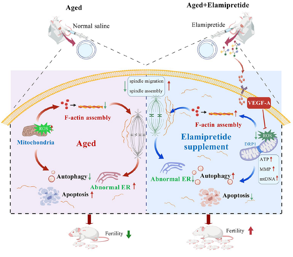
Why Male Mice Lose Anti-Cancer Immunity Earlier

A new study found that shrinking reserves of naive T cells weaken tumor recognition in male mice by middle age more than in females. Restoring thymic activity improved protection [1].

Do men lose anti-cancer defenses with age?

CD8+ T cells are immune cells that can kill infected or cancerous cells. However, an individual T cell cannot recognize every threat: its T cell receptor only recognizes particular antigens. Therefore, broad immune protection requires a diverse T cell population.

Naive T cells are those that have not yet been activated by their matching antigen. Their initial activation (priming) usually occurs in lymph nodes and depends on the appropriate T cell bumping into an antigen-presenting cell that displays its matching antigen.

This creates a problem: even if the body maintains a diverse collection of T cell receptors overall, a particular lymph node might contain too few cells to reliably include those able to recognize a nearby tumor. This might be one of the reasons why cancer incidence rises with age.

Interestingly, men have higher rates of many cancers, with the gap becoming more pronounced around middle age [2]. A new study from Massachusetts General Hospital, published in Nature Aging, investigates whether differences in how the immune system ages could help explain this disparity – in particular, whether T cell responses against tumors become less efficient in men.

Sex differences emerge

When the researchers implanted melanoma into young and middle-aged mice, tumor growth was similar between young males and females, but middle-aged males controlled it less effectively than middle-aged females did. Depleting CD8+ T cells accelerated tumor growth in both sexes, erasing the female advantage. This established that the difference depended on this particular type of immune cell.

Middle-aged female mice had more tumor-specific and activated CD8+ T cells within their tumors. The next question was where the difference in activation originated. Removing the spleen did not eliminate the female advantage, but using mice without functional lymph nodes did. This implicated lymph nodes as responsible for the stronger female response.

Female tumor-draining nodes, which receive fluid and antigens from the tumor’s location, contained more activated and tumor-specific CD8+ T cells, but there was no evidence of faster proliferation of activated cells. Antigen uptake and presentation by antigen-presenting cells also did not differ significantly between sexes. That pointed toward a different issue: the supply of T cells capable of recognizing the displayed antigen.

Counting specific cell populations indeed showed a substantial absolute loss of naive CD8+ T cells with age. Importantly, male mice started losing these cells earlier than females. Published human blood datasets that the researchers used also suggested an age-related, sex-biased depletion of naive CD8+ cells.

Human CT scans showed smaller lymph nodes in men. Mouse experiments revealed that lymph node size and cell numbers declined earlier in males. Interestingly, receptor diversity was high in both sexes, but a smaller node could retain high relative diversity while still having fewer distinct receptors in absolute terms.

Memory issues

The researchers also found an early male bias toward “virtual memory cells” [3]. When a naive T cell encounters its antigen, it undergoes rapid expansion, with some of its clones becoming cytotoxic CD8+ T cells and some becoming memory T cells. The latter linger in the body, ready to mount a fast immune response against the now familiar threat. Virtual memory cells resemble regular ones but arise from naive T cells that had never encountered an antigen that matches their receptor.

Naive T cells may be slower to respond because they require activation, but they maintain their ability to rapidly expand, while memory T cells gradually lose it with age. The paper’s overarching idea is that the male-specific bias towards naive T cells becoming virtual memory T cells begins to dampen anti-tumor responses later in life.

Indeed, the researchers found that virtual memory cells produced more immune cytokines (signaling molecules) after stimulation but proliferated less effectively. They also showed more senescence-associated gene expression.

The researchers treated young mice with an interleukin-15 (IL-15) complex. In this setting, this cytokine facilitates virtual memory formation. A brief treatment increased the number of virtual memory cells in young females but also caused them to have fewer naive CD8+ T cells seven months later. At this point, but not early after treatment, tumor control was significantly impaired.

The thymus and the testes

The team also investigated the role of the thymus, where T cells mature. With age, this crucial organ shrinks and turns into fat (thymic involution) [4], and this process is known to happen faster in males.

Human imaging and blood measurements linked poorer thymic preservation, which is more pronounced in men, with fewer naive CD8+ T cells. Markers identifying cells recently released from the thymus suggested a relationship between thymic output and the reserve of naive T cells.

The researchers then experimentally depleted immune cells in middle-aged mice. Females rebuilt their naive CD8+ population faster and had more cells that recently originated in the thymus. Thymus removal at six weeks of age eliminated the female advantage.

When challenged with melanoma in middle age, thymectomized females also lost their advantage in tumor control. This suggests that continued thymic function after early life contributes to later protection.

The final experiments asked whether the deficit could be reversed. Knowing that androgens, including testosterone, suppress thymic activity, the researchers reduced androgen signaling through surgical removal of the testes or treatment with degarelix, a drug that suppresses gonadal hormone production.

Androgen suppression increased thymic cellularity, repopulated lymph nodes, and restored naive CD8+ T cell numbers in middle-aged male mice. They had stronger tumor-specific responses and controlled melanoma more effectively, substantially reducing the sex difference.

We would like to ask you a small favor. We are a non-profit foundation, and unlike some other organizations, we have no shareholders and no products to sell you. All our news and educational content is free for everyone to read, but it does mean that we rely on the help of people like you. Every contribution, no matter if it’s big or small, supports independent ethical journalism and sustains our future.
Literature

[1] Menzel, L., Zschummel, M., O’Melia, M. J., Sandstedt, M., Zhou, H., Lei, P. J., … & Padera, T. P. (2026). Lymph node contraction links sex-biased naive CD8+ T cell decline to compromised antigen recognition during middle age Nature Aging, 1-17.

[2] Cook, M. B., Dawsey, S. M., Freedman, N. D., Inskip, P. D., Wichner, S. M., Quraishi, S. M., … & McGlynn, K. A. (2009). Sex disparities in cancer incidence by period and age Cancer Epidemiology Biomarkers & Prevention, 18(4), 1174-1182.

[3] White, J. T., Cross, E. W., Burchill, M. A., Danhorn, T., McCarter, M. D., Rosen, H. R., … & Kedl, R. M. (2016). Virtual memory T cells develop and mediate bystander protective immunity in an IL-15-dependent manner. Nature communications, 7(1), 11291.

[4] Araki, T., Nishino, M., Gao, W., Dupuis, J., Hunninghake, G. M., Murakami, T., … & Hatabu, H. (2016). Normal thymus in adults: appearance on CT and associations with age, sex, BMI and smoking. European radiology, 26(1), 15-24.

Rejuvenation Roundup October 2026

Rejuvenation Roundup October 2026

As the weather cools down, the race for functional interventions heats up, and members of our field have been looking for ways to push through bottlenecks both regulatory and scientific. Here’s what we’ve reported last month.

Interviews

Laura Minquini InterviewLaura Minquini on the Quest for Female Longevity: Laura Minquini is not a scientist, but that hasn’t stopped her from joining the front lines of the war on aging: specifically, female reproductive aging.

Advocacy and Analysis

FDA Leaders Name Longevity a Priority at ARDD: At a panel in Boston, senior officials announced plans to include aging in the agency’s regulatory science agenda and discussed how therapies could move toward approval.

Regulatory Bottleneck 1The Regulatory Bottleneck in Aging Biology: Researchers are developing biomedical repair therapies to address multiple age-related diseases at once, yet they are forced to put them through regulatory pathways built for single-disease drugs. The result is delay, duplication, and unnecessary cost precisely when speed matters the most.

ARDD 2026: Traditional Biology to AI-Driven Breakthroughs: One of the oldest and biggest longevity conferences moved this year across the Atlantic, from Copenhagen to Boston. The new location brought ARDD close to several important longevity hubs and might have contributed to this edition’s practical, entrepreneurial, and forward-oriented spirit.

Research Roundup

Brain and walkingThe Biomarkers That Predict Mortality Risk: Researchers have discovered key biomarkers that are strongly linked to all-cause mortality and identified four measurements with independent predictive power.

Declining Autophagy Impairs Senescent Cell Clearance: A new study links an age-related decline in cellular maintenance to impaired immune clearance. Activating the pathway reduced signs of senescence in aged mice.

Proteins or extracellular vesiclesUntangling Differences in Senescent Cellular Signaling: In Aging, researchers have described the differences between the secretions of cells driven senescent through a cancer gene and cells that stopped proliferating due to p16 expression.

Gut Bacteria Molecule Slows Atherosclerosis in Mice: A new study highlights a fatty acid produced by a particular genus of bacteria as a “natural statin,” suggesting a new way of treating atherosclerosis.

Inflamed lower back discsAn Anti-Senescence Gene May Fight Spinal Disc Deterioration: A team of researchers has described how a little-known gene, ZNF865, has a significant influence on back pain through the regulation of cellular senescence.

Low-Fat Vegan Diet Shows Benefits in Type 2 Diabetes: A human trial found that even without caloric restriction, a low-fat, plant-based diet improved blood sugar control, reduced liver fat, and lowered the quantity of advanced glycation end products (AGEs) in the skin.

Proteins in the brainMenopausal Serum Protein Changes May Predict Brain Health: A recent analysis of serum protein changes identified 16 proteins related to inflammatory, synaptic, metabolic, and Alzheimer’s disease processes whose levels change during the menopausal transition.

Making SIRT1 and NAD+ Therapies Work in People: Reviewers publishing in Biology have elucidated SIRT1 functional reserve as an aspect of aging, describing how its depletion is an upstream cause of age-related disorders at the cellular level and explaining why many prospective therapies that increase SIRT1 and NAD+ have not had promising results.

Bone structureBlocking STING Helps Preserve Bone in Aging Mice: A new study links STING-mediated signals from marrow fat cells to inflammation and declining NAD⁺ levels in bone-forming cells. Interrupting this pathway helped preserve aging bone.

An RNA Regulator Mitigates Senescence in Blood Vessels: Researchers publishing in Aging Cell have found that the regulatory protein MTR4 fights DNA damage in the blood vessel lining but decreases with aging.

CT Scan RobotAI Extracts Broader Health Insights From Routine CT Scans: A new study suggests that CT scans can provide information about a wide range of conditions, including some not typically diagnosed through imaging.

How Some Neuronal Cilia May Affect the Whole Body: A team of researchers has written a paper with a surprising finding: that many of the benefits of caloric restriction and increased NAD+ may be due to their effects on the cilia of hypothalamic neurons.

RNA DNA InteractionRejuvenation Factor Extends Lifespan and Healthspan in Mice: A recent study identified SRSF1, an RNA-processing and splicing factor, as a molecule with geroprotective potential.

An Astrocytic Factor Slows Whole-Body Aging in Mice: In Aging Cell, researchers have described how increasing fibroblast growth factor 13 (FGF13) in astrocytes reduces aging throughout the bodies of mice.

College for T CellsAn Anti-Senescence Vaccine Increases Mouse Lifespan: Late-life treatment with a three-element senolytic vaccine led to lower senescence markers and a 12% increase in median lifespan in female B6 mice.

Midlife time-restricted feeding preserves skeletal muscle insulin responsiveness and function during aging: Midlife time-restricted feeding preserved lean mass, muscle performance, glucose tolerance, and insulin-stimulated glucose uptake in male mice.

Vitamin D Deficiency, Supplementation, and Aging-Related Morbidity and Mortality Across Two Longitudinal Population Cohorts: These findings support severe vitamin D deficiency as an important and potentially modifiable determinant of systemic aging vulnerability and age-related multimorbidity in human populations.

The Effects of Blueberry Consumption on Cerebral Blood Flow and Brain Glutathione Levels in Older Adults: A Double-Blind Randomized, Placebo-Controlled Trial: Compared with placebo, blueberry consumption increased blood flow in the anterior cingulate cortex, a brain region relevant to age-related cognitive decline.

Resistance Training Improves Respiratory Muscle Strength and Immune Response in Older Adults: A Randomized Clinical Trial: The concomitant increase in circulating Klotho suggests that resistance training may promote biological mechanisms associated with healthy aging, supporting its implementation as a non-pharmacological strategy to preserve functional independence and respiratory health in the elderly.

When to Trust Epigenetic Clocks: Avoiding False Positives in Aging Interventions: These findings provide a framework for interpreting epigenetic clock results in anti-aging intervention studies, and suggest that some previously published single-clock findings may warrant reexamination.

Investigating the efficacy and toxicity of partial in vivo reprogramming as a rejuvenation intervention: These findings highlight tissue-specific responses to partial reprogramming and emphasize the need for larger, controlled studies to clarify physiological outcomes and safety.

H3K9me3 inhibition reverses Alzheimer’s progression by restoring synaptic and immune proteostasis across the brain–retina axis: This study identifies H3K9me3 accumulation as a shared, reversible epigenetic contributor to Alzheimer’s disease.

Synergistic reversal of mesenchymal stem cell senescence by engineered exosomal delivery of a dual miR-195/miR-188 sponge: Exosome-mediated delivery of a dual miRNA sponge represents a mechanism-driven strategy to reprogram senescence-associated gene networks and restore MSC function.

Chronic Senolytic Treatment and/or Aerobic Exercise Reduce Senescence and Improve Myocardial Function During Aging in the Heart: These findings demonstrate that long-term exercise or senolytics limit senescence at the cellular level within the myocardium and improve ventricular function.

Aged microenvironment-triggered nanoreactors rejuvenate senescent LepR+ cells to remodel aged bone marrow: Collectively, these findings suggest that the CLPM nanoreactor offers a promising approach for rejuvenation of the aging skeletal system.

Dynamic force sensing by bone delays aging and extends lifespan: This could potentially be manipulated to prevent aging, extend health, and aid those unable to exercise or with decreased mobility.

Elamipretide Rejuvenates Oocyte Quality and Restores Female Fertility During Reproductive Aging: Collectively, this study identifies a novel peptide therapeutic candidate for aging-related infertility, showing that elamipretide restores the quality of aged oocytes for fertility.

Heat-treated Lactiplantibacillus plantarum MYO Improves Muscle Strength and Modulates Gut Microbiota in Older Adults with Reduced Muscle Strength: This approach significantly improved muscle strength and muscle quality, and these muscle strength–enhancing effects may be associated with alterations in the gut microbiota.

The polycentric aging framework: epigenetic regulation as a dominant lifespan-limiting center in human aging: This paper holds that clock shifts should be interpreted against tissue-specific function, healthspan, morbidity, and mortality rather than as stand-alone evidence of rejuvenation.

Seven ways not to think carefully about aging: Accumulating data from mouse studies suggest that many or all varieties of slow-aging, long-lived mice, even as young adults, share multiple physiological and molecular traits whose effects may shed light on the pathogenesis of late-life disease, and whose origins may point to new anti-aging interventions.

Lysergic acid diethylamide extends lifespan in Caenorhabditis elegans: Together, these findings identify LSD as a tool to investigate how serotonergic signaling interfaces with conserved longevity-related pathways.

News Nuggets

Biomarkers of Aging ConsortiumGlobal Leaders in Geroscience Convene for Biomarkers of Aging: The Biomarkers of Aging Consortium has hosted its fourth annual Biomarkers of Aging Conference on October 5 and 6, 2026, at the Joseph B. Martin Conference Center at Harvard Medical School.

Evipedia Goes Hugging Face: Evipedia is a comprehensive, continuously updated online encyclopedia that provides much-needed, accurate, and up-to-date information on a wide range of health and longevity-related interventions.

Life BiosciencesLife Biosciences Announces First-in-Human Phase 1 Trial Data: Administration of ER-100 was well-tolerated through Day 56 in three participants with glaucoma, supporting dose escalation in the ongoing trial.

We would like to ask you a small favor. We are a non-profit foundation, and unlike some other organizations, we have no shareholders and no products to sell you. All our news and educational content is free for everyone to read, but it does mean that we rely on the help of people like you. Every contribution, no matter if it’s big or small, supports independent ethical journalism and sustains our future.

An Anti-Senescence Vaccine Increases Mouse Lifespan

In a new study, late-life treatment with a three-element senolytic vaccine led to lower senescence markers and a 12% increase in median lifespan in female B6 mice. However, short-lived controls complicate the picture [1].

Helping the immune system kill “zombie cells”

Cellular senescence is a double-edged sword. It plays important roles in early development, wound healing, and cancer surveillance, but when senescent cells are not cleared by the immune system and instead start to accumulate in tissues, they contribute to aging and disease.

To make the aging immune system more effective against senescent cells, scientists have experimented with anti-senescence (senolytic) vaccines [2]. The idea is simple: teach the immune system to recognize a protein associated with these cells and eliminate cells displaying it. Execution, however, has been trickier.

A new study from a group of Chinese scientists, published in Advanced Science, describes a senolytic vaccine consisting of three main elements that is aimed at dendritic cells, the immune cells that collect proteins, process them, and display fragments to T cells. The antigen is Gpnmb, a protein previously used in experimental senolytic vaccination. A special piece of CpG DNA acts as an adjuvant via the receptor TLR9, which recognizes this DNA as a microbial-like alarm signal and promotes T cell activation against the antigen. Finally, the CK5 protein scaffold packages the ingredients into nanoparticles that get absorbed by the cells.

The researchers assembled the three components into nanoparticles approximately 90 nanometers in diameter. They then generated dendritic cells from the bone marrow of young mice. Along with CK5, the team used a different scaffolding protein, K36.

Improved immune responses and senescence markers

Both nanoparticle vaccines improved antigen uptake and dendritic-cell activation compared with simpler formulations. Dendritic cells exposed to the nanoparticle vaccines stimulated greater CD8 T-cell proliferation. However, the CK5-based vaccine released Gpnmb into the dendritic cells’ cytosol more effectively than its K36-based counterpart.

This led the researchers to investigate CK5 separately. A gene expression analysis showed that treating dendritic cells with CK5 reduced expression of Aurora kinases A and B, proteins best known for regulating cell division.

Previous research has linked Aurora-kinase inhibition to reduced activity of PI3K/AKT signaling – a pathway associated with senescence [3]. This led the scientists to investigate whether CK5 can actually alleviate cellular senescence. To test the connection, they compared CK5 with K36 and danusertib, an Aurora-kinase inhibitor. D-galactose was used to produce senescence-like stress in cultured dendritic cells.

When the researchers compared the two vaccines, the CK5-based formulation reduced activation of PI3K/AKT and the senescence-associated markers p16, p21, and p53 while preserving cell viability. The treated cells also showed signs of improved metabolism: increased oxygen consumption and extracellular acidification.

The AI model AlphaFold3 predicted interactions between CK5 and each of the two Aurora kinases. However, this in itself does not prove the proposed mechanism.

A lifespan increase that comes with a caveat

The researchers first tested this vaccine’s health effects in mice fed a high-fat diet from four weeks of age, who were then vaccinated at 60 weeks. The CK5 vaccine elicited stronger responses from dendritic cells and CD8 T cells than the comparator groups, which included vaccines lacking one of the elements, and a K36-based vaccine. More CD8 T cells expressed interferon-γ and granzyme B, molecules associated with immune activation and cytotoxic capacity. The researchers then asked whether those T cells could effectively kill targets. Indeed, T cells collected from CK5-vaccinated mice killed more senescent Gpnmb-high cells in culture.

In the mice, vaccination reduced senescence markers in visceral fat and improved liver fat accumulation. The authors report approximately 50% lower senescence-associated β-galactosidase staining relative to the comparator groups (this experiment did not include untreated controls).

Importantly, glucose tolerance and insulin responses also improved. Body weight and food intake did not differ significantly between groups. This makes reduced eating or weight loss less likely explanations. Median lifespan reached 91 weeks, compared with 79-83 weeks in the comparator groups.

Then came the most important test: effect on lifespan in naturally aged mice. Normally fed mice were vaccinated at 75 weeks of age. The CK5 vaccine and the K36-plus-danusertib combination both strengthened immune responses, reduced senescence markers in fat tissue, and improved glucose regulation. Neither significantly changed food intake or body weight.

Mice that received the CK5 vaccine had a 12% longer median lifespan than untreated controls. This is a strong result considering how late in life the intervention started. However, two important caveats apply: the lifespan experiments were fairly small, with 10 mice per group, and the controls were relatively short-lived for this mouse strain (C57BL/6). Short-lived controls are a known issue in mouse lifespan experiments [4], although this does not necessarily invalidate the results. More rigorous studies are needed to test this intriguing proof of concept.

We would like to ask you a small favor. We are a non-profit foundation, and unlike some other organizations, we have no shareholders and no products to sell you. All our news and educational content is free for everyone to read, but it does mean that we rely on the help of people like you. Every contribution, no matter if it’s big or small, supports independent ethical journalism and sustains our future.
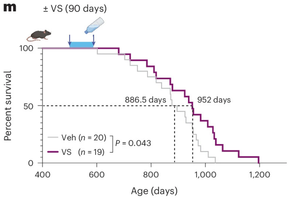
Literature

[1] Q. Song, X. Du, W. Dong, Y. Wang, X. Wang, and Q. Yin, A Bi-Adjuvant Senolytic Nanovaccine Modulating Dendritic Cell Senescence Ameliorates Aging-Related Metabolic Dysfunction. (2026). Advanced Science

[2] Wu, R., Sun, F., Zhang, W., Ren, J., & Liu, G. H. (2024). Targeting aging and age-related diseases with vaccines. Nature Aging, 4(4), 464-482.

[3] Astle, M. V., Hannan, K. M., Ng, P. Y., Lee, R. S., George, A. J., Hsu, A. K., … & Pearson, R. B. (2012). AKT induces senescence in human cells via mTORC1 and p53 in the absence of DNA damage: implications for targeting mTOR during malignancy. Oncogene, 31(15), 1949-1962.

[4] Pabis, K., Barardo, D., Gruber, J., Sirbu, O., Malavolta, M., Selvarajoo, K., … & Kennedy, B. K. (2024). The impact of short-lived controls on the interpretation of lifespan experiments and progress in geroscience–Through the lens of the “900-day rule”. Ageing Research Reviews, 101, 102512.

An Astrocytic Factor Slows Whole-Body Aging in Mice

In Aging Cell, researchers have described how increasing fibroblast growth factor 13 (FGF13) in astrocytes reduces aging throughout the bodies of mice.

A factor from a well-known family

FGFs are signaling molecules that share structural and functional commonalities and play crucial roles in biology, including neural function [1]. Some of these molecules are secreted, and others are not; one of the secreted molecules, FGF21, has been thoroughly researched as a regulator of aging processes.

However, the subject of this study, FGF13, is a non-secreted molecule with functions that are not related to the FGF receptor [2]. Previous work has found that it has beneficial effects in a mouse model of Parkinson’s [3], while its expression in endothelial cells is detrimental in a mouse model of diabetes [4]. Unlike FGF21, its role in age-related diseases has not been thoroughly explored.

Brain inflammation may spread to the body

The researchers first examined public mouse datasets and confirmed that FGF13 indeed decreases in the brains of mice; while it does not decrease in neurons, it decreases in other cells, including oligodendrocytes, astrocytes, and microglia. Interestingly, while FGF13 smoothly declined with age in the hippocampus, it increased in the cortex at 6 months of age before decreasing. While macaque data was less comprehensive, it was similar to that of mice, with a particularly strong effect in the astrocytes. Therefore, the researchers chose those cells for further experimentation.

Knocking out FGF13 in the astrocytes of aging mice exacerbated their aging. The affected mice performed even more poorly on the Morris water maze test than the aged control group did. Additionally, the affected mice had more cellular senescence in the heart, skin, liver, spleen, and kidney, even though the knockout was targeted to astrocytes, and they also had higher levels of circulating inflammatory cytokines. This led the researchers to conclude that these mice’s brains were “amplifying and transmitting aging signals to other tissues via the blood to accelerate systemic aging.”

The researchers then performed the opposite experiment, using a similar technique to increase FGF13 in the astrocytes of older mice instead. In the Morris water maze test, the treated mice performed similarly to young mice, having a substantial advantage over the untreated mice. In related spatial reasoning tests, the elevated plus maze test and the open field test, the treated mice also performed significantly better than the untreated mice, although not quite to the level of young mice.

Similar benefits were found in other tissues as well, despite this still being an astrocyte-specific intervention. Inflammatory cytokines in the bloodstream were substantially decreased, particularly IFN-γ and IL-6. Most other benefits were significant but modest, with decreases in skin, liver, spleen, and kidney senescence (although not in the heart). FGF13 overexpression in astrocytes also decreased age-related weight gain in mice.

The researchers then performed cellular experiments. They found that complement protein C3, which marks the presence of neurodegenerative diseases, is significantly increased in FGF13-deficient astrocytes. Cx43, which marks age-related neuronal injury, is increased with FGF13 deficiency. Co-culturing these deficient astrocytes with neurons found that they impaired the neurons’ synapses, and co-culturing them with microglia revealed that they induced the microglia to release inflammatory factors. FGF13 overexpression in neurons revealed benefits in all of these areas, restoring neuronal synaptic proteins, reducing microglial expression of cytokines, and reducing levels of proteins associated with injury and degeneration.

Autophagy may be the crux

RNA sequencing revealed some of the mechanisms of action behind these effects. 625 genes were upregulated and 195 were downregulated in astrocytes with the overexpression of FGF13. These genes affected multiple pathways, including lysosomal transport, inflammatory factor production, metabolism, and various signals.

FGF13 was specifically found to have a physical interaction with the protein Phafin2, a protein that is related to the cellular maintenance process of autophagy [5]. Like FGF13, Phafin2 decreases with age in mice, while increasing FGF13 also increases Phafin2. An analysis of the mice’s brain tissue found that the FGF13-overexpressing animals had increases in autophagy, as evidenced by increases in autophagosomes and autolysosomes along with the autophagy markers P62 and LC3. Similar results were found in cellular experiments.

These researchers have provided evidence that an upstream regulator in one particular brain cell has downstream ramifications throughout the body, suggesting that a treatment to increase FGF13 in the astrocytes of older people may improve multiple age-related conditions. However, this paper does not suggest any potential methods of doing so, and further work will need to be done to confirm these results and determine whether this approach is valid in non-human primates and then in people.

We would like to ask you a small favor. We are a non-profit foundation, and unlike some other organizations, we have no shareholders and no products to sell you. All our news and educational content is free for everyone to read, but it does mean that we rely on the help of people like you. Every contribution, no matter if it’s big or small, supports independent ethical journalism and sustains our future.

Literature

[1] Bollenbecker, S., Barnes, J. W., & Krick, S. (2023). Fibroblast growth factor signaling in development and disease. International Journal of Molecular Sciences, 24(11), 9734.


[2] Naderi, J., Johnson, A. K., Thakkar, H., Chandravanshi, B., Ksiazek, A., Anand, A., … & Chaurasia, B. (2025). Ceramide-induced FGF13 impairs systemic metabolic health. Cell metabolism, 37(5), 1206-1222.

[3] Song, N., Wang, X., Ha, L., Hu, L., Mei, S., Liang, Y., … & Lu, M. (2025). Neuronal FGF13 Inhibits Mitochondria‐Derived Damage Signals to Prevent Neuroinflammation and Neurodegeneration in a Mouse Model of Parkinson’s Disease. Advanced science, 12(28), 2503579.

[4] Sun, J., Guan, X., Niu, C., Chen, P., Li, Y., Wang, X., … & Cong, W. (2023). FGF13-Sensitive alteration of parkin safeguards mitochondrial homeostasis in endothelium of diabetic nephropathy. Diabetes, 72(1), 97-111.

[5] Tang, T. X., Jo, A., Deng, J., Ellena, J. F., Lazar, I. M., Davis, R. M., & Capelluto, D. G. (2017). Structural, thermodynamic, and phosphatidylinositol 3‐phosphate binding properties of Phafin2. Protein Science, 26(4), 814-823.